Abstract

Aminophenols can redox cycle through the corresponding quinone imines to generate ROS. The electrophilic quinone imine intermediate can react with protein thiols as a mechanism of immobilization in vivo. Here, we describe the previously unkown transimination of a quinone imine by lysine as an alternative anchoring mechanism. The redox properties of the condensation product remain largely unchanged because the only structural change to the redox nucleus is the addition of an alkyl substituent to the imine nitrogen. Transimination enables targeting of histone proteins since histones are lysine-rich but nearly devoid of cysteines. Consequently, quinone imines can be embedded in the nucleosome and may be expected to produce ROS in maximal proximity to the genome.
Aniline and its various ring-alkyl congeners are transformed in vivo principally to aminophenols through oxidative metabolism.1,2 This pathway is generally considered, at least with respect to carcinogenesis, as one of detoxification, but that assessment may represent a bias arising out of the understanding that N-, rather than ring-, hydroxylation of multicyclic aromatic amines is the key metabolic step in their activation to an ultimate carcinogenic form.3−5 Aminophenol formation is not necessarily the end point of oxidative metabolism if the substitution is ortho or para: it has recently been shown that these structures may be further transformed in vivo to quinone imines.6,7 Quinone imines are electrophiles and can undergo Michael addition with thiols and amines. They might, therefore, be capable of reacting with DNA nucleobases as a means of bringing about the mutagenic and carcinogenic effects observed with some alkylanilines such as 3,5-dimethylaniline,8 the focus of the present work. There is as yet no evidence for this mechanism of action.
In addition to their electrophilic character, 1,2- and 1,4-quinone imines are redox active structures that can cycle through the corresponding aminophenols to generate reactive oxygen species (ROS). As we have recently demonstrated with p-hydroxy-3,5-dimethylaniline (3,5-DMAP)9 and p-hydroxy-2,6-dimethylaniline (2,6-DMAP), aminophenols do generate ROS intracellularly.10 ROS generation in these experiments persisted for at least 24 h, indicating that the source was effectively immobilized within the cells. Immobilization could be achieved through Michael reaction with cellular thiols,11 as such products have been shown to retain their redox activity, but this is not the only mechanism available to quinone imines. They are also capable of undergoing transimination, as described previously,12,13 whereby a primary amine displaces ammonia from the quinone imine. We demonstrate here that this occurs in cells with lysine residues. Histones, which are virtually devoid of cysteine residues, were of particular interest because lysine transimination of histones places the redox-active structure in the closest possible location to oxidizable DNA nucleobases and creates a potentially potent mechanism for genetic damage by aminophenols that is distinct from covalent modification of the bases.
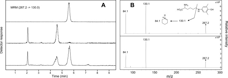
Synthesis of the lysyl transimination product of 3,5-dimethylquinone imine (3,5-DMQI) is shown in Scheme 1. 3,5-DMAP was synthesized from 2,6-dimethylphenol as described for the isomeric 2,6-DMAP.14 To make [ring-14C]-3,5-DMAP, [ring-14C]-2,6-dimethylphenol was obtained by decomposition of [ring-14C]-2,6-dimethylbenzenediazonium ion in H2O. Compound 1a was produced by reaction of 3,5-DMQI, formed transiently by PbO2 oxidation, with Nα-(t-Boc)lysine. Following the transimination reaction, product 1a was reduced with ascorbic acid to the more stable aminophenol form Nα-(t-Boc)-Nε-(4-hydroxy-3,5-dimethylphenyl)lysine (2a). With care to avoid oxidation, 2a could be isolated from the reaction mixture by preparative HPLC, and its structure was confirmed by 1D and 2D NMR and mass spectrometry. After deprotection, 2b could not similarly be isolated because its increased polarity did not permit separation from other reactants. Nevertheless, it could readily be detected and characterized by HPLC-MS (Figure 1A, upper trace, and Figure 1B, upper spectrum). Reaction of unprotected lysine with 3,5-DMQI apparently produced 1b since, after treatment with ascorbate, the product was indistinguishable from 2b by HPLC-MS.
Scheme 1. Synthesis of Nε-(4-Hydroxy-3,5-dimethylphenyl)-lysine.
Figure 1.
(A) HPLC-MS-MS analysis of synthetic 2b (top), isolate from histones in cells treated with 3,5-DMAP (bottom), and isolate from histones in untreated cells (middle). (B) CID spectra of synthetic 2b (top) and 2b isolated from 3,5-DMAP-treated cells (bottom).
Formation of Nε-(4-hydroxy-3,5-dimethylphenyl)-lysine (2b) by histones in vivo was investigated using Chinese hamster ovary AA8 cells grown in culture. Cells were treated with 25 μM 3,5-DMAP for 1 h, washed with fresh medium, and isolated by centrifugation. Histones were isolated and digested with protease in sodium acetate buffer containing 1 mM ascorbate to inhibit oxidation. After initial purification by semipreparative HPLC, 2b was analyzed by HPLC-MS using a triple-quad mass spectrometer. Data acquired in multiple reaction monitoring mode are shown in Figure 1A, which shows traces produced by synthetic 2b as well as isolates from control histones (not treated with 3,5-DMAP) and histones from treated cells. Figure 1A demonstrates cochromatography of histone isolate with synthetic 2b; Figure 1B demonstrates that the CID spectra are identical, confirming that the product isolated from histones is indeed the lysine transimination product.
As evidence that 2b originated from histone protein and not from any other protein that might have contaminated the histone preparation, cells were treated with [14C]-3,5-DMAP and the isolated histone fraction was subjected to HPLC for analysis of isotopic labeling. The chromatograpic separation described by Boyne et al. for top down MS characterization of histones15 was replicated for the present study, and the results are given in Figure 2 with identification of the histone peaks as given in the cited reference. In developing this separation, we confirmed that the indicated peaks had masses appropriate for histones but could not confirm the assignments made by Boyne because of the complexity associated with post-translational modifications. Quantitation of 14C was performed on collected fractions by accelerator mass spectrometry rather than decay counting to achieve the requisite sensitivity. Figure 2 reveals the correspondence of 14C concentration with histone fractions generally as well as two notable features that may be indicative of site-specificity in the labeling reaction. The arrow marked with an asterisk at 60.5 min indicates a region of the chromatogram where histone H2A.Z elutes. With the exception of one other fraction, this region of the chromatogram exhibits the highest levels of 14C. Histone H2A.Z appears to be the target of more extensive acetylation than other variants15 and, by implication, might be more highly targeted by 3,5-DMAP, thereby accounting for the higher degree of labeling relative to the more abundant histones. The second notable feature of Figure 2 is the 14C peak at 63.2 min that corresponds to a minor peak observed in the UV trace. The identity of this peak is unknown, so we can only speculate that it represents a hydroxydimethylphenylated histone variant. Alternatively, it might correspond to phenylated H4 whose retention on the HPLC column was increased by the modification.
Figure 2.
HPLC analysis of histones isolated from [14C]3,5-DMAP-treated cells showing UV detector response (continuous trace) and 14C concentration in collected fractions.
The transimination reaction described here, between a quinone imine and a primary amine, is not unknown but has received little attention. The analogous reaction between quinone imides, specifically, N-acetylated quinone imines, has been investigated with divergent results. 1,2-Fluorenoquinone-2-acetimide reacted with lysine in serum albumin via 1,4-addition.16 In contrast, N-acetyl-3,5-dimethyl-p-benzoquinone imine reacted with aniline to give the N-phenyl quinone imine, whereas the 2,6-isomer gave a tetrahedral adduct in which the acetamido group was retained.17 In the present work, we looked for and found no evidence by mass spectrometry for 1,4--addition of Nα-(t-Boc)lysine to 3,5-DMQI when forming 2a. 1,4-Addition is clearly not blocked by the methyl substituents, but it is directed toward the unsubstituted positions on the ring: we found that 3,5-DMQI reacts with N-acetylcysteine at the 2-position at much greater rates than the rate of transimination with Nα-(t-Boc)lysine, and similarly, N-acetyl-3,5-dimethyl-p-benzoquinone imine readily forms a Michael product with ethanethiol.17 In both instances, Michael addition takes place relative to the ketone, not the imine.
The biological consequences of quinone imine transimination may be considerable, especially insofar as they relate to hydroxyphenylation of the lysine side chains of the histones. As noted above, this reaction embeds a redox-active center in the nucleosome where it is positioned for maximum efficacy in creating genetic damage via ROS. Additionally, modification (acetylation and methylation) of certain lysine side chains is involved in epigenetic change. Their nonbiological modification through hydroxyphenylation may inhibit or replicate these changes inappropriately. The combination of two complementary modes of action may serve to endow certain monocyclic aminophenols with considerably more biological activity than has historically been attributed to them.
Acknowledgments
Radiocarbon measurements were made by Rosa G. Liberman in the MIT BEAMS laboratory. We thank Agilent Technologies for graciously providing access to the 6430 triple quadrupole mass spectrometer and the 1290 HPLC system.
Glossary
Abbreviations
- CID
collision-induced dissociation
- DMAP
dimethylaminophenol
- DMQI
dimethyl quinone imine
- MRM
multiple reaction monitoring
- ROS
reactive oxygen species
- t-Boc
tert-butyloxycarbonyl
Supporting Information Available
Experimental details and 1H NMR and mass spectral data identifying structure 2b. This material is available free of charge via the Internet at http://pubs.acs.org.
Author Present Address
§ Department of Bioscience and Technology, Chung Yuan Christian University, 200 Chung-Pei Road, Chungli, Taiwan 320.
This work was supported by NIEHS (PO1-ES006052 and P30-ES002109).
The authors declare no competing financial interest.
Funding Statement
National Institutes of Health, United States
Supplementary Material
References
- McCarthy D. J.; Waud W. R.; Struck R. F.; Hill D. L. (1985) Disposition and metabolism of aniline in Fischer 344 rats and C57BL/6 x C3H F1 mice. Cancer Res. 45, 174–180. [PubMed] [Google Scholar]
- Son O. S.; Everett D. W.; Fiala E. S. (1980) Metabolism of o-[methyl-14C]toluidine in the F344 rat. Xenobiotica 10, 457–468. [DOI] [PubMed] [Google Scholar]
- Delclos K. B., and Kadlubar F. F. (1997) Carcinogenic Aromatic Amines and Amides, in Comprehensive Toxicology (Bowden G. T., and Fischer S. M., Eds.) Vol. 12, Chemical Carcinogens and Anticarcinogens, pp 141–170, Elsevier Science, New York. [Google Scholar]
- Beland F. A., and Kadlubar F. F. (1990) Metabolic Activation and DNA Adducts of Aromatic Amines and Nitroaromatic Hydrocarbons, in Handbook of Experimental Pharmacology: Chemical Carcinogenesis and Mutagenesis (Cooper C. S., and Grover P. L., Eds.) Vol. 94/I, pp 267–325, Springer-Verlag, Berlin, Germany. [Google Scholar]
- Kadlubar F. F., and Beland F. A. (1985) Chemical Properties of Ultimate Carcinogenic Metabolites of Arylamines and Arylamides, in Polycyclic Hydrocarbons and Carcinogenesis (Harvey P. G., Ed.) ACS Symposium Series No. 283, pp 341–370, American Chemical Society, Washington, D.C. [Google Scholar]
- Jefferies P. R.; Quistad G. B.; Casida J. E. (1998) Dialkylquinonimines validated as in vivo metabolites of alachlor, acetochlor, and metolachlor herbicides in rats. Chem. Res. Toxicol. 11, 353–359. [DOI] [PubMed] [Google Scholar]
- Martínez-Cabot A.; Morató A.; Messeguer A. (2005) Synthesis and stability studies of the glutathione and N-acetylcysteine adducts of an iminoquinone reactive intermediate generated in the biotransformation of 3-(N-phenylamino)propane-1,2-diol: Implications for Toxic Oil Syndrome. Chem. Res. Toxicol. 18, 1721–1728. [DOI] [PubMed] [Google Scholar]
- Gan J.-P.; Skipper P. L.; Gago-Dominguez M.; Arakawa K.; Ross R. K.; Yu M. C.; Tannenbaum S. R. (2004) Alkylaniline–hemoglobin adducts and risk of non–smoking-related bladder cancer. J. Natl. Cancer Inst. 96, 1425–1431. [DOI] [PubMed] [Google Scholar]
- This nomenclature differs from the IUPAC system for the purpose of preserving the numbering used for the amines from which the aminophenols and quinone imines are derived.
- Chao M.-W.; Kim M. Y.; Ye W.; Ge J.; Trudel L. J.; Belanger C.; Skipper P.; Engelward B.; Tannenbaum S.; Wogan G. N. (2012) Genotoxicity of 2,6- and 3,5-dimethylaniline in cultured mammalian cells: the role of reactive oxygen species. Toxicol. Sci. 130, 48–59. [DOI] [PMC free article] [PubMed] [Google Scholar]
- Eyer P. (1994) Reactions of oxidatively activated arylamines with thiols: reaction mechanisms and biologic implications. An overview. Environ. Health Perspect. 102(6), 123–132. [DOI] [PMC free article] [PubMed] [Google Scholar]
- Yang Q.-Z.; Siri O.; Braunstein P. (2005) Tunable N-substitution in zwitterionic benzoquinonemonoimine derivatives: Metal coordination, tandemlike synthesis of zwitterionic metal complexes, and supramolecular structures. Chem.—Eur. J. 11, 7237–7246. [DOI] [PubMed] [Google Scholar]
- Lee Y.; Sayre L. M. (1995) Model reactions for the quinone-containing copper amine oxidases. Anaerobic reaction pathways and catalytic aerobic deamination of activated amines in buffered aqueous acetonitrile. J. Am. Chem. Soc. 117, 3096–3105. [Google Scholar]
- Gan J.; Skipper P. L.; Tannenbaum S. R. (2001) Oxidation of 2,6-dimethylaniline by recombinant human cytochrome P450s and human liver microsomes. Chem. Res. Toxicol. 14, 672–677. [DOI] [PubMed] [Google Scholar]
- Boyne M. T. II; Pesavento J. J.; Mizzen C. A.; Kelleher N. L. (2006) Precise characterization of human histones in the H2A gene family by top down mass spectrometry. J. Proteome Res. 5, 248–253. [DOI] [PubMed] [Google Scholar]
- Irving C. C.; Gutmann H. R. (1961) Protein binding of model quinone imides. III. Preparation of Ne-(1-hydroxy-2-acetamido-4-fluorenyl)-DL-lysine. J. Org. Chem. 26, 1859–1861. [Google Scholar]
- Fernando C. R.; Calder I. C.; Ham K. N. (1980) Studies on the mechanism of toxicity of acetaminophen. Synthesis and reactions of N-acetyl-2,6-dimethyl- and N-acetyl-3,5-dimethyl-p-benzoquinone imines. J. Med. Chem. 23, 1153–1158. [DOI] [PubMed] [Google Scholar]
Associated Data
This section collects any data citations, data availability statements, or supplementary materials included in this article.


