Abstract
Myeloid-derived suppressor cells (MDSCs) are important regulators of immune responses. These cells suppress the cytotoxic activities of natural killer (NK)-cell and T-cell effectors and promote tumor growth. We demonstrated that depleting MDSCs improve therapeutic responses to vaccination in a murine model of lung cancer. This approach may prove beneficial against tumors in which MDSC exert prominent immunosuppressive effects.
Keywords: immune suppression, immunotherapy, lung cancer, myeloid-derived suppressor cells
Immunotherapeutic approaches to lung cancer have potential. However, despite the identification of a large repertoire of tumor-associated antigens, hurdles persist that hinder the development of immunomodulatory anticancer therapies. More effective immunotherapeutic strategies will likely result from an improved understanding of the mechanisms that sustain tumor growth. Addressing the multiple immune deficits that are frequently exhibited by lung cancer patients is likely to be required for the implementation of effective immunotherapy. Indeed, the activation of the immune system alone is most often unable to mediate tumor eradication because of potent mechanisms of tumor-induced immunosuppression. Combined therapies boosting the immune system and reversing the mechanisms of immunosuppression ultimately will prove beneficial in our fight against lung cancer.
Tumor progression and local tissue invasion cause the persistence of inflammatory cellular tumor infiltrates (ICTIs). The tumor causes the cellular infiltrates to sustain dysregulated inflammation, which is immunologically unresponsive to the tumor. A significant proportion of the ICTI are myeloid-derived suppressor cells that maintain an immune suppressive environment1-3 and support tumor expansion in the surrounding tissue as well as at distant sites, by facilitating angiogenesis and metastases.4 In humans, MDSCs are identified by the surface expression of CD33, and by the lack of expression of markers that characterize mature myeloid and lymphoid cells. Thus, MDSCs typically are CD11b+CD33+CD34+CD14- cells that express to variable extents CD15, CD124, CD66 and MHC class II molecules.5 Melanoma MDSCs express CD14, CD11b and (optionally) low levels of HLA-DR,6 while CD11b+CD14+-CD15+CD33+ MDSCs with immune suppressive properties have been described in non-small cell lung carcinoma (NSCLC) patients.7 In mice, MDSCs are identified by the expression at the cell surface of Gr1 and CD11b, and the Gr1+ cell population can be further sub-categorized based on the expression of Ly6C and Ly6G. Thus, CD11b+Ly6G+Ly6Clow cells exhibiting multi-lobed nuclei and an overall granulocytic-like morphology are called granulocytic MDSCs, while CD11b+Ly6G-Ly6Chigh cells are referred to as monocytic MDSCs.4
In our recent work published in PLoS ONE,8 we investigated the contribution of MDSCs to the growth of and the therapeutic responses to weakly immunogenic murine 3LL Lewis lung cancers in C57BL/6 mice. As 3LL tumors progressed, the frequency and activity of MDSCs increased, both in the tumor and systemically, that is in the blood, spleen and bone marrow. Increased MDSC activity led to decreased intratumoral activity of antigen-presenting cell (APCs), natural killer (NK) and T cells. The generalized depletion of MDSCs, as obtained with anti-Gr1 or anti-Ly6G antibodies, restored APC, NK and T-cell activities. In line with the reactivation of immune cell effectors, we observed an increase in the frequency of apoptotic tumor cells and a concomitant reduction in tumor burden as well as in the migration of tumor cells from the primary tumor site to the lung. Following MDSC depletion, the levels of the anti-angiogenic chemokines CXCL9 and CXCL10 increased and those of the pro-angiogenic cytokines vascular endothelial growth factor α (VEGFα), angiopoietin 1and 2, CXCL2 and CXCL5 were markedly reduced. Accompanying this signature, we observed a reduction in the endothelial marker MECA32 in the tumor and an increase in the expression of T-cell activation marker CXCR3. MDSC depletion led to an influx in the tumor of interferon γ (IFNγ)-producing activated T and NK cells that promoted angiostasis by altering the balance of pro- and anti-angiogenic chemokines. This suggests that MDSC depletion not only improves APC, NK and T-cell immune activities but also promotes angiostasis leading to a more efficient control of tumor growth.
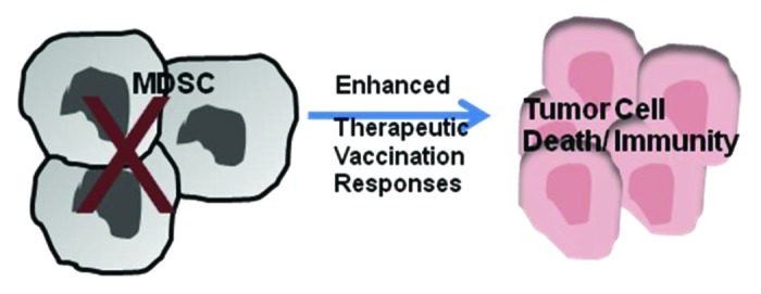
We next evaluated whether MDSC depletion would improve therapeutic vaccination responses against lung cancer. To initiate antigen-specific responses, a cellular vaccine consisting of bone marrow adherent (BMAs) cells pulsed the model antigen ovalbumin (OVA, produced by genetically modified 3LL cells) was employed. BMA cells were pulsed with the OVA antigen to allow for antigen processing and presentation and then injected s.c. on the flank contra-lateral to an established tumor, to initiate antigen-specific antitumor immune responses. Such a therapeutic vaccination decreased tumor burden but failed to completely eradicate established tumors. Conversely, the depletion of MDSCs in combination with the vaccine led to a 20-fold reduction in tumor burden in 50% of the mice and to complete tumor eradication in remaining animals. Immunological memory was induced in mice that had rejected tumors in response to the combination therapy, and these mice efficiently rejected a subsequent challenge with tumor cells of the same type. These findings reveal that an effective attack against established tumors necessitates changes in multiple components of the immune system, leading in parallel to immune cell activation and to the disruption of regulatory mechanisms that would limit antitumor immune responses. MDSCs play a major role in the suppression of T-cell activation and sustain tumor growth, proliferation and metastasis. Depletion of MDSCs reprograms the tumor niche by altering inflammatory responses, thus allowing for the elicitation of antitumor immune response as well as for the generation of immunological memory. Targeting MDSC function will be useful to control cancer growth and may be more efficient in combination with other immunomodulatory strategies. Combined approaches that downregulate MDSC-related signaling pathways, restore the activity of APCs and expand tumor-reactive T cells may be useful to improve immunomodulatory therapies against lung cancer.

Figure 1. Myeloid-derived suppressor cell (MDSC) depletion promotes therapeutic vaccination responses. MDSCs recruited and expanded in the tumor suppress natural killer (NK) and T-cell cytolytic activity. MDSC depletion enhances therapeutic vaccination responses leading to lung cancer cell death and immunological memory.
Footnotes
Previously published online: www.landesbioscience.com/journals/oncoimmunology/article/21970
References
- 1.Young MR, Wright MA, Pandit R. Myeloid differentiation treatment to diminish the presence of immune-suppressive CD34+ cells within human head and neck squamous cell carcinomas. J Immunol. 1997;159:990–6. [PubMed] [Google Scholar]
- 2.Kusmartsev SA, Ogreba VI. [Suppressor activity of bone marrow and spleen cells in C57Bl/6 mice during carcinogenesis induced by 7,12-dimethylbenz(a)anthracene] Eksp Onkol. 1989;11:23–6. [PubMed] [Google Scholar]
- 3.Kusmartsev SA, Kusmartseva IN, Afanasyev SG, Cherdyntseva NV. Immunosuppressive cells in bone marrow of patients with stomach cancer. Adv Exp Med Biol. 1998;451:189–94. doi: 10.1007/978-1-4615-5357-1_30. [DOI] [PubMed] [Google Scholar]
- 4.Condamine T, Gabrilovich DI. Molecular mechanisms regulating myeloid-derived suppressor cell differentiation and function. Trends Immunol. 2011;32:19–25. doi: 10.1016/j.it.2010.10.002. [DOI] [PMC free article] [PubMed] [Google Scholar]
- 5.Zea AH, Rodriguez PC, Atkins MB, Hernandez C, Signoretti S, Zabaleta J, et al. Arginase-producing myeloid suppressor cells in renal cell carcinoma patients: a mechanism of tumor evasion. Cancer Res. 2005;65:3044–8. doi: 10.1158/0008-5472.CAN-04-4505. [DOI] [PubMed] [Google Scholar]
- 6.Poschke I, Mougiakakos D, Hansson J, Masucci GV, Kiessling R. Immature immunosuppressive CD14+HLA-DR-/low cells in melanoma patients are Stat3hi and overexpress CD80, CD83, and DC-sign. Cancer Res. 2010;70:4335–45. doi: 10.1158/0008-5472.CAN-09-3767. [DOI] [PubMed] [Google Scholar]
- 7.Srivastava MK, Bosch JJ, Thompson JA, Ksander BR, Edelman MJ, Ostrand-Rosenberg S. Lung cancer patients’ CD4(+) T cells are activated in vitro by MHC II cell-based vaccines despite the presence of myeloid-derived suppressor cells. Cancer Immunol Immunother. 2008;57:1493–504. doi: 10.1007/s00262-008-0490-9. [DOI] [PMC free article] [PubMed] [Google Scholar]
- 8.Srivastava MK, Zhu L, Harris-White M, Kar U, Huang M, Johnson MF, et al. Myeloid suppressor cell depletion augments antitumor activity in lung cancer. PLoS One. 2012;7:e40677. doi: 10.1371/journal.pone.0040677. [DOI] [PMC free article] [PubMed] [Google Scholar]


