Figure 7.
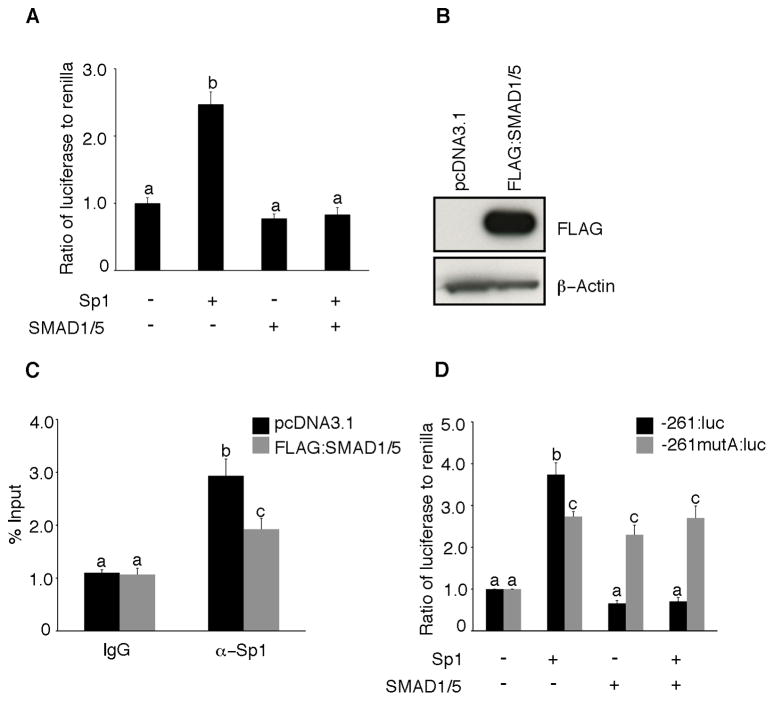
SMAD1/5 expression antagonizes Sp1 induction of PDGFA promoter. (A) COV434 cells were transiently co-transfected with 1 μg of -881:luc of the human PDGFA promoter and 0.5 μg of Sp1 and SMAD1/5 along with the renilla luciferase control plasmid (pRL-TK). Forty-eight hours after transfection, the cells were lysed and assessed for luciferase activity. Data are presented as the ratio of firefly luciferase to renilla. (B) Representative Western blot of whole cell lysates from COV434 cells transfected with either pcDNA3.1 or Flag-tagged SMAD1/5 expression plasmids and blotted with mouse anti-FLAG M2 antibody and mouse anti-β-actin antibody (loading control). (C) ChIP analysis in COV434 cells transfected with pcDNA3.1 or Flag-tagged SMAD1/5 expression plasmids. Chromatin cross-linked protein DNA complexes were immunoprecipitated with either anti-Sp1 antibody or with non-specific IgG and the PDGF-A promoter amplified by real-time PCR using locus specific primers. (D) SMAD1/5 represses the wild-type PDGFA promoter (-261:luc) but not the promoter bearing a mutation in the SMAD1/5 binding site (-261mutA: luc). COV434 cells were transiently co-transfected with 1 μg of -261:luc (control) or -261mutA: luc (mutant) and 0.5 μg of Sp1 and SMAD1/5 along with the renilla luciferase control plasmid (pRL-TK). Forty-eight hours after transfection, the cells were lysed and assessed for luciferase activity. Data are presented as the ratio of firefly luciferase to renilla. Different letters above the bars indicate statistically different means by ANOVA and post hoc analysis (n=4; P< 0.05).

