Abstract
Pancreatic ductal adenocarcinoma is believed to arise from precursor lesions termed pancreatic intraepithelial neoplasia (PanIN). Mouse models have demonstrated that targeted expression of activated K-ras to mature acinar cells in the pancreas induces the spontaneous development of PanIN lesions; implying acinar-to-ductal metaplasia (ADM) is a key event in this process. Recent studies suggest Notch signaling is a key regulator of ADM. To assess if Notch1 is required for K-ras driven ADM we employed both an in vivo mouse model and in vitro explant culture system, in which an oncogenic allele of K-ras is activated and Notch1 is deleted simultaneously in acinar cells. Our results demonstrate that oncogenic K-ras is sufficient to drive ADM both in vitro and in vivo but that loss of Notch1 has a minimal effect on this process. Interestingly, while loss of Notch1 in vivo does not affect the severity of PanIN lesions observed, the overall numbers of lesions were greater in mice with deleted Notch1. This suggests Notch1 deletion renders acinar cells more susceptible to formation of K-ras-induced PanINs.
Introduction
Pancreatic ductal adenocarcinoma (PDAC) is one of the most aggressive forms of human cancer, with a 5-year survival rate of less than 4% [1]. PDAC is believed to arise from precursor lesions termed pancreatic intraepithelial neoplasia (PanIN), which progress through defined stages ultimately leading to the development of adenocarcinoma [2]. The most commonly mutated gene in PDAC is K-ras, with greater than 90% of human cases harboring an activating mutation in this oncogene. K-ras mutations appear to occur early during the pathogenesis of PDAC, as low-grade PanIN lesions typically contain activating mutations at codon 12 [2]. Further proof that K-ras mutations represent an initiating event in PDAC comes from mouse models, in which expression of a mutant activated K-ras allele (K-rasG12D) in pancreatic epithelium is sufficient to induce the formation of both PanIN lesions and invasive pancreatic cancer, pathologically resembling the human disease [3].
The pancreas is composed of an exocrine and endocrine compartment, with the exocrine compartment consisting of acinar, ductal, and centroacinar cells. While the cell of origin for PDAC has remained elusive, recent studies utilizing mouse models have demonstrated that targeting oncogenic K-ras to mature acinar cells results in the spontaneous development of PanIN lesions, suggesting acinar cells represent the cell of origin for PDAC [4]. A feature of this model is the appearance of acinar-to-ductal metaplasia (ADM) preceding the development of PanIN lesions. Other studies have highlighted the importance of pancreatic injury in the development of PDAC. Work by Guerra and colleagues revealed that mature acinar cells expressing K-rasG12V are refractory to PanIN development unless mice are subjected to additional stimuli such as chronic chemically-induced pancreatitis [5]. Further, endocrine cells can be made susceptible to oncogenic K-ras induced transformation in the context of pancreatic injury [6]. These findings are especially relevant to human disease, in that chronic pancreatitis is a strong risk factor for the development of PDAC [7].
The Notch signaling family of proteins is composed of 4 transmembrane receptors (Notch1–4), in addition to 2 Jagged ligands, and 3 Delta-like ligands. During pancreatic development, Notch signaling is required for directing cell fate decisions and progenitor cell self renewal [8]. While the role of Notch signaling in development is well characterized, the cell types expressing Notch proteins and their function in the adult pancreas remains unclear. Recent findings indicate Notch1 plays a role in pancreatic homeostasis, since loss of Notch1 in pancreatic epithelium results in impaired acinar regeneration following acute pancreatitis [9]. Moreover, Notch signaling has been implicated in ADM in that ectopic expression of transcriptionally active forms of Notch (Nic) promote transdifferentiation in explant culture models [10]–[11]. Conversely, inhibition of Notch signaling by a γ-secretase inhibitor increases the proliferation of metaplastic exocrine cells and induces p21 expression [12]. Further work demonstrates different Notch receptors have non-overlapping functions and are expressed in unique cellular compartments, with Notch1 observed primarily in acinar cells and Notch2 expressed mainly in ductal cells [13].
Although Notch1 was originally identified as an oncogene, recent evidence indicates the Notch proteins also function as tumor suppressors in a tissue-specific manner. Conclusive evidence demonstrating Notch1 acts as a tumor suppressor came from studies in the skin, where loss of both Notch1 alleles led to development of basal cell carcinoma [14]. Subsequently, Notch receptors have been identified as tumor suppressors in hepatocellular carcinoma, chronic myelomonocytic leukaemia, and squamous cell carcinomas [15], [16], [17], [18]. Previously unknown loss of function mutations in components of the Notch pathway have been discovered in myeloid leukaemia and squamous cell carcinomas, pointing to a cell autonomous mechanism of tumor suppression for these malignancies. Alternatively, in basal cell carcinoma, Notch1 appears to function in a non-cell autonomous manner by mechanisms impacting the tumor microenvironment [19].
Previous work by our group has identified Notch1 as a tumor suppressor in a mouse model of PDAC [20]. To further investigate the mechanism of Notch1 mediated tumor suppression in pancreatic tumorigenesis we examined the effect of Notch1 deletion on acinar-to-ductal metaplasia both in vitro and in vivo. These experiments aimed to identify a cell autonomous mechanism of Notch1 mediated tumor suppression. Additionally, we investigated a potential non-cell autonomous function of Notch1 using an orthotopic transplantation tumor model.
Results
K-ras Mediated ADM does not Require Notch1 Function
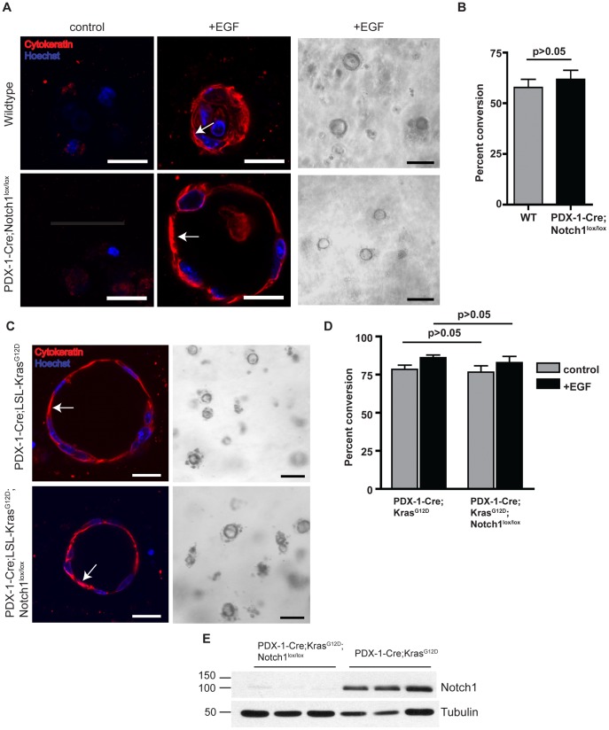
We have recently demonstrated that loss of Notch1 in a mouse model of K-ras-induced PDAC leads to increased PanIN incidence and progression [20]. To further investigate the mechanism of Notch1 mediated PanIN suppression, we examined the role of Notch1 in ADM using an in vitro explant culture model. Acinar cell clusters isolated from an adult mouse pancreas transdifferentiate to form cytokeratin-19 positive ductal cysts when embedded in a collagen matrix and treated with growth factors such as EGF or TGF-α [21]. In order to examine the effect of Notch1 deletion on cyst formation, we utilized PDX-1-Cre;Notch1lox/lox mice, which allow for conditional deletion of Notch1lox/lox alleles specifically in pancreatic epithelial cells at day 8.5 of embryonic development [22]. Acinar cells isolated from PDX-1-Cre;Notch1lox/lox mice formed ductal cysts in the presence of EGF at comparable rates to wildtype acinar cells (Figure 1A and 1B). The acinar origin of the isolated cells was verified by immunostaining for the acinar marker, amylase (Figure S1). These results indicate loss of Notch1 does not accelerate ADM and that Notch1 is not required for EGF-induced ADM in vitro.
Figure 1. Notch1 is not required for oncogenic K-ras mediated ADM in vitro.
(A) Pancreatic explants from wildtype and PDX-1-Cre;Notch1lox/lox mice embedded in collagen either untreated (control) or treated with EGF (20 µg/mL). Cells are immunostained for expression of pan-cytokeratin (red) at day 5. Nuclei are stained with Hoechst dye (blue). Scale bar, 20 µm. Arrows indicate cytokeratin-positive ductal cells. Representative brightfield images are shown at day 5 in the presence of EGF. Scale bar, 100 µm. (B) Quantitative analysis of percent ductal cyst conversion on day 5 in explants isolated from wildtype and PDX-1-Cre;Notch1lox/lox mice. n = 3 for each group. (C) Pancreatic explants from PDX-1-Cre;LSL-KrasG12D and PDX-1-Cre;LSL-KrasG12D;Notch1lox/lox mice were isolated at day 2 in the absence of EGF. Cells are immunostained for pan-cytokeratin (red) and Hoechst dye (blue). Scale bar, 20 µm. Arrows indicate cytokeratin-positive ductal cells. Representative brightfield images of cyst formation are shown. Scale bar, 100 µm. (D) Quantitative analysis of percent ductal cyst conversion at day 2 in explants isolated from PDX-1-Cre;LSL-KrasG12D (n = 5) and PDX-1-Cre;LSL-KrasG12D;Notch1lox/lox mice (n = 6). (E) Western blot analysis of Notch1 expression in acinar cells isolated from PDX-1-Cre;LSL-KrasG12D and PDX-1-Cre;LSL-KrasG12D;Notch1lox/lox mice; tubulin as loading control. Three samples are shown for each genotype.
We next examined whether loss of Notch1 in the context of activated K-ras accelerates acinar-to-ductal conversion in vitro. Recently it has been shown that K-rasG12D expression is sufficient to induce pancreatic ADM in explant cultures in the absence of exogenous growth factors [23]. Acinar cells were isolated from PDX-1-Cre;LSL-KrasG12D and PDX-1-Cre;LSL-KrasG12D;Notch1lox/lox mice. As early as 2 days after isolation, both PDX-1-Cre;LSL-KrasG12D and PDX-1-Cre;LSL-KrasG12D;Notch1lox/lox acinar explants underwent transdifferentiation to form cytokeratin positive ductal cysts in the absence of external growth factors (Figure 1C), confirming that oncogenic Kras expression is sufficient to drive ADM. Wild type acinar explants failed to undergo transdifferentiation at day 2 even in the presence of EGF (data not shown). Greater than 75% of PDX-1-Cre;LSL-KrasG12D and PDX-1-Cre;LSL-KrasG12D;Notch1lox/lox acinar explants underwent ADM conversion at day 2. Addition of EGF did not significantly increase rates of conversion in either PDX-1-Cre;LSL-KrasG12D or PDX-1-Cre;LSL-KrasG12D;Notch1lox/lox explants (Figure 1D).
Previous studies utilizing PDX-1-Cre;ROSA26R-LacZ mice have revealed a mosaic recombination pattern in the pancreas [13]. Thus, to confirm that our results do not reflect variations in recombination efficiency of the Notch1lox/lox allele, we assessed the expression of Notch1 in the explants. Western blot analysis confirmed Notch1 expression in acinar cells isolated from PDX-1-Cre;LSL-KrasG12D mice and Notch1 deletion in cells isolated from PDX-1-Cre;LSL-KrasG12D;Notch1lox/lox mice (Figure 1E) and reflect our previous findings regarding the recombination of the Notch1lox/lox in vivo [20]. Further, rates of ductal cyst formation are very similar between the different samples, indicating similar rates of recombination and activation of the LSL-KrasG12D allele. Therefore, despite the possibility of mosaic Cre expression, the variability likely does not affect the interpretation of our results.
These results further demonstrate that K-rasG12D expression is sufficient to drive ADM in vitro and that Notch1 is not required for ADM in this context.
The Role of Notch Pathway Signaling in K-ras Induced Acinar-to-ductal Metaplasia
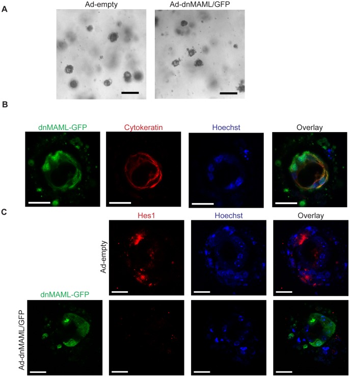
The Notch receptor family contains four members, Notch1–4. Given previous reports suggesting functional overlap between the different receptors, we investigated the effect of inhibiting multiple members of the Notch receptor family simultaneously on K-ras induced ADM in vitro. Notch signaling requires the transcriptional co-activator proteins Mastermind-like (MAML1–3) for transcription of downstream target genes. When overexpressed, a truncated form of MAML1 (aa 13–74) acts as a potent dominant-negative mutant, inhibiting Notch-mediated transcriptional activation [24]. We isolated acinar cells from PDX-1-Cre;LSL-KrasG12D mice and infected them with an adenovirus expressing DNMAML1 fused to GFP. Even in the presence of DNMAML1-GFP, PDX-1-Cre;LSL-KrasG12D acinar cells maintained the ability to transdifferentiate to cytokeratin positive ductal cysts at day 2, similar to cells expressing a control adenovirus (Figure 2A–B). In order to verify that Notch signaling is inhibited in the presence of DNMAML1, we analyzed expression of Hes1, a downstream effector of Notch signaling. Acinar cells infected with the control adenovirus express Hes1; however, Hes1 expression is reduced when cells are infected with Ad-DNMAML1-GFP (Figure 2C). To further examine the effect of globally inhibiting Notch signaling, we analyzed the effect of a gamma secretase inhibitor, DAPT, on acinar transdifferentiation. PDX-1-Cre;LSL-KrasG12D acinar cells treated with DAPT maintained the ability to form ctyokeratin positive ductal cysts (Figure S2). Overall, these results indicate that in the presence of oncogenic K-ras, Notch pathway signaling is not required for ADM in vitro.
Figure 2. DNMAML expression does not inhibit oncogenic K-ras mediated ADM in vitro.
(A) Pancreatic explants from Pdx1-Cre;LSL-KrasG12D mice at Day 2, infected with control adenovirus (Ad-empty) or Adenovirus expressing DNMAML (Ad-dnMAML/GFP). Representative brightfield images of ductal cysts shown. Scale bar, 100 µm. (B) Pdx1-Cre;LSL-KrasG12D acinar explants expressing DNMAML-GFP form cytokeratin positive cysts. Explants were stained with antibodies against GFP (green) and pan-cytokeratin (red). Nuclei are counterstained with Hoechst (blue). Images are representative of 3 separate experiments. Scale bar, 20 µm. (C) DNMAML expression inhibits Notch signaling in Pdx1-Cre;LSL-KrasG12D acinar explants. Explants were stained with antibodies against GFP (green) and Hes1 (red). Nuclei are counterstained with Hoechst (blue). Scale bar, 20 µm.
Deletion of Notch1 does not Accelerate K-ras Induced PanIN Development
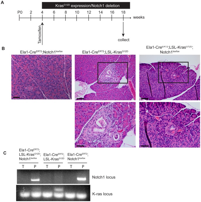
In order to investigate the role of Notch1 in acinar-to-ductal metaplasia in vivo, we utilized an Elastase1-CreERT2-driven mouse model. This transgene permits tamoxifen-inducible Cre activation specifically in adult acinar tissue [25]. Activation of oncogenic Kras in the mature acinar compartment results in the spontaneous development of PanIN lesions [4]. Extensive ADM was observed preceding the onset of PanIN lesions in this model, implying acinar cells might represent the cell of origin for PanIN development. Elastase1-CreERT2;LSL-KrasG12D and Elastase1-CreERT2;LSL-KrasG12D;Notch1lox/lox mice were generated, and treated with tamoxifen at 4 weeks of age (Figure 3A). Additionally, Elastase1-CreERT2;Notch1lox/lox mice were generated to establish the effect of Notch1 deletion on adult acinar tissue. At 3 months following tamoxifen treatment, pancreas tissue from Elastase1-CreERT2;Notch1lox/lox mice appeared grossly normal (Figure 3B). In contrast, pancreas tissue from both Elastase1-CreERT2;LSL-KrasG12D and Elastase1-CreERT2;LSL-KrasG12D;Notch1lox/lox demonstrated mild to moderate fibroplasia and fibrosis. Additionally, low-grade PanIN1A lesions were observed in 1 of 6 Elastase1-CreERT2;LSL-KrasG12D mice and 1 of 7 Elastase1-CreERT2;LSL-KrasG12D;Notch1lox/lox mice (Figure 3B, Figure S3A). Analysis by PCR demonstrated recombination at both the Kras and Notch1 gene loci in the pancerata of these mice (Figure 3C). These results demonstrate that in the context of activated Kras, deletion of Notch1 in adult acinar tissue does not accelerate spontaneous PanIN development. Additionally, the results suggest that Notch1 is not required for K-ras induced PanIN development, as an Elastase1-CreERT2;LSL-KrasG12D;Notch1lox/lox mouse developed a PanIN1A lesion. However further studies, with larger animal cohorts, are required to conclusively establish this point and to determine whether Notch1 deletion renders acinar cells more susceptible to K-ras induced transformation, in line with previous studies [20].
Figure 3. Notch1 deletion in mature acinar cells does not accelerate spontaneous PanIN development.
(A) Schematic of experimental design. Mice were treated with tamoxifen at 4 weeks of age to activate K-rasG12D and delete Notch1 expression. Pancreatic tissue was collected 3 months later. (B) Histological analysis of pancreas tissue from Elastase1-CreERT2;Notch1lox/lox, Elastase1-CreERT2;KrasG12D, and Elastase1-CreERT2;KrasG12D;Notch1lox/lox mice. Higher magnifications of the boxed areas are seen below the images. Scale bar for top images, 100 µm. Scale bar for higher magnification, 50 µm. (C) PCR analysis of Cre-mediated recombination of the LSL-KrasG12D and Notch1lox/lox loci. Genomic DNA was isolated from the tail (T) and pancreas (P) of each mouse. For the Notch1 PCR, the presence of a band indicates deletion of the floxed Notch1 gene. For the Kras PCR, the larger band represents deletion of the STOP cassette.
Notch1 Deletion does not Accelerate PanIN Development Following Acute Pancreatitis but Renders Cells More Susceptible to Formation of K-ras-induced PanINs
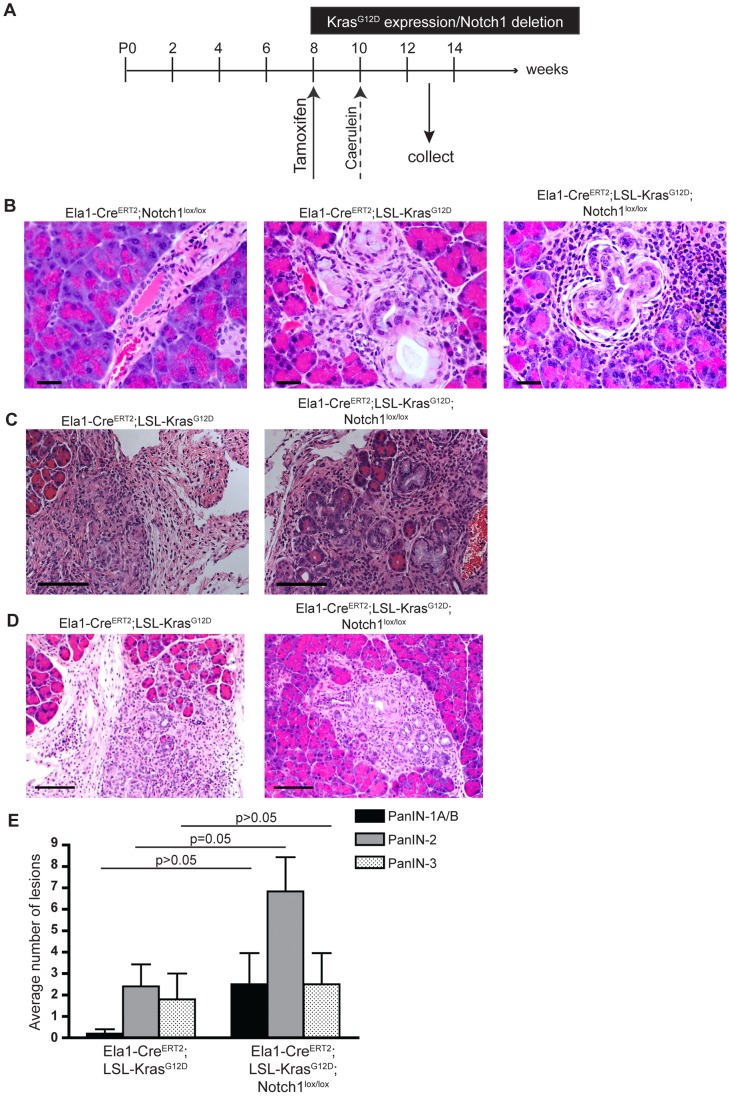
As our results demonstrated a comparatively mild phenotype in Elastase1-CreERT2;LSL-KrasG12D and Elastase1-CreERT2;LSL-KrasG12D;Notch1lox/lox mice, we proceeded to investigate the effect of caerulein-induced pancreatitis. Previous studies have demonstrated that oncogenic K-ras expression in adult acinar cells is sufficient to drive PanIN formation [4], [26], yet the process is highly inefficient. Moreover, similar mouse models have revealed that adult acinar cells are refractory to PanIN development unless mice are subjected to caerulein-induced pancreatitis [5]. Therefore, we compared pathology between Elastase1-CreERT2;Notch1lox/lox, Elastase1-CreERT2;LSL-K-rasG12D and Elastase1-CreERT2;LSL-K-rasG12D;Notch1lox/lox mice following tamoxifen induction and acute pancreatitis (Figure 4A). Three weeks following caerulein treatment, the pancreas from Elastase1-CreERT2;Notch1lox/lox mice appeared grossly normal (Figure 4B). Similar to previous models, pancreas tissue from Elastase1-CreERT2;LSL-K-rasG12D (n = 5) mice displayed a range of PanIN lesions, with PanIN-3 being the most severe (Figure 4B, Figure S3B). Additionally, moderate fibroplasia and inflammation were noted in all samples (Figure 4C), as well as atypical flat lesions (AFLs), which arise in areas of acinar-to-ductal metaplasia (Figure 4D) [27]. Similar grades of pancreatic pathology were noted in Elastase1-CreERT2;LSL-K-rasG12D;Notch1lox/lox (n = 6) mice, indicating Notch1 deletion does not accelerate PanIN formation following acute pancreatitis (Figure 4B and C). However, further analysis to determine the prevalence of the different lesions revealed that there was a trend for the Elastase1-CreERT2;LSL-K-rasG12D;Notch1lox/lox mice to develop a greater number of AFLs and PanIN lesions (Figure 4D and 4E). The average number of PanIN1-A/B, PanIN-2, and PanIN-3 lesions was greater in Elastase1-CreERT2;LSL-K-rasG12D;Notch1lox/lox mice; however, only the difference between PanIN-2 lesions was statistically significant. Additionally, only 1 of 5 Elastase1-CreERT2;LSL-K-rasG12D mice developed AFLs, while 4 of 6 Elastase1-CreERT2;LSL-K-rasG12D;Notch1lox/lox mice displayed the lesions. These results suggest Notch1 deletion renders acinar cells more susceptible to formation of K-ras-induced PanIN lesions and AFLs.
Figure 4. Notch1 deletion does not alter PanIN development following acute pancreatitis.
(A) Schematic of experimental design. Mice were treated with tamoxifen at 8 weeks of age to activate K-rasG12D and delete Notch1 expression. Two weeks later, mice were treated with caerulein for 2 consecutive days to induce acute pancreatitis. Pancreatic tissue was collected 3 weeks following the final caerulein treatment. (B) Histological analysis of pancreas tissue from Elastase1-CreERT2;Notch1lox/lox, Elastase1-CreERT2;KrasG12D, and Elastase1-CreERT2;KrasG12D;Notch1lox/lox mice following acute pancreatitis. PanIN-3 lesions are shown in Elastase1-CreERT2;KrasG12D and Elastase1-CreERT2;KrasG12D;Notch1lox/lox pancreas tissue. Scale bar, 30 µm. (C) Fibroplasia and inflammation observed in Elastase1-CreERT2;KrasG12D and Elastase1-CreERT2;KrasG12D;Notch1lox/lox pancreas tissue. Scale bar, 100 µm. (D) Atypical flat lesions (AFLs) were observed in Elastase1-CreERT2;KrasG12D and Elastase1-CreERT2;KrasG12D;Notch1lox/lox pancreas tissue. Scale bar, 100 µm. (E) Analysis of the number of PanIN lesions found per pancreas in Elastase1-CreERT2;KrasG12D (n = 5) and Elastase1-CreERT2;KrasG12D;Notch1lox/lox (n = 6) following acute pancreatitis.
Notch1 Functions in a Cell Autonomous Manner to Inhibit Tumorigenesis
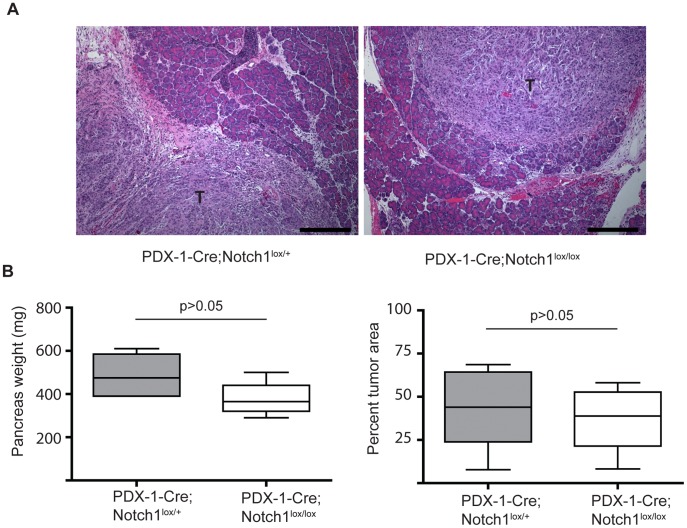
Previous studies have demonstrated that Notch1 functions as a tumor suppressor gene in the skin by mechanisms impacting the tumor microenvironment [19]. In addition, it has recently been shown that treatment with a γ-secretase inhibitor has a greater impact on the destabilization of endothelial cells rather than primary tumors in a mouse model of pancreatic cancer [28]. These studies indicate Notch proteins may function primarily in a non-cell autonomous manner in the tumor environment. To determine whether Notch1 functions in a non-cell autonomous mechanism to inhibit pancreatic tumorigenesis, we employed an orthotopic transplantation model of tumorigenesis. PDX-1-Cre;Notch1lox/lox and PDX-1-Cre;Notch1lox/+ mice were crossed to NOD scid gamma mice to generate immunodeficient hosts for transplantation. Pancreatic epithelial cell lines isolated from primary tumors of PDX-1-Cre;LSL-KrasG12D;LSL-Trp53R172H (KPC) mice [29] were injected into the pancreas of either PDX-1-Cre;Notch1lox/lox (Notch absent) or PDX-1-Cre;Notch1lox/+ (Notch present) mice. It should be noted that the PDX-1-Cre;Notch1lox/lox mice have Notch1 deleted from all pancreatic epithelial cells; however, other cells located in the tumor microenvironment, such as endothelial cells and immune cells, retain Notch1 expression. After 2 weeks, KPC cells implanted into both PDX-1-Cre;Notch1lox/lox and PDX-1-Cre;Notch1lox/+ mice formed palpable tumors (Figure 5A). Significant differences were not found in either pancreas weight or percent tumor area between the PDX-1-Cre;Notch1lox/lox and PDX-1-Cre;Notch1lox/+ hosts (Figure 5B). These results indicate Notch1 does not function in a non-cell autonomous manner to inhibit PDAC tumor maintenance.
Figure 5. Notch1 functions in a cell autonomous manner.
(A) Representative images of orthotopic tumors in Pdx1-cre;Notchlox/+ and Pdx1-Cre;Notch1lox/lox mice. Tumor cells (T) are seen adjacent to normal pancreatic tissue. Scale bar, 300 µm (B) Quantitative analysis of pancreas weight and percentage of tumor volume in Pdx1-Cre;Notchlox/+(n = 5) and Pdx1-Cre;Notch1lox/lox (n = 6) mice 2 weeks after orthotopic injections. Box plots depict median values, lower and upper quartiles, and maximum and minimum observed values.
Discussion
Evidence now suggests PanIN lesions, and ultimately PDAC, can arise from acinar to ductal metaplasia [30]. The exact molecular mechanisms controlling the conversion of normal acinar tissue to metaplastic lesions remains unclear, however activation of oncogenic K-ras appears to be a key event in the transdifferentiation process. Similar to a previous report [23], we find that expression of K-rasG12D is sufficient to induce acinar to ductal metaplasia in an in vitro model. This finding supports animal models in which activation of oncogenic K-ras in pancreatic acinar cells induces PanIN development, and provides further evidence that K-ras mutations are an initiating event in the development of PDAC.
Previous reports have suggested Notch signaling is activated in PanIN lesions and PDAC in humans [10] and in mouse models [4]. Further, inhibition of Notch signaling by treatment with a γ-secretase inhibitor (GSI) prevents tumor progression in a mouse model of PDAC [31]. Given that Notch receptors regulate cell fate decisions during pancreatic development, it is reasonable to assume Notch signaling is activated during ADM. Accordingly, ectopic expression of the activated forms of Notch1 or Notch2 (Nic) drives ADM in in vitro explant culture models [10], [11].
In contrast to these reports, previous work by our lab has demonstrated Notch1 suppresses PanIN formation in a mouse model of PDAC [20]. In the current study, we investigated the requirement for Notch1 in ADM using an explant culture model. Surprisingly, we find that Notch1 is not required for either EGF- or K-rasG12D-mediated transdifferentiation. In addition, using a dominant-negative form of MAML1 as a pan-Notch inhibitor, we show that Notch signaling is not required for ADM in vitro. However, it should be noted that MAML1 also functions as a transcriptional activator independent of the Notch signaling pathway [32]. The possibility therefore remains that Notch signaling may be functioning independent of MAML in order to regulate ADM in vitro.
One potential explanation for the differences between our results and previous reports indicating Notch can promote ADM is the fact that previous studies employed ectopic expression of activated forms of Notch receptors, leading to expression of Nic above physiological levels. Thus, the contrasting outcomes observed when Notch1 is genetically deleted versus overexpressed can be attributed to different phenotypic responses to levels of pathway activation. Indeed this has been demonstrated in studies showing that Nic expression levels affect the balance between growth-stimulating and growth-suppressive effects in mammary epithelial cultures [33]. An additional factor to consider is that many earlier studies rely on Hes1 expression as a surrogate for Notch activation, while we and others have found that inhibition of Notch signaling in the pancreas does not reduce Hes1 expression [12], [13]. Further, ectopic expression of Hes1 in an in vitro ADM assay fails to recapitulate the effects of Nic expression, implying additional downstream effectors are responsible for Notch mediated events [10]. Hence, it will be beneficial to identify all downstream effectors of Notch signaling specific to the different cellular compartments of the pancreas.
To further assess the role of Notch1 in ADM in vivo, we employed the Elastase1-CreERT2 model to drive K-rasG12D expression and Notch1 deletion. Unlike the PDX-1-Cre transgene, which induces Cre-mediated recombination during embryonic development, the Elastase1-CreERT2 model allows for the activation of Cre and recombination in mature acinar cells. Using this model we found that deletion of Notch1 in the context of activated K-ras does not accelerate the development of PanIN lesions. These results differ from our previous work using PDX-1-Cre;LSL-KrasG12D mice, where we found deletion of Notch1 accelerated PanIN formation. One reason for the differences in the two models is the timing of Notch1 deletion. In the PDX-1-Cre model, Notch1 is deleted at embryonic day 8.5 (e8.5) during a crucial stage of pancreatic development. Though Notch1 deletion at this stage does not cause defects in pancreatogenesis [34], progenitor cells may become more susceptible to oncogene induced transformation upon Notch1 deletion. Additionally, the timing of genetic events during tumor progression may be crucial. In human pancreatic tumors, K-ras mutations occur at the earliest stages of PanIN development, while loss of commonly mutated tumor suppressor genes, such as p53 and INK4a occurs later in the PanIN-PDAC lineage [7]. The chronological order in which these events occur in mouse models may be critical to tumor development and progression. Therefore it is possible that Notch1 deletion during development, concurrently with K-rasG12D activation, leads to a different phenotype than deletion of Notch1 in the mature pancreas.
Alternatively, the seeming discrepancy in these results may stem from Notch receptors being expressed in specific cellular compartments. Whereas the PDX-1-Cre allele deletes Notch1 in all pancreatic epithelial cells, the Elastase1-CreERT2 model deletes Notch1 exclusively in acinar cells. The results of both our in vitro and in vivo experiments demonstrate loss of Notch1 does not accelerate spontaneous ADM, indicating Notch1 may function in a different cellular compartment. One possible cell type to consider is centroacinar cells (CACs) which lie at the junction of the duct network and acini and which express Hes1 [35]. Recent work using a Hes1CreERT2 model identified CACs, and also a smaller fraction of ductal cells, as Notch responsive cells in the pancreas [36]. Future studies are needed to elucidate how genetic deletion of Notch1 specifically in these populations affects PanIN development and if these results support our findings in the PDX-1-Cre model.
We further investigated the effect of Notch1 deletion in the Elastase1-CreERT2;LSL-K-rasG12D model following acute pancreatitis. Recent studies indicate that acinar cell regeneration is inhibited in response to both oncogenic K-ras activation and Notch1 deletion [7], [9]. Our results suggest that Notch1 deletion in the Elastase1-CreERT2 model does not affect acinar regeneration, as pancreas tissue from Elastase1-CreERT2;Notch1lox/lox mice appeared grossly normal 3 months following caerulein treatment. However, we did not examine earlier time points after injury, when acinar regeneration is most pronounced. These opposing results may also be explained by the use of different Cre-drivers, as previous studies utilized Ptf1a+/Cre(ex1) rather than an acinar-specific promoter. As the Ptf1a+/Cre(ex1) line targets a greater number of acinar cells compared to Elastase1-CreERT2 mice, this may help explain the lack of phenotype in our model.
Following acute pancreatitis, both Elastase1-CreERT2;LSL-K-rasG12D and Elastase1-CreERT2;LSL-K-rasG12D;Notch1lox/lox mice develop high grade PanIN lesions. While the grade of PanIN lesions is similar in both cohorts, the Elastase1-CreERT2;LSL-K-rasG12D;Notch1lox/lox mice appear to have a greater abundance of PanIN lesions, as well as atypical flat lesions (AFLs), which arise in regions of ADM [27]. These results imply Notch1 deletion renders acinar cells more susceptible to K-ras induced PanIn formation, as well as AFL formation, which may represent an alternative precursor lesion to PanINs. Additional work is needed to determine the relationship between AFLs and the PanIN-PDAC progression model. Further, it remains to be determined how pancreatitis and Notch1 loss potentially synergize to promote oncogenic K-ras induced PanIN formation. However, recent studies examining the effects of oncogenic K-ras activation and Notch1 deletion individually in response to caerulein-induced pancreatitis have provided mechanistic clues. Following acute pancreatitis, acinar cells expressing oncogenic K-ras undergo a state of persistant dedifferentiation, ultimately leading to ADM and PanIN lesion formation [7]. Similarly, mice deficient for Notch1 in pancreatic epithelium also display impaired acinar regeneration following acute pancreatitis [9]. Therefore, activation of K-ras and deletion of Notch1 may cooperate to inhibit the regeneration process, resulting in a population of cells more susceptible to PanIN formation. Interestingly, both of the previous studies identified alterations in β-catenin signaling as a downstream effect of either K-rasG12D activation or Notch1 deletion.
Finally, we investigated whether Notch1 functions through a non-cell autonomous mechanism to suppress tumor maintenance using an orthotopic transplantation model. We investigated a role for Notch1 in the tumor environment by orthotopically injecting KPC tumor cells directly into the pancreas of a mouse in which Notch1 had been deleted in all epithelial cells. Our results suggest Notch1 functions in a cell autonomous manner to inhibit PanIN formation. Further work is needed determine if Notch1 can function to suppress PanIN progression in a non-cell-autonomous manner, as it has been previously shown to function in the skin [19].
In conclusion, our data reveal Notch1 is not required for pancreatic ADM. Additionally, loss of Notch1 in adult acinar cells does not accelerate ADM either in vitro or in an in vivo mouse model of PDAC, suggesting Notch1 regulates alternative molecular events in PanIN development.
Materials and Methods
Mouse Strains
The LSL-KrasG12D [37], Notch1lox/lox [38], Pdx1-Cre [3], and Elastase1-CreERT2 [25] mouse strains have been previously described. To initiate Cre-mediated recombination in Elastase1-CreERT2;LSL-KrasG12D, Elastase1-CreERT2;LSL-KrasG12D;Notch1lox/lox, and Elastase1-CreERT2;Notch1lox/lox strains, mice were treated with tamoxifen (Sigma-Aldrich) at 4 weeks of age. Mice received 5 consecutive daily intraperitoneal injections of 2 mg of tamoxifen dissolved in sunflower seed oil (Sigma-Aldrich). For pancreatitis experiments, mice were treated with tamoxifen at 8 weeks of age by oral gavage. Three doses were administered (20 mg, 20 mg, 10 mg) over the course of 1 week. Two weeks later, pancreatitis was induced by 6 hourly intraperitoneal injections of caerulein (Sigma-Aldrich), 2 µg dissolved in 0.9% NaCl, over 2 consecutive days. NOD scid gamma (NSG) mice were purchased from Jackson labs, stock number 005557. All mice used were on a mixed background. All studies were approved and conducted in compliance with Wistar Institute Institutional Animal Care and Use Committee guidelines.
Primary Acinar Cultures
Primary acinar cells were prepared as previously described [21]. Cells were maintained in RPMI1640 media (Cellgro) supplemented with Soybean Trypsin Inhibitor (Sigma-Aldrich) at 0.1 mg/mL, Dexamethazone (1 µg/mL) (Sigma-Aldrich) and Penicillin/Streptomycin (Gibco Life Technologies). When necessary, cells were treated with EGF, 20 ng/mL (BD Biosciences), DAPT, 10 µM (Sigma), or 0.1% DMSO (Sigma). Cultures were maintained at 37°C and 5% CO2 for up to 5 days. Media was changed daily. Brightfield images were captured on a Nikon TE2000 inverted microscope. For quantitative analysis, ductal cysts were counted from 20 different focal planes for each sample and divided by the total number of cell aggregates.
Immunofluorescence
Collagen disks were fixed and rehydrated as previously described [21]. Disks were blocked in PBSBT (PBS, 0.1% tritonX-100, 2%BSA) and subsequently incubated overnight in primary antibodies diluted in PBSBT. Primary antibodies used were anti-cytokeratin (Dako, 1∶500), anti-GFP-Alexa488 conjugate (Invitrogen, 1∶400), and anti-Hes1 (gift from Dr. Ben Stanger, University of Pennsylvania, 1∶100). For cytokeratin and Hes1 staining, disks were washed in PBSBT and incubated overnight in anti-rabbit Alexa568 secondary antibody (Invitrogen, 1∶500). Hoechst 33342 (Sigma-Aldrich) was used to counterstain nuclei. Images were captured on a Zeiss LSM710 Confocal microscope.
Adenoviral Infection
A cDNA coding for DNMAML-GFP fusion protein (gift from Dr. Warren Pear, University of Pennsylvania) was cloned into an Adenoviral vector. Adenoviral empty vector was used as a control. Prior to plating in collagen, acinar cell suspensions were incubated with adenovirus (MOI 20∶1) at 37°C for 1 hr with occasional rocking. Suspensions were then plated in collagen.
Western Blot Analysis
Acinar cells were homogenized in RIPA buffer, separated by SDS-PAGE and transferred to PVDF membranes (Immobilon-P, Millipore). Primary antibodies employed were: Notch1 (Epitomics, 1∶500) and Tubulin (Sigma-Aldrich, 1∶10,000). Blots were developed using horseradish peroxidase (HRP)-conjugated secondary antibodies and the ECL detection system (Amersham).
Histological Analysis and Immunohistochemistry
Formalin-fixed paraffin-embedded pancreatic tissue was processed by standard methods and stained with hematoxylin and eosin (H&E) or immunostained with an antibody for Muc-5AC Glycoprotein (Novocastra, 1∶50 dilution). Antigen retrieval was performed in 10 mM sodium citrate buffer pH 6.0. Labeling was detected using the VECTOR M.O.M. Immunodetection Kit (Vector Laboratories) following the manufacturers protocol. Slides were counterstained with hematoxylin, dehydrated, and mounted. Pancreatic pathology and scoring of lesions were performed by a board-certified veterinary pathologist. Tissue was fixed and embedded in paraffin. For each case, an H&E stained longitudinal section spanning the entire pancreas was analyzed at 20X and 60X magnification.
KrasG12D and Notch1 Allele Recombination PCR
Genomic DNA was isolated from tails and pancreas using a Genelute Mammalian Genomic DNA kit (Sigma-Aldrich). The LSL-KrasG12D allele [37] and the Notch1 allele [39] were analyzed by PCR as previously described. The primers for the LSL-KrasG12D allele were: 5′ GGG TAG GTG TTG GGA TAG CTG and 3′ TCC GAA TTC AGT GAC TAC AGA TGT ACA GAG. The primers used for the Notch1 allele were: 5′ of loxP1 CTG ACT TAG TAG GGG GAA AAC; 3′ of loxP1 AGT GGT CCA GGG TGT GAG TGT; and 3′ of loxP2 TAA AAA GCG ACA GCT GCG GAG.
Orthotopic Transplantation Experiments
KPC cells lines were derived from primary tumors of Pdx1-Cre;LSL-KrasG12D;LSL-Trp53R172H mice (gift from Dr. Robert Vonderheide, University of Pennsylvania). Mice were anesthetized in isoflurane and the pancreas was exposed by a lateral incision. 1×10⁵ cells were suspended in 50 µL DMEM (Gibco Life Technologies) and injected into the head of the pancreas using an insulin syringe. The pancreas was returned to the peritoneal cavity, and the peritoneum was closed with 4–0 chromic sutures and the skin was closed with 6–0 silk sutures (Roboz). After 2 weeks the mice were sacrificed and the pancreas was removed and weighed. Tumor to pancreas volume was assessed using ImagePro Plus (Media Cybernetics).
Statistical Analysis
Data presented is mean±standard error of mean (SEM). Statistical analysis was performed using two-tailed unpaired Student's t test. A p value of equal or less than 0.05 was considered significant.
Supporting Information
Immunofluorescence confirms acinar origin of cells. (A) Pancreatic explants from wildtype mice embedded in collagen either untreated (control) or treated with EGF (20 µg/mL). Cells are immunostained for expression of the acinar marker, amylase (green) and the ductal marker, pan-cytokeratin (red) at day 5. Scale bar, 75 µm.
(TIF)
DAPT treatment does not inhibit oncogenic K-ras mediated ADM in vitro . (A) Pancreatic explants from PDX-1-Cre;LSL-KrasG12D mice embedded in collagen either treated with 0.1% DMSO (control) or DAPT (10 µM). Cells are immunostained at Day 2 for expression of the ductal marker, pan-cytokeratin (red), and counterstained with Hoechst dye. Scale bar, 20 µm.
(TIF)
Muc-5AC staining identifies PanIN lesions. (A) Expression of Muc-5AC by immunohistochemical staining in PanIN lesions from Elastase1-CreERT2;KrasG12D and Elastase1-CreERT2;KrasG12D;Notch1lox/lox mice, 3 months following tamoxifen treatment. Scale bar, 50 µm. (B) Expression of Muc-5AC by immunohistochemical staining in PanIN lesions from Elastase1-CreERT2;KrasG12D and Elastase1-CreERT2;KrasG12D;Notch1lox/lox mice following tamoxifen treatment and caerulin-induced pancreatitis. Scale bar, 50 µm.
(TIF)
Acknowledgments
We thank A.L. Means and X. Zhao for technical assistance, and the Wistar Histology, Animal and Microscopy core facilities as well as all members of the Kissil lab.
Funding Statement
This work was supported in part by grant NIH CA124495 (J.K.). No additional external funding was received for this study. The funder had no role in study design, data collection and analysis, decision to publish, or preparation of the manuscript.
References
- 1. Bardeesy N, DePinho RA (2002) Pancreatic cancer biology and genetics. Nat Rev Cancer 2: 897–909. [DOI] [PubMed] [Google Scholar]
- 2. Maitra A, Hruban RH (2008) Pancreatic cancer. Annu Rev Pathol 3: 157–188. [DOI] [PMC free article] [PubMed] [Google Scholar]
- 3. Hingorani SR, Petricoin EF, Maitra A, Rajapakse V, King C, et al. (2003) Preinvasive and invasive ductal pancreatic cancer and its early detection in the mouse. Cancer Cell 4: 437–450. [DOI] [PubMed] [Google Scholar]
- 4. Habbe N, Shi G, Meguid RA, Fendrich V, Esni F, et al. (2008) Spontaneous induction of murine pancreatic intraepithelial neoplasia (mPanIN) by acinar cell targeting of oncogenic Kras in adult mice. Proc Natl Acad Sci U S A 105: 18913–18918. [DOI] [PMC free article] [PubMed] [Google Scholar]
- 5. Guerra C, Schuhmacher AJ, Canamero M, Grippo PJ, Verdaguer L, et al. (2007) Chronic pancreatitis is essential for induction of pancreatic ductal adenocarcinoma by K-Ras oncogenes in adult mice. Cancer Cell 11: 291–302. [DOI] [PubMed] [Google Scholar]
- 6. Gidekel Friedlander SY, Chu GC, Snyder EL, Girnius N, Dibelius G, et al. (2009) Context-dependent transformation of adult pancreatic cells by oncogenic K-Ras. Cancer Cell 16: 379–389. [DOI] [PMC free article] [PubMed] [Google Scholar]
- 7. Morris JPt, Cano DA, Sekine S, Wang SC, Hebrok M (2010) Beta-catenin blocks Kras-dependent reprogramming of acini into pancreatic cancer precursor lesions in mice. J Clin Invest 120: 508–520. [DOI] [PMC free article] [PubMed] [Google Scholar]
- 8. Apelqvist A, Li H, Sommer L, Beatus P, Anderson DJ, et al. (1999) Notch signalling controls pancreatic cell differentiation. Nature 400: 877–881. [DOI] [PubMed] [Google Scholar]
- 9. Siveke JT, Lubeseder-Martellato C, Lee M, Mazur PK, Nakhai H, et al. (2008) Notch signaling is required for exocrine regeneration after acute pancreatitis. Gastroenterology 134: 544–555. [DOI] [PubMed] [Google Scholar]
- 10. Miyamoto Y, Maitra A, Ghosh B, Zechner U, Argani P, et al. (2003) Notch mediates TGF alpha-induced changes in epithelial differentiation during pancreatic tumorigenesis. Cancer Cell 3: 565–576. [DOI] [PubMed] [Google Scholar]
- 11. Sawey ET, Johnson JA, Crawford HC (2007) Matrix metalloproteinase 7 controls pancreatic acinar cell transdifferentiation by activating the Notch signaling pathway. Proc Natl Acad Sci U S A 104: 19327–19332. [DOI] [PMC free article] [PubMed] [Google Scholar]
- 12. Rooman I, De Medts N, Baeyens L, Lardon J, De Breuck S, et al. (2006) Expression of the Notch signaling pathway and effect on exocrine cell proliferation in adult rat pancreas. Am J Pathol 169: 1206–1214. [DOI] [PMC free article] [PubMed] [Google Scholar]
- 13. Mazur PK, Einwachter H, Lee M, Sipos B, Nakhai H, et al. (2010) Notch2 is required for progression of pancreatic intraepithelial neoplasia and development of pancreatic ductal adenocarcinoma. Proc Natl Acad Sci U S A 107: 13438–13443. [DOI] [PMC free article] [PubMed] [Google Scholar]
- 14. Nicolas M, Wolfer A, Raj K, Kummer JA, Mill P, et al. (2003) Notch1 functions as a tumor suppressor in mouse skin. Nat Genet 33: 416–421. [DOI] [PubMed] [Google Scholar]
- 15. Wang NJ, Sanborn Z, Arnett KL, Bayston LJ, Liao W, et al. (2011) Loss-of-function mutations in Notch receptors in cutaneous and lung squamous cell carcinoma. Proc Natl Acad Sci U S A 108: 17761–17766. [DOI] [PMC free article] [PubMed] [Google Scholar]
- 16. Viatour P, Ehmer U, Saddic LA, Dorrell C, Andersen JB, et al. (2011) Notch signaling inhibits hepatocellular carcinoma following inactivation of the RB pathway. J Exp Med 208: 1963–1976. [DOI] [PMC free article] [PubMed] [Google Scholar]
- 17. Klinakis A, Lobry C, Abdel-Wahab O, Oh P, Haeno H, et al. (2011) A novel tumour-suppressor function for the Notch pathway in myeloid leukaemia. Nature 473: 230–233. [DOI] [PMC free article] [PubMed] [Google Scholar]
- 18. Agrawal N, Frederick MJ, Pickering CR, Bettegowda C, Chang K, et al. (2011) Exome sequencing of head and neck squamous cell carcinoma reveals inactivating mutations in NOTCH1. Science 333: 1154–1157. [DOI] [PMC free article] [PubMed] [Google Scholar]
- 19. Demehri S, Turkoz A, Kopan R (2009) Epidermal Notch1 loss promotes skin tumorigenesis by impacting the stromal microenvironment. Cancer Cell 16: 55–66. [DOI] [PMC free article] [PubMed] [Google Scholar]
- 20. Hanlon L, Avila JL, Demarest RM, Troutman S, Allen M, et al. (2010) Notch1 functions as a tumor suppressor in a model of K-ras-induced pancreatic ductal adenocarcinoma. Cancer Res 70: 4280–4286. [DOI] [PMC free article] [PubMed] [Google Scholar]
- 21. Means AL, Meszoely IM, Suzuki K, Miyamoto Y, Rustgi AK, et al. (2005) Pancreatic epithelial plasticity mediated by acinar cell transdifferentiation and generation of nestin-positive intermediates. Development 132: 3767–3776. [DOI] [PubMed] [Google Scholar]
- 22. Gu G, Brown JR, Melton DA (2003) Direct lineage tracing reveals the ontogeny of pancreatic cell fates during mouse embryogenesis. Mech Dev 120: 35–43. [DOI] [PubMed] [Google Scholar]
- 23. Scotti ML, Smith KE, Butler AM, Calcagno SR, Crawford HC, et al. (2012) Protein kinase C iota regulates pancreatic acinar-to-ductal metaplasia. PLoS One 7: e30509. [DOI] [PMC free article] [PubMed] [Google Scholar]
- 24. Maillard I, Weng AP, Carpenter AC, Rodriguez CG, Sai H, et al. (2004) Mastermind critically regulates Notch-mediated lymphoid cell fate decisions. Blood 104: 1696–1702. [DOI] [PubMed] [Google Scholar]
- 25. Desai BM, Oliver-Krasinski J, De Leon DD, Farzad C, Hong N, et al. (2007) Preexisting pancreatic acinar cells contribute to acinar cell, but not islet beta cell, regeneration. J Clin Invest 117: 971–977. [DOI] [PMC free article] [PubMed] [Google Scholar]
- 26. De La OJ, Emerson LL, Goodman JL, Froebe SC, Illum BE, et al. (2008) Notch and Kras reprogram pancreatic acinar cells to ductal intraepithelial neoplasia. Proc Natl Acad Sci U S A 105: 18907–18912. [DOI] [PMC free article] [PubMed] [Google Scholar]
- 27. Aichler M, Seiler C, Tost M, Siveke J, Mazur PK, et al. (2012) Origin of pancreatic ductal adenocarcinoma from atypical flat lesions: a comparative study in transgenic mice and human tissues. J Pathol 226: 723–734. [DOI] [PubMed] [Google Scholar]
- 28. Cook N, Frese KK, Bapiro TE, Jacobetz MA, Gopinathan A, et al. (2012) Gamma secretase inhibition promotes hypoxic necrosis in mouse pancreatic ductal adenocarcinoma. J Exp Med 209: 437–444. [DOI] [PMC free article] [PubMed] [Google Scholar]
- 29. Hingorani SR, Wang L, Multani AS, Combs C, Deramaudt TB, et al. (2005) Trp53R172H and KrasG12D cooperate to promote chromosomal instability and widely metastatic pancreatic ductal adenocarcinoma in mice. Cancer Cell 7: 469–483. [DOI] [PubMed] [Google Scholar]
- 30. Zhu L, Shi G, Schmidt CM, Hruban RH, Konieczny SF (2007) Acinar cells contribute to the molecular heterogeneity of pancreatic intraepithelial neoplasia. Am J Pathol 171: 263–273. [DOI] [PMC free article] [PubMed] [Google Scholar]
- 31.Plentz R, Park JS, Rhim AD, Abravanel D, Hezel AF, et al.. (2009) Inhibition of gamma-secretase activity inhibits tumor progression in a mouse model of pancreatic ductal adenocarcinoma. Gastroenterology 136: 1741–1749 e1746. [DOI] [PMC free article] [PubMed]
- 32. Zhao Y, Katzman RB, Delmolino LM, Bhat I, Zhang Y, et al. (2007) The notch regulator MAML1 interacts with p53 and functions as a coactivator. J Biol Chem 282: 11969–11981. [DOI] [PubMed] [Google Scholar]
- 33. Mazzone M, Selfors LM, Albeck J, Overholtzer M, Sale S, et al. (2010) Dose-dependent induction of distinct phenotypic responses to Notch pathway activation in mammary epithelial cells. Proc Natl Acad Sci U S A 107: 5012–5017. [DOI] [PMC free article] [PubMed] [Google Scholar]
- 34. Nakhai H, Siveke JT, Klein B, Mendoza-Torres L, Mazur PK, et al. (2008) Conditional ablation of Notch signaling in pancreatic development. Development 135: 2757–2765. [DOI] [PubMed] [Google Scholar]
- 35. Stanger BZ, Stiles B, Lauwers GY, Bardeesy N, Mendoza M, et al. (2005) Pten constrains centroacinar cell expansion and malignant transformation in the pancreas. Cancer Cell 8: 185–195. [DOI] [PubMed] [Google Scholar]
- 36. Kopinke D, Brailsford M, Shea JE, Leavitt R, Scaife CL, et al. (2011) Lineage tracing reveals the dynamic contribution of Hes1+ cells to the developing and adult pancreas. Development 138: 431–441. [DOI] [PMC free article] [PubMed] [Google Scholar]
- 37. Jackson EL, Willis N, Mercer K, Bronson RT, Crowley D, et al. (2001) Analysis of lung tumor initiation and progression using conditional expression of oncogenic K-ras. Genes Dev 15: 3243–3248. [DOI] [PMC free article] [PubMed] [Google Scholar]
- 38. Radtke F, Wilson A, Stark G, Bauer M, van Meerwijk J, et al. (1999) Deficient T cell fate specification in mice with an induced inactivation of Notch1. Immunity 10: 547–558. [DOI] [PubMed] [Google Scholar]
- 39. Wolfer A, Wilson A, Nemir M, MacDonald HR, Radtke F (2002) Inactivation of Notch1 impairs VDJbeta rearrangement and allows pre-TCR-independent survival of early alpha beta Lineage Thymocytes. Immunity 16: 869–879. [DOI] [PubMed] [Google Scholar]
Associated Data
This section collects any data citations, data availability statements, or supplementary materials included in this article.
Supplementary Materials
Immunofluorescence confirms acinar origin of cells. (A) Pancreatic explants from wildtype mice embedded in collagen either untreated (control) or treated with EGF (20 µg/mL). Cells are immunostained for expression of the acinar marker, amylase (green) and the ductal marker, pan-cytokeratin (red) at day 5. Scale bar, 75 µm.
(TIF)
DAPT treatment does not inhibit oncogenic K-ras mediated ADM in vitro . (A) Pancreatic explants from PDX-1-Cre;LSL-KrasG12D mice embedded in collagen either treated with 0.1% DMSO (control) or DAPT (10 µM). Cells are immunostained at Day 2 for expression of the ductal marker, pan-cytokeratin (red), and counterstained with Hoechst dye. Scale bar, 20 µm.
(TIF)
Muc-5AC staining identifies PanIN lesions. (A) Expression of Muc-5AC by immunohistochemical staining in PanIN lesions from Elastase1-CreERT2;KrasG12D and Elastase1-CreERT2;KrasG12D;Notch1lox/lox mice, 3 months following tamoxifen treatment. Scale bar, 50 µm. (B) Expression of Muc-5AC by immunohistochemical staining in PanIN lesions from Elastase1-CreERT2;KrasG12D and Elastase1-CreERT2;KrasG12D;Notch1lox/lox mice following tamoxifen treatment and caerulin-induced pancreatitis. Scale bar, 50 µm.
(TIF)


