



Background: In addition to the toxicity mediated by prion protein misfolding, mechanisms associated with its loss-of-function in genetic prion diseases are unknown.
Results: Neural cells expressing PrPC mutants associated with prion diseases present impaired laminin-mediated process outgrowth and survival.
Conclusion: PrPC mutants show loss-of-function in neural cells.
Significance: The impairment of prion protein functions may contribute to the etiology of prion diseases.
Keywords: Calcium, Cell Death, Laminin, Mutant, Prions, Loss-of-function, Prion Diseases
Abstract
Prions, the agents of transmissible spongiform encephalopathies, require the expression of prion protein (PrPC) to propagate disease. PrPC is converted into an abnormal insoluble form, PrPSc, that gains neurotoxic activity. Conversely, clinical manifestations of prion disease may occur either before or in the absence of PrPSc deposits, but the loss of normal PrPC function contribution for the etiology of these diseases is still debatable. Prion disease-associated mutations in PrPC represent one of the best models to understand the impact of PrPC loss-of-function. PrPC associates with various molecules and, in particular, the interaction of PrPC with laminin (Ln) modulates neuronal plasticity and memory formation. To assess the functional alterations associated with PrPC mutations, wild-type and mutated PrPC proteins were expressed in a neural cell line derived from a PrPC-null mouse. Treatment with the laminin γ1 chain peptide (Ln γ1), which mimics the Ln binding site for PrPC, increased intracellular calcium in cells expressing wild-type PrPC, whereas a significantly lower response was observed in cells expressing mutated PrPC molecules. The Ln γ1 did not promote process outgrowth or protect against staurosporine-induced cell death in cells expressing mutated PrPC molecules in contrast to cells expressing wild-type PrPC. The co-expression of wild-type PrPC with mutated PrPC molecules was able to rescue the Ln protective effects, indicating the lack of negative dominance of PrPC mutated molecules. These results indicate that PrPC mutations impair process outgrowth and survival mediated by Ln γ1 peptide in neural cells, which may contribute to the pathogenesis of genetic prion diseases.
Introduction
The cellular prion protein (PrPC) is a glycosylphosphatidylinositol-anchored protein whose conformationally modified isoform, PrPSc, is the major component of prions. Prions may be defined as infectious agents that cause the neurodegenerative diseases known as transmissible spongiform encephalopathies (TSEs)3 or prion diseases. These illnesses affect humans and other mammals and present sporadic, genetic, or infectious forms (1). The accumulation of the toxic, insoluble PrPSc has been described to be the most likely event responsible for neuronal death in prion diseases (2). This idea is supported by the findings that deletion of PrPC or its knockdown in early stages of the disease reverted spongiosis, neuron loss, and cognitive and behavioral deficits as well as impaired neurophysiological function in mice (3–5).
On the other hand, neuronal loss may be observed even in the absence of characteristic PrPSc deposits in sporadic, genetic, and iatrogenic forms of prion diseases (6–8). Small, punctate synaptic deposits of PrPSc correlate with neuronal loss, whereas the larger, focal types of deposits show an inverse correlation with neuronal counts (9). These findings could be related to the generation of oligomers of low molecular weight, present in brains of sporadic Creutzfeldt-Jakob (CJD) disease patients, that control the progression rate of the disease (10). Remarkably, neurotoxicity was associated not only with PrPC interaction with PrPSc but also with other β-sheet-rich conformers of different origin in which toxicity was independent of prions replication (11). Whether toxic signaling is due to gain-of-function of PrPC or by the impairment of a normal signaling is unknown. Indeed, it is possible that PrPC loss-of-function may also play a major role in the etiology of these maladies (12–14).
Interestingly, PrPC-null mice do not have an obvious phenotype, implying that PrPC may not have an essential function or that its loss may be compensated by other molecules (15, 16). However, the expression of mutated PrPC proteins carrying various deletions result in diverse phenotypic abnormalities that parallel neurodegenerative processes. These findings are consistent with the existence of domains within the PrPC protein that have physiologically relevant functions (17). In fact, PrPC domains have been shown to be involved with some neurotrophic functions (14). Remarkably, several disease-associated mutations in PrPC have been localized to these domains, and although these mutations account for only a small fraction of prion diseases, mutated PrPC proteins provide us with an ideal model system to study PrPC loss-of-function.
The genetic human spongiform encephalopathies, including CJD, fatal familial insomnia, and Gerstmann-Straussler-Scheinker disease, are among the TSEs that have been linked to specific mutations in the PRNP gene that encodes PrPC (18, 19). A common human PrPC polymorphism at amino acid residue 129, which may be either a methionine or a valine, specifies the disease phenotype associated with an aspartate to glutamine mutation at position 178 (D178N). Specifically, the Met-129/Asn-178 haplotype is linked to fatal familial insomnia, whereas the Val-129/Asp-178 haplotype causes CJD (20). These disorders are autosomal dominant, fully penetrant inherited conditions, and the age of disease onset is around 50 years (OMIM® entry 176640).
In the last decade a diverse range of functions has been attributed to the native PrPC protein, such as neuroprotection against cellular and systemic insults, neuritogenesis, neuronal plasticity and excitability, and memory formation and consolidation (21–33). Noteworthy, PrPC mutations associated to CJD, fatal familial insomnia, and Gerstmann-Straussler-Scheinker disease decrease or abolish the anti-bax function in primary human neurons and breast cancer cell lines promoting programmed cell death (34).
A vast number of ligands and signaling pathways have also been associated with PrPC functions (78), and PrPC has been proposed to organize a dynamic cell surface platform for the assembly of signaling modules (13). In addition, the neurotrophic functions attributed to PrPC may reside in its ability to drive the assembly of multicomponent complexes at the cell surface (14). Laminin (Ln), an extracellular matrix protein, is one of the high affinity ligands for PrPC (24). The mouse PrPC-Ln binding sites map to amino acids 1575–1584 (RNIAEIIKDI) in the γ1 chain region of Ln (Ln γ1; Ref. 24) and to amino acids 173–182 in PrPC (22). Administration of the Ln γ1 peptide, which represents the PrPC binding site, induces PrPC-dependent neuritogenesis and reproduces the neuronal maturation phenotype that is dependent upon the binding of Ln to PrPC (24). The PrPC-Ln γ1 interaction requires the activity of the group I metabotropic glutamate receptors, mGluR1 and mGluR5, to promote neuritogenesis through activation of phospholipase C and intracellular Ca2+ mobilization (35). The formation of a PrPC-Ln-mGluR1/5 signaling complex is consistent with the scaffold properties of PrPC and its possible role in allosteric regulation of signal transduction (36). In agreement with the idea that the PrPC-Ln γ1 peptide interaction induces physiological signals in the nervous system, the activity of this complex has been shown to enhance memory consolidation (22).
In the present report we determined whether PrPC mutations associated with genetic prion diseases corrupt PrPC-Ln-dependent signaling pathways. The wild-type mouse PrPC as well as mouse PrPC carrying mutations at codons Leu-101, Leu-104, Val-116, Asn-177, Ile-179, and Lys-199 (equivalent to human mutated proteins Leu-102, Leu-105, Val-117, Asn-178, Ile-180, and Lys-200, respectively) were expressed in cells immortalized from primary neural cultures derived from PrPC-null mice. The exogenous wild-type and mutant PrPC proteins were evaluated for cell membrane expression and proteinase K resistance. The ability to interact with the Ln-γ1 chain peptide upon Ca2+ signaling to effect process outgrowth and induction of protective response was also determined. Our findings contribute to the identification of cellular mechanisms associated with PrPC loss-of-function.
EXPERIMENTAL PROCEDURES
Peptides and Chemicals
The Ln γ1 peptide (RNIAEIIKDI) linked to BSA and Ln γ1 scrambled (SCR) peptide (IRADIEIKID) were synthesized by GenScript Corp. and NeoMPS SA (Strasbourg, France). Laminin was purified as previously described in Ref. 37. The group I metabotropic glutamate receptor agonist (S)-3,5-dihydroxyphenylglycine (DHPG) and mGluR1 and -5 antagonists LY367385 and 2-methyl-6-(phenylethynyl)-pyridine (MPEP) were purchased from Tocris Biosciences (Ellisville, MO). Mitomycin C was purchased from Sigma. TO-PRO-3 Iodite was purchased from Invitrogen. The antibodies used were mAb 3F4 (Dako), mouse polyclonal anti-PrPC antibody (33), mouse monoclonal anti-MAP2 (Sigma), rabbit anti-cleaved caspase 3 (Cell Signaling), goat anti-mouse Alexa Fluor 488, goat anti-rabbit Alexa Fluor 488 and goat anti-mouse Alexa Fluor 546 (Molecular Probes), and anti-mouse IgG conjugated to R-phycoerythrin (Dako).
Cell Culture
CF10, a PrPC-null neural cell line derived from 129/Ola Prnp0/0 mice (16) and immunoreactive for the neuroectodermal stem cell marker nestin (35, 38, 39, 40), was used for the reconstitution of PrPC expression. CF10 cells were cultured in DMEM (Invitrogen) containing glutamine (2 mm; Invitrogen), penicillin (100 IU), streptomycin (100 μg/ml; Invitrogen) and supplemented with 10% fetal bovine serum and cultured at 37 °C and 5% CO2.
Transfection, Selection, and Sorting
We used a site-directed mutagenesis kit (Stratagene #200518) to make the following amino acid substitutions in the PrPC protein: P101L, P104L, A116V, D177N, V179I, and E199K using a pcDNA3 PrP3F4 wild-type vector (41). The sequences of the primers used for the mutagenesis are shown in Table 1. PCR was carried out according to the manufacturer's recommendations: 1 μl (20 units) of DpnI (New England Biolabs) was added to the plasmid DNA and incubated at 37 °C for 1 h to digest methylated DNA. All constructs were sequenced to confirm the presence of the desired mutations and the integrity of the rest of the sequence and were used to transform competent Escherichia coli JM109 cells. CF10 cells were transfected using Lipofectamine 2000 (Invitrogen) according to the manufacturer's instructions using a 1:3 ratio of DNA to Lipofectamine. After transfection, cells were selected with G418 (2 mg/ml; Invitrogen) for 15 days, and resistant cells were subjected to magnetic cell sorting using 3F4 antibody (Dako), magnetic micro beads, and magnetic separation columns (Miltenyi Biotec). Cells were maintained as a non-clonal population. DNAs from all cell lines were also sequenced to confirm the presence of the desired mutations and the integrity of the rest of the PrPC sequence.
TABLE 1.
List of primer sequences
Primers used to perform the site-directed substitutions of amino acids P101L, P104L, A116V, D177N, V179I, and E199K on pcDNA3 PrP3F4 wild-type PrPC (128 M). Letters in bold italics denote the substituted nucleotides.
| Primers | Sequences |
|---|---|
| Forward, P101L | 5′-CAG TGG AAC AAG CTC AGC AAA CCA AAA-3′ |
| Reverse, P101L | 5′-TTT TGG TTT GCT GAG CTT GTT CCA CTG-3′ |
| Forward, P104L | 5′-AAG CCC AGC AAA CTA AAA ACC AAC CTC-3′ |
| Reverse, P104L | 5′-GAG GTT GGT TTT TAG TTT GCT GGG CTT-3′ |
| Forward, A116V | 5′-GCA GGG GCT GCG GTA GCT GGG GCA GTA-3′ |
| Reverse, A116V | 5′-TAC TGC CCC AGC TAC CGC AGC CCC TGC-3′ |
| Forward, D177N | 5′-AAC TTC GTG CAC AAC TGC GTC AAT ATC-3′ |
| Reverse, D177N | 5′-GAT ATT GAC GCA GTT GTG CAC GAA GTT-3′ |
| Forward, V179I | 5′-GTG CAC GAC TGC ATC AAT ATC ACC ATC-3′ |
| Reverse, V179I | 5′-GAT GGT GAT ATT GAT GCA GTC GTG CAC-3′ |
| Forward, E199K | 5′-GAG AAC TTC ACC AAG ACC GAT GTG AAG-3′ |
| Reverse, E199K | 5′-CTT CAC ATC GGT CTT GGT GAA GTT CTC-3′ |
Flow Cytometry
To analyze PrPC expression on the plasma membrane, transfected CF10 cells and non-transfected controls were incubated with blocking solution (0.5% BSA in PBS) plus 10 μg/ml 3F4 antibody followed by anti-mouse IgG conjugated to R-phycoerythrin (1:200), both for 1 h at 4 °C. Analyses were performed using a FACSCalibur flow cytometer (BD Biosciences), and data acquired from 10,000 events were analyzed using CellQuest software (BD Biosciences).
Immunofluorescence
Cells were plated on glass coverslips, fixed with 4% paraformaldehyde in 0.12 m sucrose in PBS, and permeabilized with 0.2% Triton X-100 in PBS for 5 min at room temperature. After rinsing with PBS, cells were blocked with PBS plus 5% BSA and incubated with 1 μg/ml 3F4 antibody or mouse monoclonal anti-MAP2 (1:200) for 16 h. After washes in PBS, cells were incubated with the secondary antibodies (1:1000), anti-mouse Alexa Fluor 488 for microtubule-associated protein 2 (MAP2) and 546 for PrPC, and also nuclear staining with 5 μm TO PRO-3 (Invitrogen) for 1 h. Coverslips were mounted on slides using Fluorsave Reagent (Calbiochem). Immunolabeled cells were imaged using confocal microscopy (Leica TCS SP5 II).
Western Blotting
Cell extracts were prepared by homogenizing the cell pellet in lysis buffer (100 mm Tris, pH 7.4, 150 mm NaCl, 1 mm EDTA, 1% Triton X-100, 0.1% SDS, and 1% sodium deoxycholate) containing a protease inhibitor mixture (complete protease inhibitor tablets; Roche Diagnostics). Proteins were resolved using SDS-PAGE, transferred to nitrocellulose membranes (Millipore), and incubated with 1 μg/ml 3F4 antibody followed by HRP-anti-mouse IgG. Immunoreactivity was revealed by enhanced chemiluminescence (ECL Plus; GE Healthcare).
Proteinase K Resistance
Transfected CF10 and control cells were washed twice with cold PBS, scraped from the plate, pelleted by centrifugation, and lysed in cold buffer (0.5% Triton X-100, 0.5% sodium deoxycholate in PBS, 10 mm Tris-HCl, 10 mm NaCl, and 10 mm EDTA). For proteolysis experiments, lysates were shared (25% for control and 75% for digestion) and incubated for 30 min at 4 °C with proteinase K (PK) (Roche Diagnostics) at the concentrations indicated. Samples were precipitated with methanol. One volume of sample was mixed with 2.5 volumes of methanol. After 2 h at −80 °C, the mix was centrifuged at 10000 × g for 30 min at 4 °C, the supernatant was discarded, and pellets were air-dried. The dried pellets were dissolved in resuspension buffer (150 mm NaCl, 50 mm Tris-HCl, 50 mm EDTA). The presence of residual, proteinase K-resistant PrPC was evaluated by Western blotting using mouse anti-PrPC antibody (1:1000; 33). To quantification, the values were relativized by the extract concentration.
Calcium Signaling and Data Analysis
CF10 cells (transfected or non-transfected) were plated onto coverslips and serum-starved for 48 h before the experiments. Cells were loaded with 10 μm intracellular Ca2+ indicator Fluo3 AM (Invitrogen) for 30 min at 37 °C in DMEM (Invitrogen) supplemented with 2 mm CaCl2. Cells were washed 3 times with PBS and resuspended in Krebs buffer (124 mm NaCl, 4 mm KCl, 25 mm HEPES, 1.2 mm MgSO4, 10 mm glucose) supplemented with 2 mm CaCl2. Ca2+-free experiments were performed in Krebs buffer without CaCl2 plus 2 mm EGTA. Cells were preincubated in the presence or absence of mGluR1 and/or mGluR5 inhibitors LY367385 (50 μm) or MPEP (5 μm), respectively, for 30 min followed by treatment with Ln-γ1 peptide (37 μm). In some experiments, CF10 cells were treated with Ln-γ1 peptide (37 μm) followed by the mGluR1/mGluR5 agonist DHPG (100 μm). In control experiments, cells were also treated with the sarco/endoplasmic reticulum Ca2+-ATPase inhibitor thapsigargin (1 μm). All experiments were done at 37 °C. Image acquisition was performed using confocal microscopy. The fluorescent signal was normalized as F1/F0 (F1, maximal fluorescence after drug addition; F0, basal fluorescence before drug addition). Software-based analysis (WCIF ImageJ) measured the change in the fluorescent signal in the selected cell as function of time. Experiments were carried out in at least three distinct cell cultures, and 40–50 cells were monitored in each experiment. Traces represent typical single cell responses.
Process Outgrowth Assay
CF10 cells (transfected and non-transfected) were pretreated with 10 μg/ml mitomycin C for 3 h at 37 °C in a 5% CO2 atmosphere. After washing, cells were detached with 0.02% EDTA in PBS and plated onto coverslips (12 mm2) coated with 5 μg/ml poly-l-lysine (PL) plus 37 μm BSA-Ln-γ1 peptide, 37 μm BSA-SCR peptide or laminin, and incubated for 48 h at 37 °C and 5% CO2 atmosphere. Cells were fixed with 4% paraformaldehyde in 0.12 m sucrose in PBS for 20 min at room temperature, washed 3 times with PBS, and processed for immunocytochemistry. Morphometric analysis was performed using Image J software (National Institute of Health) and the Neuron J plug-in. The analyzed parameters consisted of the percentage of cells with cytoplasmic processes. A total of 200 cells were analyzed per sample.
Cell Death Assay
The effect of mitomycin C upon cell death was evaluated after 3 or 48 h of treatment using trypan blue exclusion or by activation of caspase 3, respectively. For cell death protection assays, non-transfected CF10 cells and cells transfected with wild-type PrPC (PrP3F4) or with the PrPC mutants Leu-101, Leu-104, Val-116, Asn-177, Ile-179, and Lys-199 were cultured on 5 μg/ml PL plus 37 μm Ln γ1 SCR peptide or 5 μg/ml PL plus 37 μm Ln γ1 peptide for 32 h followed by incubation with 100 nm staurosporine (nonselective protein kinase inhibitor) for 16 h. Cell cultures were fixed for 20 min with 4% paraformaldehyde plus 0.12 m sucrose in PBS, pH 7.4. The staurosporine-induced cell death was detected by immunofluorescence in permeabilized cells using anti-active caspase-3 antibody (1:300) and DAPI. Coverslips were mounted on slides using Fluorsave Reagent (Calbiochem). Immunolabeled cells were imaged using Nikon TE2000U microscope.
Statistical Analysis
The statistical analyses were performed using GraphPad Prism 5 (GraphPad, San Diego, CA). Results are represented as the means ± S.E., and the number of experiments performed in each experiment is stated in the respective figure legend. Data were compared by one-way analysis of variance and Dunnett's post test. Differences were considered significant at p < 0.05.
RESULTS
CF10 Cells Transfected with Wild-type and Mutated Murine PrPC Express Similar Levels of Proteins with Localization to the Cell Surface
PrPC-null CF10 cells (38, 39) were transiently transfected with cDNAs encoding a 3F4-tagged full-length murine wild-type PrPC (PrP3F4) or the 3F4-tagged PrPC mutants, Leu-101, Leu-104, Val-116, Asn-177, Ile-179, and Lys-199, which correspond to the disease-associated human mutations at amino acids 102, 105, 117, 178, 180, and 200, respectively. All the transfected PrPC proteins carried a methionine at amino acid 128. Transfected CF10 cells were selected for similar expression of either wild-type or mutated PrPC molecules, maintained as a non-clonal population, and verified by flow cytometry in non-permeabilized cells using 3F4 antibody (Fig. 1A), immunofluorescence of permeabilized cells (Fig. 1B), and Western blotting (Fig. 1C). Wild-type and mutated PrPC expression was properly reconstituted in CF10 cells, and similar amounts of the proteins are present at the plasma membrane (Fig. 1A). However, as described previously (42, 43), the mutant, but not the wild-type, proteins displayed an intracellular accumulation (Fig. 1B). Western blot analysis also demonstrated the expression of di-, mono-, and non-glycosylated forms of PrPC, with a molecular mass ranging from 24 to 38 kDa (Fig. 1C). As expected, no endogenous PrPC expression was present in non-transfected CF10 cells (Fig. 1, A–C). The localization of these proteins at the cell surface is of major importance to this study, as PrPC distribution at the membrane is crucial for laminin binding (24, 35).
FIGURE 1.

Expression of wild-type and mutant forms of PrPC in PrPC-null neural cells. The PrPC-null neuronal cell line CF10 was transfected with an expression vector encoding wild-type PrPC (PrP3F4) or PrPC mutants Leu-101, Leu-104, Val-116, Asn-177, Ile-179, and Lys-199 and maintained as a non-clonal population. PrPC expression was detected using the anti-3F4 and R-phycoerythrin conjugated secondary antibodies. A, shown is flow cytometry of non-permeabilized cells expressing wild-type or PrPC mutants. B, immunofluorescence for PrPC in permeabilized cells using anti-3F4 antibody (red) and DAPI (blue) is shown. C, Western blotting of cell extracts using the anti-3F4 antibody is shown.
PrPC Mutants Show Minor Resistance to Proteinase K Digestion
The TSEs are characterized by the conversion of the protease-sensitive prion protein into aggregates of protease-resistant isoform associated with the neuropathogenic process in vivo (44). Our results show that the proteinase K (PK) resistance of the Lys-101, Val-116, Asn-177, and Lys-199 mutants was essentially identical to that of wild-type PrPC. The Lys-104 and Ile-179 mutants have a slight increase in resistance to PK-mediated degradation compared with wild-type PrPC (Fig. 2). However, it is also possible that the apparent proteinase resistance of these mutants compared with wild-type proteins is caused by their relative insolubility in Triton X-100. These results are in agreement with similar findings for pathological PrPC mutants in different cell models (45–48). However, it is important to note that the PK concentrations used in the current study are at least 5 times lower than those used to show resistance of the prion isoform associate with infection (49–51), thus suggesting that a putative function of these proteins may not be affected by aggregation.
FIGURE 2.

PrPC mutants show minor resistance to proteinase K digestion. Extracts from CF10 cells expressing wild-type (PrP3F4) or PrPC mutants were treated or not with increasing concentrations of PK (0, 0.5, 1, and 4 μg/ml) for 30 min at 4 °C. The condition where cells were treated with PK had three times more total protein than the undigested condition. The remaining undigested PrPC was detected by Western blotting (anti-PrPC antibody). A, the relative levels of PrPC represent the ratio between the levels of PrPC after PK treatment and the total load of PrPC (control). Relative values are represented as the mean ± S.E. (n = 3). B, representative Western blots contain 3 times more total protein in the PK digestion conditions than in non-digested ones. The asterisk on the left side of the panel is to shown the migration of the molecular marker of 30 kDa.
The Ectopic Expression of Wild-type PrPC Reconstitutes Ln γ1 Peptide Signaling in CF10 Cells
Our previous results demonstrated that the interaction of PrPC with the Ln γ1 peptide in primary neuronal cultures leads to the mobilization of Ca2+ from intracellular stores followed by Ca2+ influx and the activation of PKC and ERK1/2 (35). This PrPC-mediated transmembrane signaling is dependent on the activity of group I mGluR (mGluR1 and/or mGluR5) receptors, which were found to associate with PrPC (35).
A pharmacological approach was conducted to evaluate the activity of mGluR1 and/or mGluR5 in CF10 cells. The treatment with DHPG, a group I mGluR agonist, causes an increase in cytoplasmic calcium levels in non-transfected CF10 cells and those expressing wild-type PrPC (Figs. 3, A and B, respectively). This effect was partially impaired by LY367385 and MPEP, which are specific inhibitors of mGluR1 and mGluR5, respectively (Figs. 3, F and H), and DHPG-mediated calcium increases were completely abolished upon treatment with both inhibitors (Fig. 3J). Thus, non-transfected and wild-type PrPC-transfected CF10 cells (PrP3F4) express functional mGluR1 and mGluR5.
FIGURE 3.
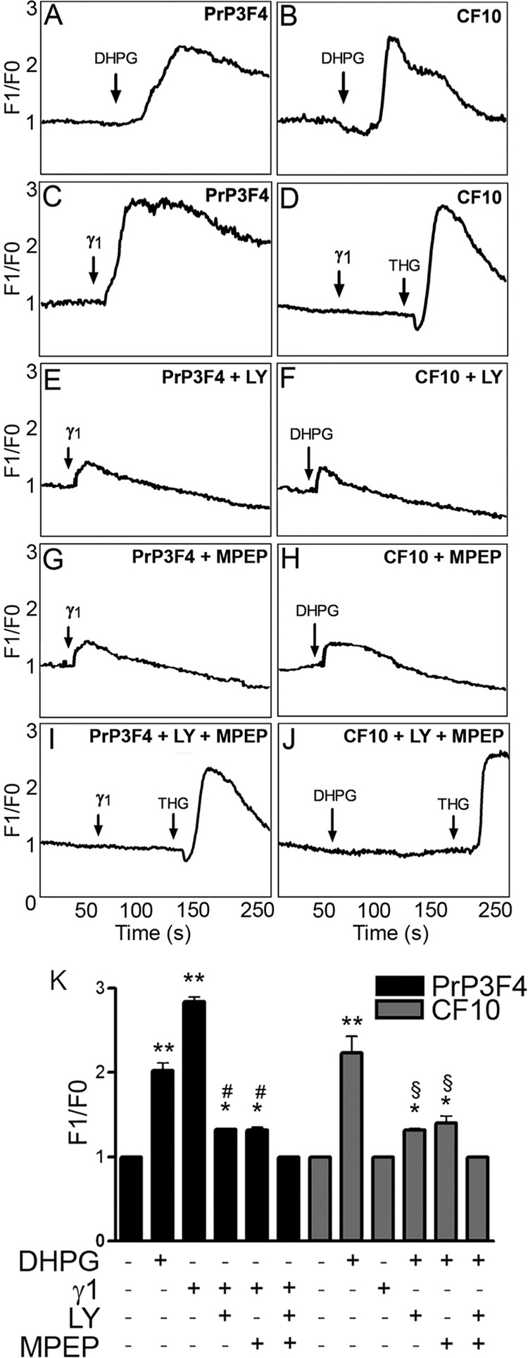
mGluR1 and mGluR5 mediate intracellular calcium increase upon PrPC-Ln γ1 peptide binding. A and B, CF10 cells expressing PrPC wild-type (PrP3F4) and their PrPC-null counterpart (CF10) were loaded with 10 μm Fluo3 AM and treated with the mGluR agonist DHPG (100 μm). C and D, shown is Ln γ1 peptide (37 μm) or thapsigargin (THG) (1 μm) in a medium supplemented with 2 mm CaCl2. Cells were also treated with Ln γ1 peptide or DHPG in the presence of mGluR1 antagonist (LY367385, 50 μm) (E and F), in the presence mGluR5 antagonist (MPEP, 5 μm) (G and H), or in the presence of both 50 μm LY367385 and 5 μm MPEP (I and J). K, relative intracellular calcium levels are expressed as the mean ± S.E. n = 3. *, p < 0.05 versus control; **, p < 0.001 versus control; #, p < 0.001 versus PrP3F4 treated with Ln γ1 peptide; §, p < 0.001 versus CF10 treated with DHPG.
When wild-type PrPC-transfected CF10 (PrP3F4) cells were treated with the Ln γ1 peptide, a significant increase in cytoplasmic calcium levels was observed (Fig. 3C). Conversely, no increases in intracellular Ca2+ were induced by Ln γ1 application in non-transfected PrP-null CF10 cells. These cells have normal levels of intracellular store calcium, as evidenced by their response to thapsigargin (Fig. 3D). Treatment with LY367385 (Fig. 3E) or MPEP (Figs. 3G) partially inhibited intracellular Ca2+ mobilization by Ln γ1 peptide in wild-type PrPC-transfected cells, whereas a combination of the two inhibitors completely abolished the Ln γ1 peptide-induced increases in the calcium signal (Fig. 3I). Quantitative analyses of these data are presented in Fig. 3K. These results are in accordance with our previous findings in primary hippocampal neurons (35) and indicate that the expression of wild-type PrPC in PrPC-null neuronal CF10 cells is able to reconstitute the cellular signaling modulated by the PrPC- Ln γ1-mGluR1/5 pathway.
CF10 Cells Expressing Mutant Prion Proteins Present Impaired Ln γ1-induced Calcium Signaling
Because calcium is one of the major intracellular signals triggered by PrPC-Ln γ1 peptide mGluR1/5, we then examined whether disease-related mutations in the PrPC protein cause disturbances in Ln γ1 peptide-induced intracellular calcium signaling. As previously demonstrated (Fig. 3C), the expression of wild-type PrPC, PrP3F4, rescued Ca2+ signaling triggered by Ln γ1 peptide (Fig. 4A) in CF10 cells. Interestingly, the expression of PrPC proteins carrying TSE-related mutations only partially rescued Ln γ1 peptide-induced Ca2+ signaling in CF10 cells when compared with cells expressing the wild-type protein (Fig. 4, C–H and I). In addition, when treated with DHPG, all cells bearing mutant proteins show an increment in calcium signaling similar to those expressing wild-type protein (Fig. 4J), indicating that they have similar levels and activity of mGluR1/5. These data indicate that TSE-associated PrPC mutations result in deficiencies in intracellular calcium mobilization triggered by Ln γ1 peptide.
FIGURE 4.

Partial impairment in calcium signaling mediated by Ln γ1 peptide in CF10 expressing mutant PrPC. CF10 cells transfected with wild-type PrPC (PrP3F4) (A), non-transfected (CF10) (B), or transfected with the PrPC mutants Lys-101 (C), Lys-104 (D), Val-116 (E), Asn-177 (F), Ile-179 (G), and Lys-199 (H) were loaded with 10 μm Fluo-3 AM and treated with Ln γ1 peptide (37 μm) and thapsigargin (THG) (1 μm) in medium supplemented with 2 mm CaCl2. I, relative intracellular calcium levels are expressed as the mean ± S.E. n = 7; *, p < 0.05; **, p < 0.01. J, shown is intracellular calcium concentration in cells treated or not with DHPG. Calcium levels at the untreated condition (white bars) were considered = 1, and the values after DHPG treatment (gray bars) were relative to it. Values represent the mean ± S.E. n = 3.
PrPC Mutations Impaired Process Outgrowth Triggered by Ln γ1 Peptide
As previously demonstrated (35), the association of PrPC and the Ln γ1 peptide co-opts mGluR1/5 to promote neuronal differentiation and neuritogenesis in primary neuronal cultures through the mobilization of intracellular Ca2+. CF10 cells are strongly positive for nestin (36) and neuron-specific enolase but are negative for βIII tubulin and NeuN (data not shown); thus, it is likely that they are neural progenitor cells. In addition, CF10 cells were immortalized via expression of the SV40 large T antigen and are, therefore, unable to arrest in the cell cycle, preventing the possibility of morphological alterations such as process outgrowth that resembles cell polarization and neurite extension observed in post-mitotic neurons (52, 53). Even the treatment with the full-length Ln, a powerful molecule associated with neurite extension (54), was unable to promote process outgrowth in CF10 cells with (PrP3F4) or without PrPC expression (Fig. 5A). To promote cell cycle arrest, CF10 cells were treated with mitomycin C, a well known inhibitor of cell proliferation in vitro (55–57). We found that at the concentration used, mitomycin C impaired cell growth (Fig. 5B) without affecting cell viability (Fig. 5C). Morphometric analyses showed that mitomycin C-treated CF10 cells either non-transfected or transfected with wild-type PrPC (PrP3F4) extended robust cytoplasmic processes when plated onto full-length laminin (Fig. 5D). Nonetheless, these cells do not express βIII tubulin and NeuN (data not shown) after this treatment. Yet, under specific experimental conditions, these cells are able to grow neurite-like processes; in addition, when full-length Ln is present, other receptors besides PrPC, probably integrins, may contribute to the phenotype. In contrast, process outgrowth was observed when wild-type PrPC-expressing cells (PrP3F4), but not the non-transfected CF10 one, was plated onto Ln γ1 peptide (Fig. 5D). These results confirm that the outgrowth of neurite-like processes, mediated by the PrPC-Ln γ1 interaction, can be rescued in CF10 cells expressing the wild-type PrPC.
FIGURE 5.

Process outgrowth mediated by the Ln γ1-peptide can be rescued after the reconstitution of PrPC expression in CF10 cells. A, CF10 cell lines with and without transfections of wild-type PrPC (PrP3F4) were cultured 48 h on coverslips pretreated with 10 μg/ml full-length laminin or 5 μg/ml poly-l-lysine (PL). Cells were fixed and labeled, and process outgrowth was quantified. B and C, PrP3F4- and non-transfected CF10 cells were treated with 10 μg/ml mitomycin C or vehicle for 3 h, washed, and plated on PL for 48 h to assess. The total cell number (proliferation) after 48 h (B) or cell death (using trypan blue) (C) after 3 h of mitomycin treatment. Values represent the mean ± S.E. (n = 3). *, p < 0.05 versus control. D, PrP3F4- and non-transfected CF10 cells pretreated with 10 μg/ml mitomycin C for 3 h were cultured on 5 μg/ml PL, PL plus laminin (2.5 or 5 μg/ml), or PL plus Ln γ1 peptide (Pep g1) (16.8 or 37 μm) for 48 h. Values represent the mean ± S.E. n = 3. *, p < 0.05 compared with the control PrP3F4 or CF10 in PL; **, p < 0.05 compared with the control PrP3F4 in PL.
In CF10 cells that express mutated PrPC, mitomycin C also inhibited cell growth (Fig. 6A) without affecting viability measured by trypan blue exclusion (Fig. 6B). Cell death induced by mitomycin C 48 h after treatment was also evaluated by activation of caspase 3. Contrary to the positive control, staurosporine, mytomycin C did not induce cell death (Fig. 6C). These cells together with non-transfected and wild-type PrPC-expressing CF10 cells (PrP3F4) were also evaluated for process outgrowth mediated by the Ln γ1 peptide. Fig. 6E shows representative images of the morphology of these cells after Ln γ1 peptide treatment and immunolabeling with MAP2. CF10 cells transfected with wild-type PrPC (PrP3F4) presented long cytoplasmic processes (arrows in the first panel; upper left). Remarkably, the addition of the Ln γ1 peptide was unable to promote process outgrowth in CF10 cells expressing mutated PrPC molecules (quantification in Fig. 6D). These results indicate that CF10 cells lacking PrPC or expressing mutated PrPC are unable to extend cytoplasmic processes upon Ln γ1 peptide treatment.
FIGURE 6.

Process outgrowth induced by Ln γ1 is impaired in cells expressing PrPC mutants. PrP3F4- and non-transfected CF10 cells as well as CF10 cells expressing the PrPC mutants Leu-101, Leu-104, Val-116, Asn-177, Ile-179, and Lys-199 were treated with 10 μg/ml mitomycin C or vehicle for 3 h washed and plated on poly-l-lysine for 48 h. A, shown is total cell number (proliferation). *, p < 0.05 versus control. B, shown is the percentage of cell death (using trypan blue) measured after 3 h of mitomycin C treatment. *, p < 0.05 versus control. C, shown is the percentage of cell death (capase-3 activation) using staurosporine (STS) as the positive control after 48 h of mitomycin C treatment. *, p < 0.05 versus control; #, p < 0.05 PrP3F4 STS treatment versus all others STS treatment. D, cells were treated with 10 μg/ml mitomycin C for 3 h, washed, plated on poly-l-lysine, and treated with 37 μm Ln γ1 scrambled (SCR) peptide or 37 μm Ln γ1 peptide (γ1) for 48 h. The percentage of cells with process outgrowth was evaluated (*, p < 0.05 versus al others). E, shown are representative images of CF10 cells expressing wild-type (PrP3F4) and PrPC mutants treated with mitomycin C followed by treatment with 37 μm Ln γ1 peptide. The arrows show the structures considered as typical process outgrowth. For all experiments values represent the mean ± S.E. of at least three independent experiments.
PrPC Mutations Impaired Protection against Cell Death Triggered by Ln γ1 Peptide
PrPC has been involved in neuronal protection against cell death, and consensus results from different groups showed that neurons from PrPC-null mice presented a higher sensitivity to agents that induce cell death, including staurosporine, than neurons from wild-type animals (for review, see Ref. 13). Remarkably, our results (Fig. 6C) demonstrated that CF10 cells or cells expressing mutated PrPC also presented a higher sensitivity to staurosporine than cells that were reconstituted with wild-type PrPC.
We then tested whether Ln γ1 chain peptide induces protection against staurosporine-induced cell death. Ln γ1 peptide, but not the SCR peptide, was able to protect PrP3F4 cells against staurosporine-induced cell death. However, any protective effect was observed when CF10 or CF10 cells expressing PrPC mutants were cultured on Ln γ1 peptide (Fig. 7A).
FIGURE 7.
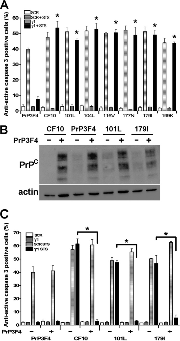
Cell survival promoted by Ln γ1 is impaired in cells expressing PrPC mutants. PrP3F4- and non-transfected CF10 cells as well as CF10 cells expressing PrPC mutants Leu-101, Leu-104, Val-116, Asn-177, Ile-179, and Lys-199 were plated on 5 μg/ml poly-l-lysine plus 37 μm Ln γ1 peptide (γ1) or 37 μm Ln γ1 SCR peptide and treated or not with 100 nm staurosporine (STS). A, quantification of active caspase 3-positive cells is shown. *, p < 0.01 Ln γ1 peptide plus STS treated CF10 and PrPC mutants versus Ln γ1 peptide plus STS treated PrP3F4. B, PrP3F4, CF10 cells, and CF10 cells expressing the PrPC mutants Lys-101 and Ile-179 were co-transfected with wild-type PrPC (PrP3F4) and immunoblotted for PrPC. C, cells were plated on 5 μg/ml poly-l-lysine plus 37 μm Ln γ1 peptide or 37 μm Ln γ1 SCR peptide and treated or not with 100 nm STS. Active caspase 3 positive cells were quantified. *, p < 0.01.
Finally, some experiments were conducted to evaluate if mutant PrPC molecules present dominant negative or prion effects upon wild-type PrPC. CF10, PrP3F4, and CF10 cells expressing the mutants Lys-101 and Ile-179 were transiently transfected with PrP3F4 (Fig. 7B) and tested for Ln γ1 protection against cell death induced by staurosporine. The expression of PrP3F4 was able to rescue the neuroprotective effects of Ln γ1 even when in the presence of mutated PrPC molecules (Fig. 7C), thus indicating that these mutants do not have a dominant negative or prion effect upon wild-type PrPC and may have lost their function triggered by engagement with Ln γ1 peptide.
DISCUSSION
The goal of this study was to investigate the consequences of PrPC loss-of-function that result from disease-associated mutations in the PrPC protein. To model the function of PrPC we expressed wild-type and mutant PrPC proteins on a PrPC-null background neural cell line (CF10). Our results show that substitutions within the PrPC protein decrease calcium signaling and impair process outgrowth and protective response that are mediated by the interaction between PrPC and Ln γ1 chain peptide.
The effects of PrPC and PrPC mutants expression have been previously investigated at the cellular level using neuroblastoma, breast cancer, fibroblasts, and Fischer rat thyroid cell lines (43, 58–61). However, data regarding the cellular and biochemical properties of prion proteins are controversial; disparities between groups may reflect differences in the cell types in which these proteins have been expressed (21, 27, 62–64). In the current application, we have addressed this question in non-tumor-derived neural cells; a model may more faithfully replicate the effects of prion proteins in neurons. Remarkably, the reconstitution of wild-type PrPC in PrPC-null neural CF10 cells was able to reproduce all the effects that have been observed in primary neuronal cultures after the interaction of PrPC with stress-inducible protein 1 (STI1) (39) and with Ln γ1 chain peptide (33).
The current study also demonstrates the utility of the CF10 cell lines as a model for the study of exogenously-expressed prion mutated proteins. We found that these cells expressed PrPC at the cell surface, where it should be to interact with ligands at the membrane or at the extracellular matrix (including Ln) and whose interaction modulates cellular functions (13). In addition, transfected CF10 cells express similar levels of the wild-type or mutated PrPC at the cell membrane, although some intracellular accumulation of the mutant PrPC proteins was detected. Moreover, only a minor PK resistance was observed in two of six PrPC mutants, suggesting that aggregation may not interfere with the function of these proteins.
PrPC binds to the carboxy-terminal domain of the Ln γ1 chain, a region where no other laminin receptors have been mapped (24). Thus, the use of the Ln γ1 peptide allows to specifically examinate phenotypes associated with the PrPC-Ln interaction (24). We recently demonstrated that the Ln γ1 peptide induces PrPC-dependent neuritogenesis via increases in intracellular Ca2+ and the activation of PKC and ERK1/2 in a manner dependent upon the participation of mGluR1/5, which also interact with PrPC (35).
Our data show that calcium signaling mediated by the Ln γ1 peptide and mGLUR1/5 is partially impaired in cells expressing the Leu-101, Leu-104, Val-116, Asn-177, Ile-179, and Lys-199 PrPC mutants compared with wild-type PrPC. Remarkably, in the course of TSE disease, it has been demonstrated that prion infection modifies Ca2+ responses (65) and impairs mGluR1/PLC/PKC pathway signaling in neurons derived from a murine model of BSE as well as humans with sporadic Creutzfeldt-Jakob disease (66, 67). Together, these results suggest that modifications in the PrPC protein impact type 1 mGluR-mediated cell signaling pathways and that these alterations play a role in both sporadic and genetic prion diseases.
Although only a partial decrease on the Ln γ1-mediated Ca2+ response was observed in cells expressing mutant PrPC proteins, we found that Ln γ1-induced process outgrowth was completely abolished in the presence of mutant PrPC molecules. One simple explanation is that the increment in Ca2+ levels did not achieve the threshold necessary to promote this phenotype in these cells. It is important to note that the Asn-177 and Ile-179 mutants are in the vicinity of the Ln γ1 binding site on the PrPC molecule, amino acids 173–182 (22). Thus, these alterations may abrogate the interaction between PrPC and laminin. On the other hand, we show similar results with PrPC molecules carrying disease-association mutations in the amino-terminal domain (Lys-101, Lys-104, and Val-116) or in the vicinity of the third α-helix region (Lys-199). It is possible that these mutations instead affect the interaction between PrPC and mGluR1/5; however, the binding site between these molecules has not yet been identified.
PrPC was also pointed to protect neurons against injury both in vitro and in vivo (13, 19, 25). PrPC protein is itself a receptor for the secreted form of STI1, an interaction that has been shown to promote neuroprotection and neuronal differentiation (25, 68, 69). Our present data indicate that Ln γ1 peptide impaired staurosporine-induced cell death in PrP3F4 cells, whereas Ln γ1 peptide fails to rescue CF10 and CF10 cells expressing PrPC mutants from death. Indeed, the results are consistent with the participation of the Ln γ1 peptide-PrPC complex in neuroprotection and that mutations within PrPC abolish its neuroprotective function. Furthermore, when the wild-type PrPC is co-expressed with mutant PrPC molecules, Ln γ1 peptide rescues staurosporine-induced cell death. This seems to show the absence of a dominant-negative effect, which is consistent with previous data showing that neurodegeneration mediated by expression of PrPC deletion mutants (some of the deleted sites include amino acids mutated here) could be reversed by expression of the wild-type protein (70–72).
The results of the current study may provide support to the idea that PrPC plays an important role as a scaffolding protein that has allosteric functions and dysfunctions (36). The interaction between PrPC and STI1 or between PrPC and the STI1 peptide 230–245, which mimics the PrPC binding site, has been shown to alter the structure of both components, potentially impacting the ability of either protein to interact with other ligands (73) and to organize a functional signaling platform (13). The conformational alterations imposed by the PrPC-Ln γ1 interaction remain unknown. However, some PrPC mutants have been shown to destabilize the native PrPC structure, possibly by increasing the stability of partially folded intermediate species (74–77). The conformational alterations imposed by these mutations may alter the ability of PrPC to interact with ligands such as laminin or mGluR1/5 and may even impair the interactions of these ligands with other proteins. Depending on the multicomplex formed, a specific cellular event could be partially impaired, as is observed for Ca2+ signaling, or completely blocked, as is the case for process outgrowth, by mutations in PrPC.
PrPC-mutated molecules may lose the ability to modulate Ln γ1-mediated neuronal plasticity and survival, thus representing the loss of a physiological function associated with the PrPC. This study points toward the necessity of further evaluation of the role of native and mutated PrPC proteins in neuronal plasticity in animal models of genetic prion diseases even before the onset of clinical symptoms.
Acknowledgments
We are also grateful to Roger Morris (Molecular Neurobiology Group, MRC Centre for Developmental Neurobiology, King's College London Guy's Campus) who kindly provided us with the pcDNA3 plasmid expressing wild-type PrPC and Suzette A. Priola (Rocky Mountain Laboratories, Laboratory of Persistent Viral Diseases, NIAID, National Institutes of Health) who provided the CF10 cells.
This work was supported by Fundação de Amparo à Pesquisa do Estado de São Paulo (FAPESP 2009/01427-2), the Howard Hughes Medical Institute, and the National Institute of Tanslational Neuroscience-Conselho Nacional de Desenvolvimento Científico e Tecnológico/Ministério da Ciência e Tecnologia (INNT-CNPq/MCT) and Ludwig Institute for Cancer Research. This work was also supported by the following fellowships from Fundação de Amparo à Pesquisa do Estado de São Paulo: 06/61251-7 (to C. F. M.); 09/51653–9 (T. G. S.); 08/04776-5 (to D. B.).
- TSE
- transmissible spongiform encephalopathies
- Ln
- laminin
- SCR
- scrambled
- DHPG
- (S)-3,5-dihydroxyphenylglycine
- MPEP
- 2-methyl-6-(phenylethynyl)-pyridine
- PK
- proteinase K
- STI1
- stress-inducible protein 1
- CJD
- Creutzfeldt-Jakob disease
- PL
- poly-l lysine.
REFERENCES
- 1. Weissmann C. (2004) The state of the prion. Nat. Rev. Microbiol. 2, 861–871 [DOI] [PubMed] [Google Scholar]
- 2. Prusiner S. B. (1998) The prion diseases. Brain Pathol. 8, 499–513 [DOI] [PMC free article] [PubMed] [Google Scholar]
- 3. Mallucci G., Dickinson A., Linehan J., Klöhn P. C., Brandner S., Collinge J. (2003) Depleting neuronal PrP in prion infection prevents disease and reverses spongiosis. Science 302, 871–874 [DOI] [PubMed] [Google Scholar]
- 4. Mallucci G. R., White M. D., Farmer M., Dickinson A., Khatun H., Powell A. D., Brandner S., Jefferys J. G., Collinge J. (2007) Targeting cellular prion protein reverses early cognitive deficits and neurophysiological dysfunction in prion-infected mice. Neuron 53, 325–335 [DOI] [PubMed] [Google Scholar]
- 5. White M. D., Farmer M., Mirabile I., Brandner S., Collinge J., Mallucci G. R. (2008) Single treatment with RNAi against prion protein rescues early neuronal dysfunction and prolongs survival in mice with prion disease. Proc. Natl. Acad. Sci. U.S.A. 105, 10238–10243 [DOI] [PMC free article] [PubMed] [Google Scholar]
- 6. Dorandeu A., Wingertsmann L., Chrétien F., Delisle M. B., Vital C., Parchi P., Montagna P., Lugaresi E., Ironside J. W., Budka H., Gambetti P., Gray F. (1998) Neuronal apoptosis in fatal familial insomnia. Brain Pathol. 8, 531–537 [DOI] [PMC free article] [PubMed] [Google Scholar]
- 7. Gray F., Adle-Biassette H., Chrétien F., Ereau T., Delisle M. B., Vital C. (1999) Neuronal apoptosis in human prion diseases. Bull. Acad. Natl. Med. 183, 305–320 [PubMed] [Google Scholar]
- 8. Gray F., Chrétien F., Adle-Biassette H., Dorandeu A., Ereau T., Delisle M. B., Kopp N., Ironside J. W., Vital C. (1999) Neuronal apoptosis in Creutzfeldt-Jakob disease. J. Neuropathol. Exp. Neurol. 58, 321–328 [DOI] [PubMed] [Google Scholar]
- 9. Faucheux B. A., Morain E., Diouron V., Brandel J. P., Salomon D., Sazdovitch V., Privat N., Laplanche J. L., Hauw J. J., Haïk S. (2011) Quantification of surviving cerebellar granule neurones and abnormal prion protein (PrPSc) deposition in sporadic Creutzfeldt-Jakob disease supports a pathogenic role for small PrPSc deposits common to the various molecular subtypes. Neuropathol. Appl. Neurobiol. 37, 500–512 [DOI] [PubMed] [Google Scholar]
- 10. Kim C., Haldiman T., Surewicz K., Cohen Y., Chen W., Blevins J., Sy M. S., Cohen M., Kong Q., Telling G. C., Surewicz W. K., Safar J. G. (2012) Small protease sensitive oligomers of PrPSc in distinct human prions determine conversion rate of PrP(C). PLoS. Pathog. 8, e1002835. [DOI] [PMC free article] [PubMed] [Google Scholar]
- 11. Resenberger U. K., Harmeier A., Woerner A. C., Goodman J. L., Müller V., Krishnan R., Vabulas R. M., Kretzschmar H. A., Lindquist S., Hartl F. U., Multhaup G., Winklhofer K. F., Tatzelt J. (2011) The cellular prion protein mediates neurotoxic signalling of β-sheet-rich conformers independent of prion replication. EMBO J. 30, 2057–2070 [DOI] [PMC free article] [PubMed] [Google Scholar]
- 12. Hetz C., Maundrell K., Soto C. (2003) Is loss of function of the prion protein the cause of prion disorders? Trends Mol. Med. 9, 237–243 [DOI] [PubMed] [Google Scholar]
- 13. Linden R., Martins V. R., Prado M. A., Cammarota M., Izquierdo I., Brentani R. R. (2008) Physiology of the prion protein. Physiol. Rev. 88, 673–728 [DOI] [PubMed] [Google Scholar]
- 14. Martins V. R., Beraldo F. H., Hajj G. N., Lopes M. H., Lee K. S., Prado M. M., Linden R. (2010) Prion protein. Orchestrating neurotrophic activities. Curr. Issues Mol. Biol. 12, 63–86 [PubMed] [Google Scholar]
- 15. Büeler H., Aguzzi A., Sailer A., Greiner R. A., Autenried P., Aguet M., Weissmann C. (1993) Mice devoid of PrP are resistant to scrapie. Cell 73, 1339–1347 [DOI] [PubMed] [Google Scholar]
- 16. Manson J. C., Clarke A. R., Hooper M. L., Aitchison L., McConnell I., Hope J. (1994) 129/Ola mice carrying a null mutation in PrP that abolishes mRNA production are developmentally normal. Mol. Neurobiol. 8, 121–127 [DOI] [PubMed] [Google Scholar]
- 17. Baumann F., Pahnke J., Radovanovic I., Rülicke T., Bremer J., Tolnay M., Aguzzi A. (2009) Functionally relevant domains of the prion protein identified in vivo. PLoS One 4, e6707. [DOI] [PMC free article] [PubMed] [Google Scholar]
- 18. Parchi P., Petersen R. B., Chen S. G., Autilio-Gambetti L., Capellari S., Monari L., Cortelli P., Montagna P., Lugaresi E., Gambetti P. (1998) Molecular pathology of fatal familial insomnia. Brain Pathol. 8, 539–548 [DOI] [PMC free article] [PubMed] [Google Scholar]
- 19. Prusiner S. B., Scott M. R. (1997) Genetics of prions. Annu. Rev. Genet. 31, 139–175 [DOI] [PubMed] [Google Scholar]
- 20. Goldfarb L. G., Petersen R. B., Tabaton M., Brown P., LeBlanc A. C., Montagna P., Cortelli P., Julien J., Vital C., Pendelbury W. W. (1992) Fatal familial insomnia and familial Creutzfeldt-Jakob disease. Disease phenotype determined by a DNA polymorphism. Science 258, 806–808 [DOI] [PubMed] [Google Scholar]
- 21. Bounhar Y., Zhang Y., Goodyer C. G., LeBlanc A. (2001) Prion protein protects human neurons against Bax-mediated apoptosis. J. Biol. Chem. 276, 39145–39149 [DOI] [PubMed] [Google Scholar]
- 22. Coitinho A. S., Freitas A. R., Lopes M. H., Hajj G. N., Roesler R., Walz R., Rossato J. I., Cammarota M., Izquierdo I., Martins V. R., Brentani R. R. (2006) The interaction between prion protein and laminin modulates memory consolidation. Eur. J. Neurosci. 24, 3255–3264 [DOI] [PubMed] [Google Scholar]
- 23. Coitinho A. S., Lopes M. H., Hajj G. N., Rossato J. I., Freitas A. R., Castro C. C., Cammarota M., Brentani R. R., Izquierdo I., Martins V. R. (2007) Short-term memory formation and long-term memory consolidation are enhanced by cellular prion association to stress-inducible protein 1. Neurobiol. Dis. 26, 282–290 [DOI] [PubMed] [Google Scholar]
- 24. Graner E., Mercadante A. F., Zanata S. M., Forlenza O. V., Cabral A. L., Veiga S. S., Juliano M. A., Roesler R., Walz R., Minetti A., Izquierdo I., Martins V. R., Brentani R. R. (2000) Cellular prion protein binds laminin and mediates neuritogenesis. Brain Res. Mol. Brain Res. 76, 85–92 [DOI] [PubMed] [Google Scholar]
- 25. Lopes M. H., Hajj G. N., Muras A. G., Mancini G. L., Castro R. M., Ribeiro K. C., Brentani R. R., Linden R., Martins V. R. (2005) Interaction of cellular prion and stress-inducible protein 1 promotes neuritogenesis and neuroprotection by distinct signaling pathways. J. Neurosci. 25, 11330–11339 [DOI] [PMC free article] [PubMed] [Google Scholar]
- 26. McLennan N. F., Brennan P. M., McNeill A., Davies I., Fotheringham A., Rennison K. A., Ritchie D., Brannan F., Head M. W., Ironside J. W., Williams A., Bell J. E. (2004) Prion protein accumulation and neuroprotection in hypoxic brain damage. Am. J. Pathol. 165, 227–235 [DOI] [PMC free article] [PubMed] [Google Scholar]
- 27. Roucou X., Guo Q., Zhang Y., Goodyer C. G., LeBlanc A. C. (2003) Cytosolic prion protein is not toxic and protects against Bax-mediated cell death in human primary neurons. J. Biol. Chem. 278, 40877–40881 [DOI] [PubMed] [Google Scholar]
- 28. Sakudo A., Lee D. C., Li S., Nakamura T., Matsumoto Y., Saeki K., Itohara S., Ikuta K., Onodera T. (2005) PrP cooperates with STI1 to regulate SOD activity in PrP-deficient neuronal cell line. Biochem. Biophys. Res. Commun. 328, 14–19 [DOI] [PubMed] [Google Scholar]
- 29. Sakurai-Yamashita Y., Sakaguchi S., Yoshikawa D., Okimura N., Masuda Y., Katamine S., Niwa M. (2005) Female-specific neuroprotection against transient brain ischemia observed in mice devoid of prion protein is abolished by ectopic expression of prion protein-like protein. Neuroscience 136, 281–287 [DOI] [PubMed] [Google Scholar]
- 30. Shyu W. C., Lin S. Z., Chiang M. F., Ding D. C., Li K. W., Chen S. F., Yang H. I., Li H. (2005) Overexpression of PrPC by adenovirus-mediated gene targeting reduces ischemic injury in a stroke rat model. J. Neurosci. 25, 8967–8977 [DOI] [PMC free article] [PubMed] [Google Scholar]
- 31. Weise J., Crome O., Sandau R., Schulz-Schaeffer W., Bähr M., Zerr I. (2004) Up-regulation of cellular prion protein (PrPc) after focal cerebral ischemia and influence of lesion severity. Neurosci. Lett. 372, 146–150 [DOI] [PubMed] [Google Scholar]
- 32. Weise J., Sandau R., Schwarting S., Crome O., Wrede A., Schulz-Schaeffer W., Zerr I., Bähr M. (2006) Deletion of cellular prion protein results in reduced Akt activation, enhanced postischemic caspase-3 activation, and exacerbation of ischemic brain injury. Stroke 37, 1296–1300 [DOI] [PubMed] [Google Scholar]
- 33. Zanata S. M., Lopes M. H., Mercadante A. F., Hajj G. N., Chiarini L. B., Nomizo R., Freitas A. R., Cabral A. L., Lee K. S., Juliano M. A., de Oliveira E., Jachieri S. G., Burlingame A., Huang L., Linden R., Brentani R. R., Martins V. R. (2002) Stress-inducible protein 1 is a cell surface ligand for cellular prion that triggers neuroprotection. EMBO J. 21, 3307–3316 [DOI] [PMC free article] [PubMed] [Google Scholar]
- 34. Jodoin J., Laroche-Pierre S., Goodyer C. G., LeBlanc A. C. (2007) Defective retrotranslocation causes loss of anti-Bax function in human familial prion protein mutants. J. Neurosci. 27, 5081–5091 [DOI] [PMC free article] [PubMed] [Google Scholar]
- 35. Beraldo F. H., Arantes C. P., Santos T. G., Machado C. F., Roffe M., Hajj G. N., Lee K. S., Magalhães A. C., Caetano F. A., Mancini G. L., Lopes M. H., Américo T. A., Magdesian M. H., Ferguson S. S., Linden R., Prado M. A., Martins V. R. (2011) Metabotropic glutamate receptors transduce signals for neurite outgrowth after binding of the prion protein to laminin γ1 chain. FASEB J. 25, 265–279 [DOI] [PubMed] [Google Scholar]
- 36. Linden R., Cordeiro Y., Lima L. M. (2012) Allosteric function and dysfunction of the prion protein. Cell. Mol. Life Sci. 69, 1105–1124 [DOI] [PMC free article] [PubMed] [Google Scholar]
- 37. Paulsson M., Aumailley M., Deutzmann R., Timpl R., Beck K., Engel J. (1987) Laminin-nidogen complex. Extraction with chelating agents and structural characterization. Eur. J. Biochem. 166, 11–19 [DOI] [PubMed] [Google Scholar]
- 38. Greil C. S., Vorberg I. M., Ward A. E., Meade-White K. D., Harris D. A., Priola S. A. (2008) Acute cellular uptake of abnormal prion protein is cell type and scrapie-strain independent. Virology 379, 284–293 [DOI] [PMC free article] [PubMed] [Google Scholar]
- 39. Caetano F. A., Lopes M. H., Hajj G. N., Machado C. F., Pinto Arantes C., Magalhães A. C., Vieira Mde P., Américo T. A., Massensini A. R., Priola S. A., Vorberg I., Gomez M. V., Linden R., Prado V. F., Martins V. R., Prado M. A. (2008) Endocytosis of prion protein is required for ERK1/2 signaling induced by stress-inducible protein 1. J. Neurosci. 28, 6691–6702 [DOI] [PMC free article] [PubMed] [Google Scholar]
- 40. Caetano F. A., Beraldo F. H., Hajj G. N., Guimaraes A. L., Jürgensen S., Wasilewska-Sampaio A. P., Hirata P. H., Souza I., Machado C. F., Wong D. Y., De Felice F. G., Ferreira S. T., Prado V. F., Rylett R. J., Martins V. R., Prado M. A. (2011) Amyloid-beta oligomers increase the localization of prion protein at the cell surface. J. Neurochem. 117, 538–553 [DOI] [PubMed] [Google Scholar]
- 41. Sunyach C., Jen A., Deng J., Fitzgerald K. T., Frobert Y., Grassi J., McCaffrey M. W., Morris R. (2003) The mechanism of internalization of glycosylphosphatidylinositol-anchored prion protein. EMBO J. 22, 3591–3601 [DOI] [PMC free article] [PubMed] [Google Scholar]
- 42. Campana V., Sarnataro D., Fasano C., Casanova P., Paladino S., Zurzolo C. (2006) Detergent-resistant membrane domains but not the proteasome are involved in the misfolding of a PrP mutant retained in the endoplasmic reticulum. J. Cell Sci. 119, 433–442 [DOI] [PubMed] [Google Scholar]
- 43. Schiff E., Campana V., Tivodar S., Lebreton S., Gousset K., Zurzolo C. (2008) Coexpression of wild-type and mutant prion proteins alters their cellular localization and partitioning into detergent-resistant membranes. Traffic 9, 1101–1115 [DOI] [PubMed] [Google Scholar]
- 44. Aguzzi A., Baumann F., Bremer J. (2008) The prion's elusive reason for being. Annu. Rev. Neurosci. 31, 439–477 [DOI] [PubMed] [Google Scholar]
- 45. Kiachopoulos S., Bracher A., Winklhofer K. F., Tatzelt J. (2005) Pathogenic mutations located in the hydrophobic core of the prion protein interfere with folding and attachment of the glycosylphosphatidylinositol anchor. J. Biol. Chem. 280, 9320–9329 [DOI] [PubMed] [Google Scholar]
- 46. van der Kamp M. W., Daggett V. (2009) The consequences of pathogenic mutations to the human prion protein. Protein Eng. Des. Sel. 22, 461–468 [DOI] [PMC free article] [PubMed] [Google Scholar]
- 47. van der Kamp M. W., Daggett V. (2010) Pathogenic mutations in the hydrophobic core of the human prion protein can promote structural instability and misfolding. J. Mol. Biol. 404, 732–748 [DOI] [PMC free article] [PubMed] [Google Scholar]
- 48. Watanabe Y., Hiraoka W., Shimoyama Y., Horiuchi M., Kuwabara M., Inanami O. (2008) Instability of familial spongiform encephalopathy-related prion mutants. Biochem. Biophys. Res. Commun. 366, 244–249 [DOI] [PubMed] [Google Scholar]
- 49. McKinley M. P., Bolton D. C., Prusiner S. B. (1983) A protease-resistant protein is a structural component of the scrapie prion. Cell 35, 57–62 [DOI] [PubMed] [Google Scholar]
- 50. Pastrana M. A., Sajnani G., Onisko B., Castilla J., Morales R., Soto C., Requena J. R. (2006) Isolation and characterization of a proteinase K-sensitive PrPSc fraction. Biochemistry 45, 15710–15717 [DOI] [PubMed] [Google Scholar]
- 51. Thackray A. M., Hopkins L., Bujdoso R. (2007) Proteinase K-sensitive disease-associated ovine prion protein revealed by conformation-dependent immunoassay. Biochem. J. 401, 475–483 [DOI] [PMC free article] [PubMed] [Google Scholar]
- 52. Ali S. H., DeCaprio J. A. (2001) Cellular transformation by SV40 large T antigen. Interaction with host proteins. Semin. Cancer Biol. 11, 15–23 [DOI] [PubMed] [Google Scholar]
- 53. Galderisi U., Jori F. P., Giordano A. (2003) Cell cycle regulation and neural differentiation. Oncogene 22, 5208–5219 [DOI] [PubMed] [Google Scholar]
- 54. Esch T., Lemmon V., Banker G. (1999) Local presentation of substrate molecules directs axon specification by cultured hippocampal neurons. J. Neurosci. 19, 6417–6426 [DOI] [PMC free article] [PubMed] [Google Scholar]
- 55. Blacker K. L., Williams M. L., Goldyne M. (1987) Mitomycin C-treated 3T3 fibroblasts used as feeder layers for human keratinocyte culture retain the capacity to generate eicosanoids. J. Invest. Dermatol. 89, 536–539 [DOI] [PubMed] [Google Scholar]
- 56. Ponchio L., Duma L., Oliviero B., Gibelli N., Pedrazzoli P., Robustelli della Cuna G. (2000) Mitomycin C as an alternative to irradiation to inhibit the feeder layer growth in long-term culture assays. Cytotherapy. 2, 281–286 [DOI] [PubMed] [Google Scholar]
- 57. Blake D. A., Sahiner N., John V. T., Clinton A. D., Galler K. E., Walsh M., Arosemena A., Johnson P. Y., Ayyala R. S. (2006) Inhibition of cell proliferation by mitomycin C incorporated into P(HEMA) hydrogels. J. Glaucoma 15, 291–298 [DOI] [PubMed] [Google Scholar]
- 58. Diarra-Mehrpour M., Arrabal S., Jalil A., Pinson X., Gaudin C., Piétu G., Pitaval A., Ripoche H., Eloit M., Dormont D., Chouaib S. (2004) Prion protein prevents human breast carcinoma cell line from tumor necrosis factor α-induced cell death. Cancer Res. 64, 719–727 [DOI] [PubMed] [Google Scholar]
- 59. Jodoin J., Misiewicz M., Makhijani P., Giannopoulos P. N., Hammond J., Goodyer C. G., LeBlanc A. C. (2009) Loss of anti-Bax function in Gerstmann-Sträussler-Scheinker syndrome-associated prion protein mutants. PLoS One 4, e6647. [DOI] [PMC free article] [PubMed] [Google Scholar]
- 60. Petersen R. B., Parchi P., Richardson S. L., Urig C. B., Gambetti P. (1996) Effect of the D178N mutation and the codon 129 polymorphism on the metabolism of the prion protein. J. Biol. Chem. 271, 12661–12668 [DOI] [PubMed] [Google Scholar]
- 61. Roucou X., Giannopoulos P. N., Zhang Y., Jodoin J., Goodyer C. G., LeBlanc A. (2005) Cellular prion protein inhibits proapoptotic Bax conformational change in human neurons and in breast carcinoma MCF-7 cells. Cell Death Differ. 12, 783–795 [DOI] [PubMed] [Google Scholar]
- 62. Fioriti L., Dossena S., Stewart L. R., Stewart R. S., Harris D. A., Forloni G., Chiesa R. (2005) Cytosolic prion protein (PrP) is not toxic in N2a cells and primary neurons expressing pathogenic PrP mutations. J. Biol. Chem. 280, 11320–11328 [DOI] [PubMed] [Google Scholar]
- 63. Ma J., Lindquist S. (2002) Conversion of PrP to a self-perpetuating PrPSc-like conformation in the cytosol. Science 298, 1785–1788 [DOI] [PubMed] [Google Scholar]
- 64. Ma J., Wollmann R., Lindquist S. (2002) Neurotoxicity and neurodegeneration when PrP accumulates in the cytosol. Science 298, 1781–1785 [DOI] [PubMed] [Google Scholar]
- 65. Kristensson K., Feuerstein B., Taraboulos A., Hyun W. C., Prusiner S. B., DeArmond S. J. (1993) Scrapie prions alter receptor-mediated calcium responses in cultured cells. Neurology 43, 2335–2341 [DOI] [PubMed] [Google Scholar]
- 66. Rodríguez A., Freixes M., Dalfó E., Martín M., Puig B., Ferrer I. (2005) Metabotropic glutamate receptor/phospholipase C pathway. A vulnerable target to Creutzfeldt-Jakob disease in the cerebral cortex. Neuroscience 131, 825–832 [DOI] [PubMed] [Google Scholar]
- 67. Rodríguez A., Martín M., Albasanz J. L., Barrachina M., Espinosa J. C., Torres J. M., Ferrer I. (2006) Group I mGluR signaling in BSE-infected bovine-PrP transgenic mice. Neurosci. Lett. 410, 115–120 [DOI] [PubMed] [Google Scholar]
- 68. Lima F. R., Arantes C. P., Muras A. G., Nomizo R., Brentani R. R., Martins V. R. (2007) Cellular prion protein expression in astrocytes modulates neuronal survival and differentiation. J. Neurochem. 103, 2164–2176 [DOI] [PubMed] [Google Scholar]
- 69. Roffé M., Beraldo F. H., Bester R., Nunziante M., Bach C., Mancini G., Gilch S., Vorberg I., Castilho B. A., Martins V. R., Hajj G. N. (2010) Prion protein interaction with stress-inducible protein 1 enhances neuronal protein synthesis via mTOR. Proc. Natl. Acad. Sci. U.S.A. 107, 13147–13152 [DOI] [PMC free article] [PubMed] [Google Scholar]
- 70. Li A., Christensen H. M., Stewart L. R., Roth K. A., Chiesa R., Harris D. A. (2007) Neonatal lethality in transgenic mice expressing prion protein with a deletion of residues 105–125. EMBO J. 26, 548–558 [DOI] [PMC free article] [PubMed] [Google Scholar]
- 71. Baumann F., Tolnay M., Brabeck C., Pahnke J., Kloz U., Niemann H. H., Heikenwalder M., Rülicke T., Bürkle A., Aguzzi A. (2007) Lethal recessive myelin toxicity of prion protein lacking its central domain. EMBO J. 26, 538–547 [DOI] [PMC free article] [PubMed] [Google Scholar]
- 72. Flechsig E., Hegyi I., Leimeroth R., Zuniga A., Rossi D., Cozzio A., Schwarz P., Rülicke T., Götz J., Aguzzi A., Weissmann C. (2003) Expression of truncated PrP targeted to Purkinje cells of PrP knockout mice causes Purkinje cell death and ataxia. EMBO J. 22, 3095–3101 [DOI] [PMC free article] [PubMed] [Google Scholar]
- 73. Romano S. A., Cordeiro Y., Lima L. M., Lopes M. H., Silva J. L., Foguel D., Linden R. (2009) Reciprocal remodeling upon binding of the prion protein to its signaling partner hop/STI1. FASEB J. 23, 4308–4316 [DOI] [PubMed] [Google Scholar]
- 74. Apetri A. C., Surewicz K., Surewicz W. K. (2004) The effect of disease-associated mutations on the folding pathway of human prion protein. J. Biol. Chem. 279, 18008–18014 [DOI] [PubMed] [Google Scholar]
- 75. Liemann S., Glockshuber R. (1999) Influence of amino acid substitutions related to inherited human prion diseases on the thermodynamic stability of the cellular prion protein. Biochemistry 38, 3258–3267 [DOI] [PubMed] [Google Scholar]
- 76. Rossetti G., Cong X., Caliandro R., Legname G., Carloni P. (2011) Common structural traits across pathogenic mutants of the human prion protein and their implications for familial prion diseases. J. Mol. Biol. 411, 700–712 [DOI] [PubMed] [Google Scholar]
- 77. Swietnicki W., Petersen R. B., Gambetti P., Surewicz W. K. (1998) Familial mutations and the thermodynamic stability of the recombinant human prion protein. J. Biol. Chem. 273, 31048–31052 [DOI] [PubMed] [Google Scholar]
- 78. Linden R., Martins V. R., Prado M. A. (2011) UCSD Nature Molecule Pages, doi: 10.1038/mp.a003935.01 [DOI] [Google Scholar]