Abstract
Objectives
The various types of cone beam CT (CBCT) differ in several technical characteristics, notably their spatial resolution, which is defined by the acquisition voxel size. However, data are still lacking on the effects of voxel size on the metric accuracy of three-dimensional (3D) reconstructions. This study was designed to assess the effect of isotropic voxel size on the 3D reconstruction accuracy and reproducibility of CBCT data.
Methods
The study sample comprised 70 teeth (from the Institut d’Anatomie Normale, Strasbourg, France). The teeth were scanned with a KODAK 9500 3D® CBCT (Carestream Health, Inc., Marne-la-Vallée, France), which has two voxel sizes: 200 µm (CBCT 200 µm group) and 300 µm (CBCT 300 µm group). These teeth had also been scanned with the KODAK 9000 3D® CBCT (Carestream Health, Inc.) (CBCT 76 µm group) and the SCANCO Medical micro-CT XtremeCT (SCANCO Medical, Brüttisellen, Switzerland) (micro-CT 41 µm group) considered as references. After semi-automatic segmentation with AMIRA® software (Visualization Sciences Group, Burlington, MA), tooth volumetric measurements were obtained.
Results
The Bland–Altman method showed no difference in tooth volumes despite a slight underestimation for the CBCT 200 µm and 300 µm groups compared with the two reference groups. The underestimation was statistically significant for the volumetric measurements of the CBCT 300 µm group relative to the two reference groups (Passing–Bablok method).
Conclusions
CBCT is not only a tool that helps in diagnosis and detection but it has the complementary advantage of being a measuring instrument, the accuracy of which appears connected to the size of the voxels. Future applications of such measurements with CBCT are discussed.
Keywords: cone-beam computed tomography, X-ray microtomography, three-dimensional imaging
Introduction
Cone beam CT (CBCT) allows the hard tissues of the maxillofacial region to be assessed in three dimensions.1-3 Several CBCT systems are currently on the market. They differ in several technical characteristics, notably their spatial resolution, which is defined by the size of the acquisition voxel.1-5 The clinical applications differ according to the size of the field of view (FOV) of the CBCT scanner. The smaller the FOV, the better the spatial resolution and the smaller the voxel size.
CBCT shows great promise of becoming a useful tool for both patient management and research.6 However, if it is to be relied on, the accuracy of the three-dimensional (3D) reconstructions coming from 3D images needs to be clearly established. Accuracy has already been assessed7 by comparing reconstructions using CBCT with those provided by the standard in 3D dental research, micro-CT.8-12 Similar volumetric measurements were obtained using CBCT with an isotropic voxel size of 76 µm and the reference method, micro-CT, with an isotropic voxel size of 41 µm.7 The important influence of voxel size on the quality of CBCT images and on scanning and reconstruction times is already acknowledged.13 However, data are still lacking on the effects of voxel size on the metric accuracy of reconstructions.
The aim of this study was to assess the influence of isotropic voxel size on 3D reconstruction accuracy and reproducibility. We assessed the accuracy of CBCT units by comparing volumetric measurements reconstructed with two isotropic voxel sizes against the references of the smaller voxel size CBCT and micro-CT measurements.
Materials and methods
Sample
The sample used ten mandibles from children who had died in the late nineteenth and early twentieth centuries and whose bodies were donated to science at the Institut d’Anatomie Normale, Strasbourg, France. French law does not require additional institutional review board approval to be obtained for research on existing human specimens. Table 1 details the gender (six females, four males) and age at death (14–64 months) of the children from whom the specimens were obtained. The sample comprised 70 developing germs of permanent teeth in all. Each mandible contained two to eight tooth germs according to the stage of development (Table 1).
Table 1. Sample (n = 70 teeth) investigated in this study. Teeth are labelled following the International Dental Federation nomenclature.
| Specimen number | Age (months) | Sex | Permanent germ or tooth (n) |
| 202 | 42 | F | 31–32–33–36–41–42–43–46 (8) |
| 210 | 54 | F | 31–32–33–36–41–42–43–46 (8) |
| 211 | 36 | F | 31–32–33–36–41–42–43–46 (8) |
| 213 | 30 | M | 31–32–33–41–42–43 (6) |
| 214 | 30 | M | 31–32–33–36–41–42–43–46 (8) |
| 318 | 14 | F | 36–46 (2) |
| 319 | 48 | F | 31–32–33–36–41–42–43–46 (8) |
| 383 | 64 | M | 31–32–33–36–41–42–43–46 (8) |
| 512 | 54 | M | 31–32–33–41–42–43 (6) |
| 574 | 64 | F | 31–32–33–36–41–42–43–46 (8) |
The size of the study sample was calculated by considering the confidence interval (CI) of the intraclass correlation coefficient (ICC)14 found from the results of a previous study,7 which was 0.997 (95% CI 0.996 to 0.998). We expected to observe an ICC greater than 0.9. It was necessary to include 70 teeth in each group to estimate an ICC with a 95% CI of ±5% between the volumes reconstructed using the different CBCT systems.
Data acquisition
Each mandible was scanned in 2010 using a CBCT scanner (KODAK 9500 3D; Carestream Health, Inc., Marne-la-Vallée, France) with two isotropic voxel sizes: 200 µm and 300 µm, two FOVs: 90×150 mm and 180×200 mm, a tube potential of 90 kV and a tube current of 10 mA. Other acquisitions were also included in this sample. These acquisitions were made with two devices: a CBCT scanner [KODAK 9000 3D® (Carestream Health, Inc.), isotropic voxel size 76 µm, FOV 50×37 mm, tube potential 85 kV, tube current 2 mA] and a high-resolution peripheral micro-CT scanner [XtremeCT (SCANCO Medical, Brüttisellen, Switzerland), isotropic voxel size 41 µm, FOV 126×150 mm, tube potential 60 kV, tube current 1 mA].
Data exporting, segmentation and 3D reconstruction
Data were exported in DICOM (digital imaging and communications in medicine) format and then converted to TIFF (tagged image file) format for the subsequent tooth-of-interest segmentation with the AMIRA® software package (v. 5, Visualization Sciences Group, Burlington, MA, http://www.amiravis.com). As there is no fully automatic segmentation method available yet, we carried out semi-automatic segmentation of the micro-CT and CBCT data sets. After segmentation, the 3D triangle-based surface of each tooth was reconstructed in PLY (polygon) format without smoothing to preserve its raw volume measurement.
The volumetric measurements obtained were divided into four groups:
CBCT 200 µm group: volumetric measurements obtained from CBCT 9500 3D (KODAK) acquisitions with a voxel size of 200 µm
CBCT 300 µm group: volumetric measurements obtained from CBCT 9500 3D (KODAK) acquisitions with a voxel size of 300 µm
CBCT 76 µm group: volumetric measurements obtained from CBCT 9000 3D (KODAK) acquisitions with a voxel size of 76 µm
micro-CT 41 µm group: volumetric measurements obtained from micro-CT Xtreme CT acquisitions with a voxel size of 41 µm.
The volumetric measurements of the CBCT 200 µm and 300 µm groups were compared with those of the CBCT 76 µm and micro-CT 41 µm groups.
Reproducibility of measurements
CBCT 200 µm and CBCT 300 µm groups
Data for slices of a random sample of 20 teeth were re-examined 1 week after the initial examination to test intraexaminer consistency using the ICC.15 The ICC was also calculated to assess interexaminer reproducibility, again using a random sample of 20 teeth. Two observers (DM and AP) were trained on AMIRA before any segmentation.
CBCT 76 µm and micro-CT 41 µm groups
The intra- and interexaminer reproducibility between the CBCT (9000 3D KODAK) and the micro-CT (XtremeCT) has been calculated previously.7
Comparison of volumes
All data were analysed with two statistical software packages, R.2.10.0 (r-project.org) and MedCalc (medcalc.org). Potential relationships between the volumetric measurements were highlighted by Pearson correlation analysis and the ICC. The agreement between the two techniques was assessed by comparing the results by means of the Bland–Altman method in which the difference between measurements is plotted against their mean (considered to be the best estimate of the true values).16 The limits of agreement represent the deviation of the CBCT 200 µm or 300 µm group values from those of the CBCT 76 µm or micro-CT 41 µm group.
The Passing–Bablok method was used for regression analysis and p-values of ≤0.05 were taken to indicate significance.17 This is a non-parametric method for estimating the slope of the relationship (Slope b) between two measurements that are compared and the ordinate of the relationship at the origin (Intercept a). If the 95% CI of Slope b includes 1 and that of Intercept a includes the value 0, there is no statistically significant difference.17
Results
Study sample
The four groups (CBCT 200 µm, CBCT 300 µm, CBCT 76 µm, micro-CT 41 µm) were composed of 70 volumetric measurements each.
Reproducibility of measurements
The intraexaminer reproducibility of 20 volume measurements was very high, with ICCs of 0.998 and 0.999 for CBCT 200 µm and CBCT 300 µm, respectively. The interexaminer reproducibility of 20 volume measurements was also very high, with ICCs of 0.999 and 0.988 for CBCT 200 µm and CBCT 300 µm, respectively.
With the groups CBCT 76 µm and micro-CT 41 µm, intra- and interexaminer reproducibility were also very high.7
Comparison of volumes for CBCT 200 µm group vs CBCT 76 µm group or micro-CT 41 µm group
A strong correlation was found for all comparisons (Table 2).
Table 2. Relationships between data compared with Pearson correlation coefficient and Passing–Bablok method, sample size for each group: n = 70.
| Comparison devices (size of voxel) | r2 | Intercept a (95% CI) | Slope b (95% CI) |
| CBCT 9500 (200 µm), CBCT 9000 (76 µm) | 0.9997 | −0.377 (−1.62 to 0.709) | 1.002 (0.994–1.009) |
| CBCT 9500 (200 µm), micro-CT (41 µm) | 0.9997 | −0.624 (−2.158 to 0.411) | 0.996 (0.988–1.006) |
| CBCT 9500 (300 µm), CBCT 9000 (76 µm) | 0.9996 | −1.925 (−3.318 to −0.851) | 1.009 (1.002–1.018) |
| CBCT 9500 (300 µm), micro-CT (41 µm) | 0.9998 | −1.883 (−3.008 to −1.129) | 1.004 (0.998–1.012) |
CI, confidence interval; CBCT, cone beam CT.
The CBCT 200 µm group and the CBCT 76 µm group (r2 = 0.9997, p < 0.0001), ICC = 0.998 (95% CI 0.996–0.999).
The CBCT 200 µm group and the micro-CT 41 µm group (r2 = 0.9997, p < 0.0001), ICC = 0.997 (95% CI 0.994–0.998).
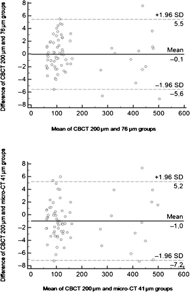
We compared the degree of agreement with the Bland–Altman method. About 95% of the errors on measurements estimated by the difference between the two groups lay within the limits of agreement (Figure 1). The volume reconstructions for the CBCT 200 µm group were not graphically different from those obtained in the CBCT 76 µm and micro-CT 41 µm groups. We observed that the CBCT 200 µm slightly underestimated volumetric measurements compared with the CBCT 76 µm and the micro-CT (Figure 1).
Figure 1.

Bland–Altman plot of volumes of teeth between cone beam CT (CBCT) 200 µm and 76 µm groups (top) and CBCT 200 µm and micro-CT 41 µm groups (bottom). Negative values indicate smaller volumes calculated from the CBCT 200 µm group than with the CBCT 76 µm (top) and micro-CT 41 µm groups (bottom). SD, standard deviation
The Passing–Bablok regression showed no statistically significant difference between the CBCT 200 µm and CBCT 76 µm or micro-CT 41 µm volume measurements (Table 2).
Comparison of volumes for the CBCT 300 µm group vs the CBCT 76 µm group or micro-CT 41 µm group
A strong correlation was found for all comparisons (Table 2).
The CBCT 300 µm group and the CBCT 76 µm group (r2 = 0.9996, p < 0.0001), ICC = 0.997 (95% CI 0.995–0.998).
The CBCT 300 µm group and the micro-CT 41 µm group (r2 = 0.9998, p < 0.0001), ICC = 0.998 (95% CI 0.996–0.999).
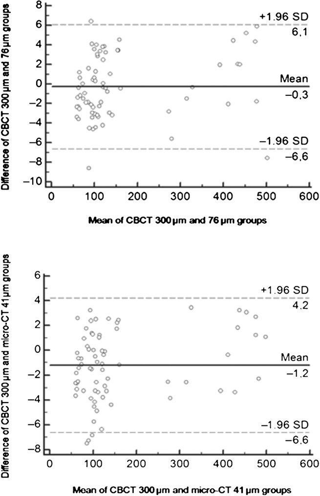
We compared the degree of agreement with the Bland–Altman method. About 95% of the errors on measurements estimated by the difference between the two groups lay within the limits of agreement (Figure 2). We observed that the CBCT 300 µm underestimated volumetric measurements compared with the CBCT 76 µm and the micro-CT 41 µm (Figure 2). Based on the Passing–Bablok regression, the hypothesis that the Intercept a = 0 was not accepted (Table 2).
Figure 2.

Bland–Altman plot of volumes of teeth between cone beam (CBCT) 300 µm and 76 µm groups (top) and CBCT 300 µm and micro-CT 41 µm groups (bottom). Negative values indicate smaller volumes calculated from the CBCT 300 µm group than with the CBCT 76 µm (top) and micro-CT 41 µm groups (bottom). SD, standard deviation
Discussion
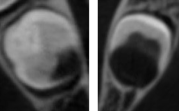
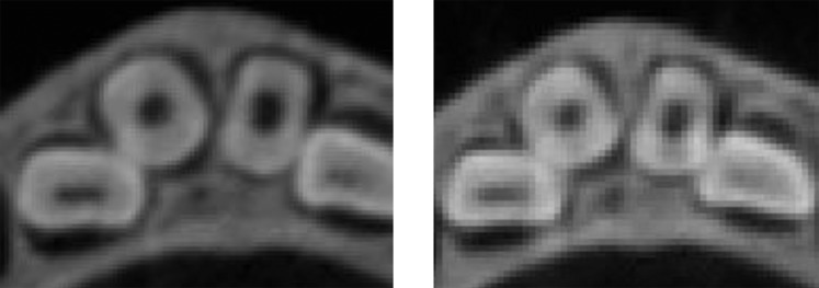
The reconstructions from volumetric measurements at 200 µm and 300 µm were compared with those obtained with CBCT, using a voxel size of 76 µm, and micro-CT, voxel size of 41 µm, considered as references. We showed in an earlier study that the volumes obtained with CBCT 76 µm were similar to those from micro-CT 41 µm.7 The Kodak 9000 3D provided submillimetre isotropic voxel resolution that was the closest to that given by micro-CT. The influence of voxel size can thus be assessed using these two reference systems. Segmentation requires thorough knowledge of the images to be analysed and of the information to be extracted subsequently. The prior training of users (DM and AP) in displaying the segmentation of the images before starting to build up the database was an indispensable stage. Good knowledge of the situation enables the user to separate artefacts and noise from what is of interest. Nevertheless, the subjectivity involved in this method can lead to different results being found by two users or by a single user between two segmentations of the same tooth. The visualization among the different voxel sizes (i.e. 41 µm; 76 µm; 200 µm; and 300 µm voxel size devices) is shown (Figure 3). With the CBCT, the signal-to-noise ratio and the contrast were lower than for the micro-CT. Also, the segmentation of a tooth becomes more difficult when it is adjacent to cortical bone (Figure 4), or to an another tooth (Figure 5), a difficulty already reported.18 It is thus indispensable to include inter- and intraindividual reproducibility calculations in the statistical analysis plan. Both intra- and interexaminer reproducibility were very high in the 200 µm and 300 µm CBCT groups.
Figure 3.
Visualization of images relative to voxel size. The same tooth is shown (Tooth 36, Specimen Number Emb 383) with a voxel size of (a) 41 µm; (b) 76 µm; (c) 200 µm; (d) 300 µm
Figure 4.
Cone beam CT images with a voxel size of 200 µm (left, Tooth 36, Specimen Number Emb 574; right, Tooth 46, Specimen Number Emb 202). The tooth and the adjacent cortical bone are very close, illustrating the difficulty with segmentation
Figure 5.
Cone beam CT images with a voxel size of 200 µm (left) and 300 µm (right) (teeth 31, 32, 41, 42; specimen number Emb 383). Note that the tooth of interest and another are very close, the distinction between two teeth is difficult
In a study of the relationships existing between volumetric measurements, the comparison of two series of these values requires various statistical techniques.19 With the Bland–Altman method, the tooth volumes were slightly underestimated by the CBCT 200 µm compared with the CBCT 76 µm or micro-CT 41 µm groups. With the Passing–Bablok method, the null hypothesis of the Intercept a and the Slope b were accepted. No statistically significant differences were found between the 200 µm and 76 µm or 41 µm volumetric measurements.
Some differences between groups were apparent in the results when 300 µm CBCT and 76 µm CBCT or 41 µm micro-CT data were compared. The Bland–Altman method indicated that the tooth volumes were slightly underestimated by CBCT 300 µm. Even though highly significant linear correlation was indicated by the Pearson correlation coefficient, the Passing–Bablok method rejected the null hypothesis. The CBCT 300 µm significantly underestimated volumetric measurements compared with the CBCT 76 µm and the micro-CT 41 µm. This measurement underestimation can be explained by the partial volume effect, a consequence of the spatial resolution. The spatial resolution is affected by the partial volume effect, which has repercussions on the image quality.20,21 CBCT devices that have a high spatial resolution are less affected by partial volume effect as their voxel sizes are smaller.21 Compared with micro-CT, CBCT underestimates volumetric measurements. This slight underestimation does not appear to be significant for voxel sizes of 200 µm and 76 µm.7 The underestimation increases and becomes statistically significant for a voxel size of 300 µm. Moreover, the images become less sharp as the voxel size increases and certain features, like tooth fissures, connected with the post-mortem dehydration process are less visible on images obtained with the CBCT at 300 µm. This is in agreement with the results found in the literature.22 Increasing the acquisition resolution of CBCT improves the detection of anatomical structures, such as the mesiobuccal canals in maxillary molars.22
The use of CBCT is increasing in dental practice. However, CBCT is not just a tool that helps in diagnosis and detection; it has the complementary advantage of being a measuring instrument.23,24 The accuracy of the measurement and its clinical significance must be considered in the context of each study. It should be noted that the contrast, signal-to-noise-ratio and sharpness of the images obtained from CBCT acquisitions still need to be improved to make segmentation easier. The perspective of segmenting zones of interest in routine clinical practice using CBCT as a measuring instrument would require a fast method that could be carried out automatically. Obtaining volumetric measurements from CBCT acquisitions is interesting in the context of age estimation.25-27 Star et al27 presented a dental age estimation methodology using 3D calculations on CBCT scans of fully developed single-rooted teeth from living individuals by assessing pulp–tooth volume ratios on clinically acquired CBCT images. The improvements, and suitable separating and segmenting software, will enable CBCT results to be obtained faster in the near future.27 Finally, the prospect of a clinical application of CBCT as a measuring tool is of interest in endodontics.28 3D analysis of the canal network and volumetric measurements of the pulp would provide complementary information for the new engine-driven endodontic instrument, the self-adjusting file, which adapts to the shape of the root canal in the three dimensions, including its cross-section.29-31
In conclusion, volumetric measurements made with CBCT are all similar for voxel sizes up to 200 µm despite a slight tendency towards underestimation, which increases with voxel size. At 300 µm and beyond, the underestimation of the measurements becomes statistically significant. It would be interesting to clarify the significance limit by studying reconstructions made using voxel sizes between 200 µm and 300 µm.
Acknowledgments
We thank S Becker for language revision, and L Braak (Institut de Médecine et de Physiologie Spatiales, Toulouse, France), E Legrand (Pau, France) and V Fayat (Toulouse, France) for the data acquisition.
References
- 1.Scarfe WC, Farman AG, Sukovic P. Clinical applications of cone-beam computed tomography in dental practice. J Can Dent Assoc 2006;72:75–80 [PubMed] [Google Scholar]
- 2.Patel S, Dawood A, Ford TP, Whaites E. The potential applications of cone beam computed tomography in the management of endodontic problems. Int Endod J 2007;40:818–830 [DOI] [PubMed] [Google Scholar]
- 3.Dawood A, Patel S, Brown J. Cone beam CT in dental practice. Br Dent J 2009;207:23–28 [DOI] [PubMed] [Google Scholar]
- 4.Hatcher DC. Operational principles for cone-beam computed tomography. J Am Dent Assoc 2010;141:3S–6S [DOI] [PubMed] [Google Scholar]
- 5.Watanabe H, Honda E, Tetsumura A, Kurabayashi T. A comparative study for spatial resolution and subjective image characteristics of a multi-slice CT and a cone-beam CT for dental use. Eur J Radiol 2011;77:397–402 [DOI] [PubMed] [Google Scholar]
- 6.Pinsky HM, Dyda S, Pinsky RW, Misch KA, Sarment DP. Accuracy of three-dimensional measurements using cone-beam CT. Dentomaxillofac Radiol 2006;35:410–416 [DOI] [PubMed] [Google Scholar]
- 7.Maret D, Molinier F, Braga J, Peters OA, Telmon N, Treil J, et al. Accuracy of 3D reconstructions based on cone beam computed tomography. J Dent Res 2010;89:1465–1469 [DOI] [PubMed] [Google Scholar]
- 8.Peters OA, Laib A, Ruegsegger P, Barbakow F. Three-dimensional analysis of root canal geometry by high-resolution computed tomography. J Dent Res 2000;79:1405–1409 [DOI] [PubMed] [Google Scholar]
- 9.Hannig C, Krieger E, Dullin C, Merten HA, Attin T, Grabbe E, et al. Volumetry of human molars with flat panel based volume CT in vitro. Clin Oral Investig 2006;10:253–257 [DOI] [PubMed] [Google Scholar]
- 10.Olejniczak AJ, Tafforeau P, Smith TM, Temming H, Hublin JJ. Technical note: compatibility of microtomographic imaging systems for dental measurements. Am J Phys Anthropol 2007;134:130–134 [DOI] [PubMed] [Google Scholar]
- 11.Kato A, Ohno N. Construction of three-dimensional tooth model by micro-computed tomography and application for data sharing. Clin Oral Investig 2009;13:43–46 [DOI] [PubMed] [Google Scholar]
- 12.Someda H, Saka H, Matsunaga S, Ide Y, Nakahara K, Hirata S, et al. Age estimation based on three-dimensional measurement of mandibular central incisors in Japanese. Forensic Sci Int 2009;185:110–114 [DOI] [PubMed] [Google Scholar]
- 13.Kamburoglu K, Kursun S. A comparison of the diagnostic accuracy of CBCT images of different voxel resolutions used to detect simulated small internal resorption cavities. Int Endod J 2010;43:798–807 [DOI] [PubMed] [Google Scholar]
- 14.Streiner DL, Norman GR. Health measurement scales: a practical guide to their development and use. Oxford, UK: Oxford University Press; 2003 [Google Scholar]
- 15.Ferrante L, Cameriere R. Statistical methods to assess the reliability of measurements in the procedures for forensic age estimation. Int J Legal Med 2009;123:277–283 [DOI] [PubMed] [Google Scholar]
- 16.Bland JM, Altman DG. Statistical methods for assessing agreement between two methods of clinical measurements. Lancet 1986;1:307–310 [PubMed] [Google Scholar]
- 17.Passing H, Bablok W. A new biometrical procedure for testing the equality of measurements from two different analytical methods. Applications of linear regression procedures for method comparison studies in clinical chemistry. J Clin Chem Clin Biochem 1983;21:709–720 [DOI] [PubMed] [Google Scholar]
- 18.Liu Y, Olszewski R, Alexandroni ES, Enciso R, Xu T, Mah JK. The validity of in vivo tooth volume determinations from cone-beam computed tomography. Angle Orthodontist 2010;80:160–166 [DOI] [PMC free article] [PubMed] [Google Scholar]
- 19.Bonnet N, Laroche N, Vico L, Dolleans E, Courteix D, Benhamou CL. Assessment of trabecular bone microarchitecture by two different x-ray microcomputed tomographs: a comparative study of the rat distal tibia using Skyscan and Scanco devices. Med Phys 2009;36:1286–1297 [DOI] [PubMed] [Google Scholar]
- 20.Chakeres DW. Clinical significance of partial volume averaging of the temporal bone. AJNR Am J Neuroradiol 1984;5:297–302 [PMC free article] [PubMed] [Google Scholar]
- 21.Molen AD. Considerations in the use of cone-beam computed tomography for buccal bone measurements. Am J Orthod Dentofacial Orthop 2010;137Suppl. 1:S130–S135 [DOI] [PubMed] [Google Scholar]
- 22.Bauman R, Scarfe W, Clark S, Morelli J, Scheetz J, Farman A. Ex vivo detection of mesiobuccal canals in maxillary molars using CBCT at four different isotropic voxel dimensions. Int Endod J 2011;44:752–758 [DOI] [PubMed] [Google Scholar]
- 23.Suomalainen A, Vehmas T, Kortesniemi M, Robinson S, Peltola J. Accuracy of linear measurements using dental cone beam and conventional multislice computed tomography. Dentomaxillofac Radiol 2008;37:10–17 [DOI] [PubMed] [Google Scholar]
- 24.Stratemann SA, Huang JC, Maki K, Miller AJ, Hatcher DC. Comparison of cone beam computed tomography imaging with physical measures. Dentomaxillofac Radiol 2008;37:80–93 [DOI] [PubMed] [Google Scholar]
- 25.Yang F, Jacobs R, Willems G. Dental age estimation through volume matching of teeth imaged by cone-beam CT. Forensic Sci Int 2006;159:78–83 [DOI] [PubMed] [Google Scholar]
- 26.Maret D, Peters OA, Dedouit F, Telmon N, Sixou M. Cone-beam computed tomography: a useful tool for dental age estimation? Med Hypotheses 2011;76:700–702 [DOI] [PubMed] [Google Scholar]
- 27.Star H, Thevissen P, Jacobs R, Fieuws S, Solheim T, Willems G. Human dental age estimation by calculation of pulp-tooth volume ratios yielded on clinically acquired cone beam computed tomography images of monoradicular teeth. J Forensic Sci 2011;56Suppl. 1:S77–S82 [DOI] [PubMed] [Google Scholar]
- 28.Michetti J, Maret D, Mallet JP, Diemer F. Validation of cone beam computed tomography as a tool to explore root canal anatomy. J Endod 2010;36:1187–1190 [DOI] [PubMed] [Google Scholar]
- 29.Metzger Z, Teperovich E, Zary R, Cohen R, Hof R. The self-adjusting file (SAF). Part 1: respecting the root canal anatomy–a new concept of endodontic files and its implementation. J Endod 2010;36:679–690 [DOI] [PubMed] [Google Scholar]
- 30.Metzger Z, Zary R, Cohen R, Teperovich E, Paqué F. The quality of root canal preparation and root canal obturation in canals treated with rotary versus self-adjusting files: a three-dimensional micro-computed tomographic study. J Endod 2010;36:1569–1573 [DOI] [PubMed] [Google Scholar]
- 31.Paqué F, Peters OA. Micro-computed tomography evaluation of the preparation of long oval root canals in mandibular molars with the self-adjusting file. J Endod 2011;37:517–521 [DOI] [PubMed] [Google Scholar]


