Figure 2.
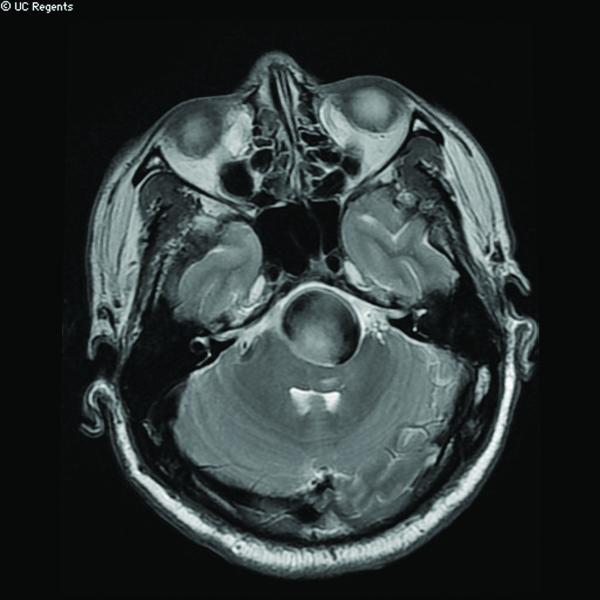
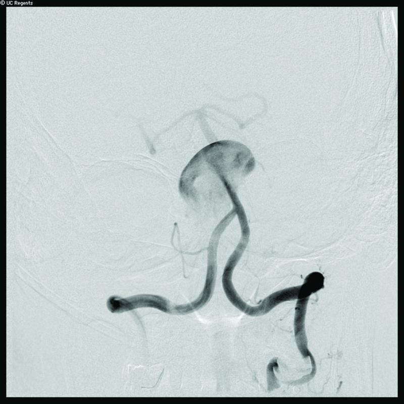
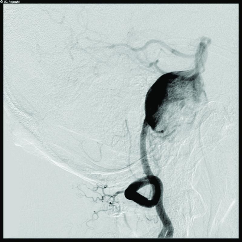
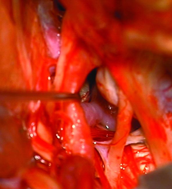
Case 14. (A) Axial T2-weighted magnetic resonance images demonstrated a giant basilar trunk aneurysm with marked pontine compression in this 52 year-old man. The aneurysm had dolichoectatic morphology with separation of afferent and efferent basilar artery, as seen on digital subtraction angiography (left vertebral artery injection, (B) anteroposterior and (C) lateral views). (D) The distal aneurysm was exposed with an orbitozygomatic-pterional craniotomy and dissection through the carotid-oculomotor triangle. (E) A radial artery graft was anastomosed to the P2 segment, (F) as part of an MCA-PCA bypass. Note the proximal donor anastomosis in the Sylvian fissure. (G) The aneurysm was distal occluded with an aneurysm clip. (H) Postoperative angiography demonstrated slow antegrade filling of the aneurysm and brisk filling of the basilar apex through the bypass (right ICA injection, anteroposterior view). Gradual aneurysm thrombosis also occluded basilar perforators and the patient died. This case demonstrates that post-surgical aneurysm thrombosis can occlude perforators, even with distal aneurysm occlusion that preserves antegrade flow.

