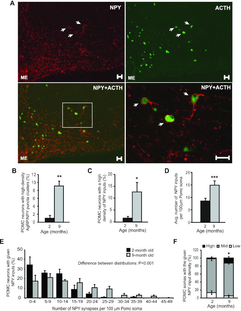
Fig. 4.
POMC neurons are among the targets of age-dependent increase in AgRP/NPY innervation. A, Wide-field image from a middle-aged mouse (9 months) depicting POMC neurons in the arcuate nucleus receiving a dense degree of NPY inputs. NPY immunoreactivity is shown in red and ACTH (POMC product) is in green. Scale bar, 25 μm. B, Percentage of POMC neurons that were surrounded by clusters containing a high density of AgRP/NPY puncta in young adult (2 months) and middle-aged (9 months) mice. C, Confocal quantification of the percentage of POMC neurons that received a high density of NPY inputs. D, Average number of NPY puncta per 100 μm POMC soma in young and middle-aged mice. E and F, Distribution plots for the percentage of POMC cells receiving a given density of NPY inputs in 2-month-old and 9-month-old mice. C–E, n = 3–5, n ≥ 16. Error bars denote sem. *, P < 0.05, **, P < 0.01, ***, P ≤ 0.001 compared with 2 months.

