Abstract
In a study visualizing ventilation with hyperpolarized 3He magnetic resonance imaging (MRI) in elite breath hold divers, the dynamic MRI images in one subject exhibited an apparent alternation of the image intensity between left and right lung. We hypothesized that the alternation resulted from alternating variations in inspiratory flow rate to left and right lungs. Analysis showed the alternation was not due to random uncorrelated temporal fluctuations of intensity (p < 0.001). The frequency of alternation was approximately 56 min−1, suggesting a cardiac origin. Similar alternation of ventilation was confirmed retrospectively in 4 of 6 additional subjects. These observations are consistent with previous studies showing cardiogenic mixing of gas in the lung. We speculate that cardiogenic pendelluft, possibly from ballistic lateral motion of the beating heart, could cause alternating variations of inspiratory flow to the lungs.
Keywords: pulmonary ventilation, magnetic resonance imaging, hyperpolarized helium, cardiogenic mixing
1. Introduction
In a previous study of lung mechanics in elite breath-hold divers (Sun et al., 2009), where ventilation was visualized with hyperpolarized 3He magnetic resonance imaging (MRI), we noticed that the dynamic MRI images in one subject exhibited an apparent alternation of the image intensity between left and right lung. A review of similar dynamic images from two other divers and four healthy subjects from two unrelated studies revealed similar alternation of intensity. To explore this phenomenon further, we determined in all subjects whether the apparent alternation could be attributed simply to random fluctuations in image intensity or whether it was systematic throughout the recording. We found significant left-right alternation in inspiratory flow in 5 of the 7 subjects, which we call “ventilatory alternans”, and propose a likely mechanism involving known cardiopulmonary interactions.
2. Methods
2.1 General methods
The HIPAA-compliant research protocol of this study was approved by the local Institutional Review Board, and written informed consent was obtained from all subjects. Data from seven healthy subjects including three competitive breath-hold divers (D1–D3; 2 males) and 4 healthy male control subjects from a previous study (C1–C4) were analyzed. A fifth control subject was excluded because of a central airway abnormality. Dynamic projection ventilation MRI scans were performed using hyperpolarized 3He with a Fast Gradient Echo pulse sequence acquiring coronal projection images with the following parameters: 46 cm FOV, 0.75 PhaseFOV, 128! 256 matrix, TE/TR 1.2 ms/5 ms. Sequential images were acquired at 2.5 Hz for 14 s during inhalation of a 1 liter mixture of 33% 3He-67% N2 from functional residual capacity (FRC). Subjects were imaged in the supine position. The inhalation began with the start of scanning and typically lasted about 15 sec, with 35 frames acquired for each run.
2.2 Data analysis
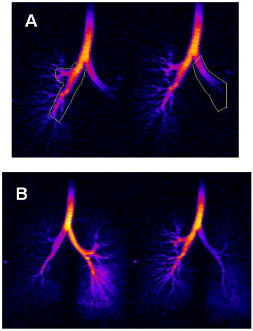
Two regions of interest (ROI), fixed in area and common to all 35 images, were drawn to include portions of the left or right lung using ImageJ. ROIs included the main stem bronchi and a few generations of central airways (Fig. 1A) and were large enough to include the central airways as they moved throughout inspiration without requiring operator manipulation between images.
Fig 1.
Dynamic MR images in subject D1. A) Regions of interest for right and left lungs in a single scan. B) Sequential images (frames 5 and 6) temporally separated by 400 msec, during inspiration of hyperpolarized 3He from FRC.
Mean intensities within each ROI were recorded for each frame were denoted as R(t) and L(t) for the respective right and left lung regions. To test the possibility that frame to frame changes in R(t) and L(t) were coupled, we tested the significance of the degree to which the direction of intensity change was concordant or discordant between R(t) and L(t). A common increase or decrease could be associated with any number of factors, but opposite or discordant changes would reflect ventilatory alternation between the two lungs. We computed the fraction of sequential image pairs with discordant changes in intensity. If these were due to uncorrelated noise, they would occur with probability 0.5; if alternation were present, they would occur with a probability significantly greater than 0.5. We tested these data by comparing the observed frequency of discordance against the null hypothesis quantified by the cumulative binomial distribution for a ”fair coin” in the sense that concordant and discordant image pairs would occur with equal probability. Specifically, we counted the number of sequential frame pairs that showed L/R intensity changes that were of opposite sign; the probability that discordant counts would be equal to or greater than the observed count is given by the upper tail of the cumulative binomial distribution. These are the probabilities given in Table 1. Note that these estimates of the significance of alternating intensities are very conservative; due to the fall of intensity due to e.g. repeated RF interrogations, whatever alternation does exist would be superposed on a continuously falling intensity in both lungs. This in turn would bias the analysis towards having a majority of image pairs showing concordant intensity changes.
Table 1.
| Subject | Probability that the observed level of alternation occurred by chance. | Inferred HR (min−1) |
|---|---|---|
| D1 | 0.000745 | 56 (or 94)* |
| D2 | 0.020695 | 66 (or 84) |
| D3 | 0.612793 | |
| C1 | 0.000659 | 52 (or 98) |
| C2 | 0.971313 | |
| C3 | 0.000002 | 61 (or 89) |
| C4 | 0.004521 | 56 (or 94) |
The higher heartrates in (), which are consistent with the peak frequencies observed, cannot be distinguished from the lower rates with this sampling frequency (2.5 Hz). The lower rates shown are more likely in these young healthy subjects resting supine.
Frames at the end of the run typically showed a significant falloff of intensity; those with intensities <20% of maximum were discarded as being below the noise floor. Finally, in those cases where there was significant alternation, we determined the period of alternation by performing a Fourier analysis of the R(t) and L(t) differences from the mean over a sequence of the first 32 frames.
3. Results
3.1 Left-right alternation of image intensity
Dynamic MRI cines revealed a left-right alternation of image intensity in 5 of our 7 subjects (see Online Supplement). Fig. 1B shows two sequential dynamic MR images, separated by 400 ms, obtained during inhalation of 1 liter of 3He mixture from FRC in subject D1. These frames provide visualization of the inspired gas as it passes through the trachea, main stem bronchus and more distal airways. Note the alternation of intensity between the left and right central airways from frame to frame; this alternation often continued throughout the entire inspiration of the 1 liter of 3He gas (see Online Supplement). An estimate of the rate of 3He polarization loss in the absence of inspiratory flow was obtained from data acquired in subject D2, who inspired the 3He bolus rapidly over about 8 seconds and then stopped inspiring (see data supplement). The abrupt quasi-exponential decay of intensity caused by the RF pulses had a time constant of approximately 0.4 s or about one frame to frame interval. This implies that the spatial distribution of intensity in each frame is strongly dependent on the flow rate of fresh 3He into the airway during the preceding interval, the intensity fluctuating with fluctuating inspiratory flow to right and left lung.
3.2 Discordance of left-right changes in intensity
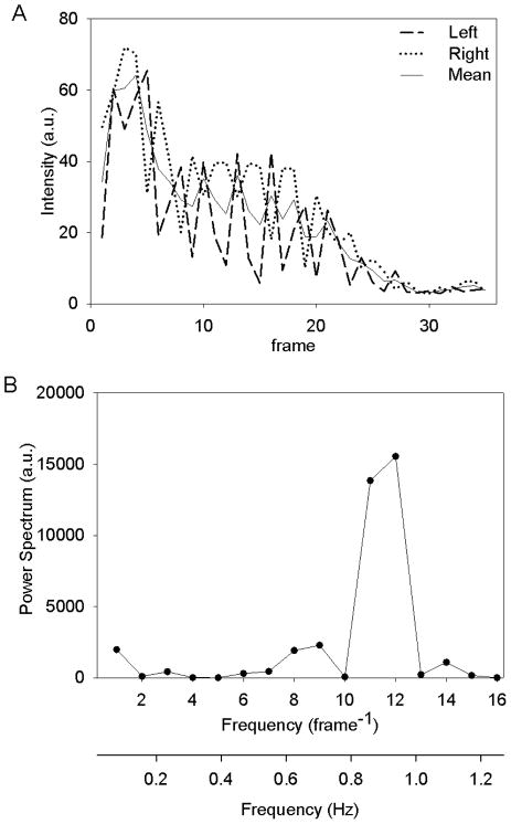
Right and left ROI intensities plotted in Fig 2A for subject D1 show a progressive decrease in R(t) and L(t) over many seconds. This progressive decrease implies a progressive decrease in average inspiratory flow rate of 3He gas during the scan sequence. Note also the striking degree of discordance in left and right intensities; increases in the left ROI are usually associated with decreases in the right ROI and vice versa; this is evidence of ventilatory alternans. In this subject, R(t) and L(t) changed in opposite directions in 18 of 21 frame to frame intervals despite the slowly progressive decrease in intensity of both signals. The probability of ≥18 occurrences of alternation out of 21 frame pairs occurring due to independent chance alone, is less than 10−3. The ventilatory alternans was statistically significant in 5 of 7 subjects (Table 1).
Fig. 2.
Frame by frame intensities of 3He signals in right and left ROIs (A), and power spectrum of the variation of the intensity about the mean (B) in subject D1. Frames were acquired at 2.5 Hz.
3.4 Frequency of alternation
The power spectrum obtained from Fourier analysis of R(t) and L(t) is shown in Fig 2B for the same subject D1. There is a strong peak at a frequency of 11–12 frames−1 or 0.97 Hz, which suggests a cardiac origin of alternation with a heart rate of 56 in this highly trained athlete. (Because the images are acquired at only 2.5 Hz, it is not possible to distinguish a heart rate of 56 from 94, but the latter is less likely in this subject.) Table 1 shows similar frequency peaks in the other 4 subjects with significant ventilatory alternans.
4. Discussion
4.1 Previous observations
Using hyperpolarized 3He MRI to visualize air flow in the airways dynamically during inspiration, we observed an alternation of gas flow into the right and left lung that is most likely a manifestation of cardiogenic flow that Engel et al. described after sampling gas concentrations within 3–19 mm airways in anesthetized dogs (Engel et al., 1973). In that study, concentrations of alveolar gas fluctuated at the heart rate during inspiration in living dogs but not in dogs after cardiac arrest, implying that motion of the heart plays a role in gas mixing and regional fluctuations in flow during respiration. Cardiogenic mixing has been extensively described in animals and has also been shown to improve oxygenation during breath holding (Kelly et al., 1987) and to augment gas exchange during tracheal insufflation of oxygen (Burwen et al., 1986). However, those “pendelluft” phenomena are most likely occurring at the lobar or sublobar level. To our knowledge, the existence of such cardiogenic pendelluft leading to alternating fluctuations of inspiratory flow rate between lungs has not been previously reported.
4.2 Variation among subjects
Not all subjects showed statistical evidence of alternation. Subjects D3 and C2 inhaled most of the 3He bolus early in the scan sequence. Thus, the brief rapid inspiration may have overwhelmed small cardiogenic alternation in flow rate early in the scan and also resulted in very low flow rates and intensities in the later part of the scan, decreasing the data suitable for analysis. In general, the non-parametric test for discordance between R(t) and L(t) is biased against a finding of discordance by the concordant changes in intensity caused by increasing or decreasing inspiratory flow rate and magnetization loss. In this respect, our analysis is conservative. Finally, to the extent that the alternans is of cardiac origin, significant peak frequencies in the Fourier analyses require a relatively constant heart rate causing alternating flow rates over most of the scan sequence; this was not apparent in the brief inspirations in D3 and C2.
4.3 Limitations of the study
As the data reported here are incidental findings from studies unrelated to ventilatory alternans, the subjects’ heart rates were not recorded. Although the Fourier analysis suggests heart rates consistent with young healthy males and elite breath-hold divers, we can only infer that the alternation is of cardiac origin. The frequency resolution and statistical significance of the Fourier analysis was limited by the frequency of image acquisition (2.5 Hz) and total number of images (34–35). Nevertheless, in those subjects with significant discordance, the characteristic peak power frequencies are compatible with a cardiogenic origin of the alternation observed.
Although alternation of flow is apparent, we cannot determine its magnitude with certainty. The hyperpolarization of 3He decays rapidly during the scan sequence due to the RF pulses as well as the admixture of oxygen in the lungs and the close proximity of airway walls. This rapid but unknown decay rate results in a non-linear and uncertain dependence of absolute image intensity on inspiratory flow rate. However, the alternation observed is suggestive of left-right variations in flow of a magnitude comparable to the inspiratory flow rates, which range up to approximately 125 ml/s in some subjects. We used a simulation program based on inspiratory flow rate, decay rate of hyperpolarization estimated from data of D2, and intensity data observed all subjects to estimate the magnitude of cardiogenic flow from one side to the other. We found that a pendelluft volume of approximately 20 ml per frame-frame interval was sufficient to reproduce the behavior in subject D1, and lesser volumes in other subjects.
4.4 Conclusion
We observed a striking alternation of 3He intensity between left and right lungs in young healthy subjects and elite divers, which we interpret as alternating inspiratory flow rate, a phenomenon we term “ventilatory alternans”. Spectral analysis shows that the alternation is commensurable with heart rate, and strongly suggests a side-to-side pendelluft phenomenon of cardiac origin, possibly the result of ballistic motion of the heart laterally during each heartbeat.
Supplementary Material
Highlights.
Inspiratory flow was visualized using MRI-cine with hyperpolarized 3He
Image intensity alternated between right and left central airways in 5 of 7 subjects
Alternation was consistent with lateral cardiac movements causing asymmetric flow
Acknowledgments
This work was supported in part by National Aeronautics and Space Administration (NAG9- 1469) and National Institutes of Health (R33 EB001689 and R01 HL52586).
Footnotes
Publisher's Disclaimer: This is a PDF file of an unedited manuscript that has been accepted for publication. As a service to our customers we are providing this early version of the manuscript. The manuscript will undergo copyediting, typesetting, and review of the resulting proof before it is published in its final citable form. Please note that during the production process errors may be discovered which could affect the content, and all legal disclaimers that apply to the journal pertain.
References
- Burwen DR, Watson J, Brown R, Josa M, Slutsky AS. Effect of cardiogenic oscillations on gas mixing during tracheal insufflation of oxygen. J Appl Physiol. 1986;60:965–971. doi: 10.1152/jappl.1986.60.3.965. [DOI] [PubMed] [Google Scholar]
- Engel LA, Wood LD, Utz G, Macklem PT. Gas mixing during inspiration. J Appl Physiol. 1973;35:18–24. doi: 10.1152/jappl.1973.35.1.18. [DOI] [PubMed] [Google Scholar]
- Kelly SM, Brancatisano AP, Engel LA. Effect of cardiogenic gas mixing on arterial O2 and CO2 tensions during breath holding. J Appl Physiol. 1987;62:1453–1459. doi: 10.1152/jappl.1987.62.4.1453. [DOI] [PubMed] [Google Scholar]
- Sun Y, Butler JP, Lindholm P, Walvick RP, Loring SH, Gereige J, Ferrigno M, Albert MS. Marked pericardial inhomogeneity of specific ventilation at total lung capacity and beyond. Respir Physiol Neurobiol. 2009;169:44–49. doi: 10.1016/j.resp.2009.07.024. [DOI] [PMC free article] [PubMed] [Google Scholar]
Associated Data
This section collects any data citations, data availability statements, or supplementary materials included in this article.


