Abstract
Background:
There have been several publications from different countries on the relationship between the placental weight and birth weight of the neonate. However, such reports from Nigeria are lacking in literature. The objective of this study was to determine the relationship between the placental weight and birth weight of the neonate at term pregnancy in a Nigerian hospital.
Materials and Methods:
It was a cross-sectional study conducted at Usmanu Danfodiyo University Teaching Hospital Sokoto between 1st October 2008 and 31st March 2009. Data gestational age at delivery (in weeks), parity, mode of delivery, fetal birth weight, placental weight, fetal gender, presence or absence of maternal medical diseases were obtained from 1009 singleton term deliveries who met the inclusion criteria for the study. The data was processed using EPI-INFO version 2005 and statistical analysis performed using one-way analysis of variance. A probability of 0.05 was set for statistical significance.
Results:
The placental birth weight ranged from 300 to 890 g with a mean of 590±82 g while the birth weight of the neonate ranged from 2030 to 5020 g with an average of 3275±469 g. The mean gestational age at delivery was 38.8±1.1 weeks while the mean placental birth weight ratio was 18.2±2.4 Increase in birth weight of the neonate was associated with corresponding increase in placental weight. However, as the gestational age at term advances the proportion of increase in the former was greater than that of the latter.
Conclusions:
There is a positive correlation between placental weight and birth weight of the neonate. However, the ratio of the placental and neonatal birth weights at term decreases with advancing gestational age. Thus, prolongation of pregnancy at term may adversely affect the fetus.
Keywords: Birth weight, placenta, relationship
INTRODUCTION
The ability of the fetus to grow and thrive in utero depends on the placental function and the average weight of the placenta at term is 508 g.1 The ratio between placenta weight and birth weight of the newborn is 1:6.1 However, methods of measurement vary widely particularly due to differences in placental preparations.2 Placental weight and its relationship to infant size at birth have been studied for more than a century.3 Past studies indicated that placental weight was associated with pregnancy outcome. High placenta weight was associated with a poor perinatal outcome, a low Apgar score, respiratory distress syndrome and perinatal death; whereas a low placental weight was associated with medical complications in the mother.4 Barker et al reported that altered growth of the placenta was a predictor of maternal medical diseases including cardiovascular disease, hypertension and diabetes mellitus.5 Other factors such as race and socioeconomic status also affect the placental weight.6
Careful examination of the placenta can provide insight regarding the in utero environment of the fetus before delivery. Two standard references are endorsed by the College of American Pathologists: absolute placental weight and fetal/placental weight (F/P) ratio.4,7,8 Clinical associations with placental weights and F/P ratio have been documented. For example, small placentas may be associated with trisomies, whereas large placentas may be associated with maternal diabetes. Disproportionately large placentas (low F/P ratio) could reflect acute placental injury resulting in villous edema or a chronic process requiring placental overgrowth, such as maternal anemia or malnutrition. Disproportionately small placentas (high F/P ratio) may be seen in maternal hypertension, and may result in fetal distress or low Apgar scores.4,8–11 Recent birth weight tables show fetal birth weights at term have increased over time.9 There is a positive correlation between fetal weight and placental weights.4 The standard method of weighing the placenta, after trimming the placental disk of membranes and umbilical cord, may also merit simplification. Leary et al suggested that the fetal weight/placental weight correlation does not change when placentas are weighed trimmed compared to when they are weighed untrimmed.12 The placenta can be weighed with membranes and cord attached, but the standard approach since its proposal by Benirschke in the early 1960s is to weigh the placenta after the extra placental membranes and the umbilical cord are trimmed from the disk.13 This limits the measurement to the weight of the placental disk, the actual nutrient exchange part of the placenta. However, Leary et al suggested that trimmed and untrimmed placental weights are exchangeable, based on their high correlation.12 Paucity of report in literature on placental weight and its relationship to the weight of the neonate at birth in Nigeria, informed this study.
MATERIALS AND METHODS
The study was a cross-sectional study between 1st October 2008 to 31st of March 2009 at Usmanu Danfodiyo University Teaching Hospital (UDUTH) Sokoto. UDUTH is situated in Sokoto Metropolis, the capital of Sokoto State. The State is located in the north-western part of Nigeria and has a population of 6 million people. The land area measures 28232.37 square kilometers and is situated 900 m above the sea level. Usmanu Danfodiyo University Teaching Hospital has a 500-bed space and an annual delivery rate of approximately 2900. One thousand and nine (1009) women who met the selection criteria were recruited for the study. The selection criteria were singleton delivery at term (37–42 weeks) and mothers’ willingness to participate in the study after explanation. The exclusion criteria included retained placenta, multiple pregnancies, morbidly adherent placenta, placenta, abruption placenta and mothers’ refusal to participate in the study. The information obtained from this study population included gestational age at delivery (in weeks), maternal age, parity, mode of delivery, birth weight, freshly delivered untrimmed placental weight, foetal gender and presence/absence of maternal medical diseases (e.g., hypertensive disorders and diabetes mellitus). The gestational age was estimated using last menstrual period (LMP). However when the LMP was unknown, the gestational age was estimated via ultrasound. All placentae were weighed shortly after delivery on table top beam weighing scale together with the membranes and the cord after removing obvious blood clots. The placental-birth weight ratio (PBWR) was calculated as ratio of placental weight to neonatal weight multiplied by 100.6
The weights of the newborn babies were recorded to the nearest gram. Weight measurements were made by the nursing staff on duty or the attending doctor using the same table top Beam weighing scale. The data was processed using the EPI info version 2005 and statistical analysis performed using one-way analysis of variance (ANOVA). A probability of 0.05 was set for statistical significance.
RESULTS
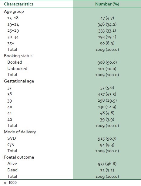
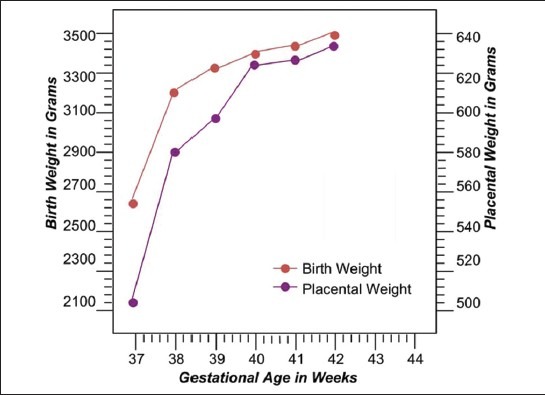
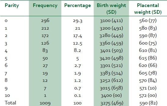
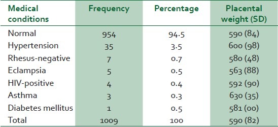
Out of a total of 1451 total deliveries within the study period, 1009 mothers met the inclusion criteria (69.5%). Their ages ranged from 16 to 38 years with a mean of 26.2±4.1 years. The mean parity of the subject was 1.9±2 with a range of 0–10. The average gestational age at delivery was 38.8±1.1 weeks with a range of 37- 42 weeks as shown in Table 1. The mean neonatal birth weight was 3275±469 g with a range of 2030–5020 g while the average placental weight was 590±82 g with a range of 300–890 g. The mean placental birth weight ratio (PBWR) was 18.2±2.4 with a range of 10.1–28.8 as depicted in Table 2. Both the placental and neonatal birth weights increase with advancing gestation (with the latter increasing more than the former). The PBWR decreases slightly with increasing gestation. This relationship is depicted in Figure 1. The correlation coefficient (r) between the placental weight and fetal birth weight was 0.33 (reflecting a positive but weak correlation). Table 3 shows the distribution of mean birth weight and placental weight by parity. There is a slight increase in mean placental weight with increasing parity with a decrease at parity of six and above. The sex ratio (male to female) was 1.04:1. The distribution of birth weight by gender is shown in Table 4. The mean birth weight of male babies (3299 g) was higher than that of their female counterpart (3250 g). However, it was not statistically significant (P>0.05). Table 5 depicts the distribution of mean placental weight in relation to maternal medical conditions. There is no significant effect of maternal medical conditions on mean birth weight (P>0.05). Ninety four patients (9.3%) were delivered by caesarean section giving a caesarean section rate of 9.3%. The mean birth weight and placenta weight from caesarean section were comparable to those from vaginal delivery (3269 g vs 3334 g) and (599.3 g vs 589.8 g), respectively.
Table 1.
Maternal characteristics and perinatal outcome

Table 2.
Mean neonatal birth weight (g), placental weight (g), and placental-birth weight ratio by gestational age at birth

Figure 1.

Mean birth weight and placental weight by gestation
Table 3.
Mean birth weight (g), placental weight (g) by parity

Table 4.
Mean birth weight (g) and placental weight (g) by fetal gender

Table 5.
Placental weight (g) in relation to maternal medical conditions

DISCUSSION
Placental weight and birth weight of the neonate are widely available measures. The ratio of these two variables is a useful marker of foetal nutrition and utero-placental function.1 The mean placental weight of 590 g in this study is lower than 643 and 630 g reported in western Europe and eastern Nigeria, respectively.5,14 However, it is more than 588 and 470 g reported in Asia and Ukraine, respectively.6,15 The variations in the mean weight of the placenta may be due to variations in the methodology of preparing and weighing the placenta together with cord clamping time.16 It has also been reported that ethnicity and some unknown factors may affect the placental weight.12 The mean birth weight of the neonate in this study ( 3275 g) is lower than 3425, 3382, and 3400 g reported in Ukraine, western Europe, and eastern Nigeria respectively; but higher than 3036 g in Asia and 3103 g in the Afro-Caribbean region.5,6,14,15,17 These differences in mean birth weight may be due to altitude; maternal nutrition and maternal diseases.18 However, mean birth weight of 3065 g was reported earlier in this institution.19 This was lower than the present study perhaps due to a relatively small sample size in the previous study.
The mean PBWR of 18.2% in this study is lower than 20.0% and 19.5% reported in western Europe and Asia but higher than 17.08% and 13.9 % in Thailand and Ukraine, respectively.5,6,12,17 The positive correlation ( weak) between the placental weight and neonatal birth weight noted in this study has been observed by previous authors.8,13,15,20,21 The decline in PBWR with increasing gestational age at term observed in the present study has been reported by previous authors.8,12,20 An abnormally high PBWR (i.e., low foetal weight relative to the placental weight) is thought to indicate an abnormal placenta with impaired function as in human immune deficiency virus infection, obesity, maternal anemia, cigarettes smoking and low socioeconomic status.6,22–25 Infants with such abnormal ratios are at increased risk of perinatal death.26 Conversely, an abnormally low PBWR indicates fetuses with presumed reduced placental reserves.21 Such fetuses tend to show asymmetric growth restriction, suggesting that the small placenta limits optimal foetal growth.27,28
The age range of the subjects in this study 16–38 years is similar to that of participants in the previous studies.6,21 The mean gestational age in the study (38.8 weeks) is lower than 39.7 weeks and 39.9 weeks reported in western Europe and eastern Nigeria, respectively, but same as that reported in Italy.14,20,29 Most of the women enrolled for the study were booked patients (90%) because most of the unbooked patients did not satisfy the inclusion criteria for the study. The mean birth weight of babies delivered by caesarean section in this study was lower than those delivered vaginally while the placental weight was higher in the former than later. Similar observation had been made in previous studies.30,31 Differences in cord clamping time have been suggested to explain these differences since the umbilical cords are frequently clamped relatively late in vaginal delivery (so as to optimize blood transfer to the fetus) while in caesarean section early clamping of the cord is usually the rule.30 Furthermore, due to the absence of uterine contractions during caesarean section, the intervillous space in the placenta is more expansive and likely to contain more maternal blood than in vaginal deliveries where maternal blood is squeezed out of the placenta by contraction of the uterus.30 Relative increase in both placental and neonatal birth weight associated with increasing parity in this study is in agreement with Kloosterman's previous report that the placentae and babies from multiparous women from 32 weeks onwards are heavier than those from primiparous women.31
In conclusion, this study has also shown that there is a positive correlation between the placental and infants weight. In addition, the ratio of the placenta and foetal weights at term decreases with advancing gestational age. Thus, prolongation of pregnancy at term may adversely affect the fetus.
Footnotes
Source of Support: Nil
Conflict of Interest: None declared.
REFERENCES
- 1.Cunningham FG, Leveno KJ, Bloom SL, Hauth JC, Gilstrap LC, III, Wenstrom KD. Williams Obstetrics. 2nd ed. New York: McGraw- Hill; 2005. Implantation, embryogenesis and placental development; pp. 39–90. [Google Scholar]
- 2.Thomson AM, Billewicz WZ, Hytten FE. The weight of the placenta in relation to birthweight. J Obstet Gynaecol Br Commonw. 1969;76:865–72. doi: 10.1111/j.1471-0528.1969.tb15722.x. [DOI] [PubMed] [Google Scholar]
- 3.Adair FL, Thelander H. A study of the weight and dimensions of the human placenta in its relation to the weight of the newborn infant. Am J Obstet Gynecol. 1925;10:172–205. [Google Scholar]
- 4.Naeye RL. Do placental weights have clinical significance? Hum Pathol. 1987;18:387–91. doi: 10.1016/s0046-8177(87)80170-3. [DOI] [PubMed] [Google Scholar]
- 5.Barker DJ, Bull AR, Osmond C, Simmonds SJ. Fetal and placental size and risk of hypertension in adult life. BMJ. 1990;301:259–62. doi: 10.1136/bmj.301.6746.259. [DOI] [PMC free article] [PubMed] [Google Scholar]
- 6.Perry IJ, Beevers DG, Whincup PH, Bareford D. Predictor of ratio of placental weight to fetal weight in multiethnic community. BMJ. 1995;310:436–9. doi: 10.1136/bmj.310.6977.436. [DOI] [PMC free article] [PubMed] [Google Scholar]
- 7.Langston C, Kaplan C, Macpherson T, Manci E, Peevy K, Clark B, et al. Practice guideline for the examination of the placenta : developed by the Placental Pathology Practice Guideline Development Task Force of the College of American Pathologists. Arch Pathol Labor Med. 1997;121:449–76. [PubMed] [Google Scholar]
- 8.Molteni RA, Stys SJ, Battaglia FC. Relationship of fetal and placental weight in human beings: fetal/placental weight ratios at various gestational ages and birth weight distributions. J Reprod Med. 1978;21:327–34. [PubMed] [Google Scholar]
- 9.Robertson CM, Svenson LW, Kyle JM. Birth weight by gestational age for Alberta liveborn infants, 1985 through 1998. J Obstet Gynaecol Can. 2002;24:138–48. doi: 10.1016/s1701-2163(16)30295-x. [DOI] [PubMed] [Google Scholar]
- 10.Fox GE, Van Wesep R, Resau JH, Sun CJ. The effect of immersion formaldehyde fixation on human placental weight. Arch Pathol Lab Med. 1991;115:726–8. [PubMed] [Google Scholar]
- 11.Eriksson J, Forsen T, Tuomilehto J, Osmond C, Barker D. Foetal and childhood growth and hypertension in adult life. Hypertension. 2000;36:790–4. doi: 10.1161/01.hyp.36.5.790. [DOI] [PubMed] [Google Scholar]
- 12.Leary SD, Godfrey KM, Greenaway LJ, Davill VA, Fall CH. Contribution of the umbilical cord and membranes to untrimmed placental weight. Placenta. 2003;24:276–8. doi: 10.1053/plac.2002.0888. [DOI] [PubMed] [Google Scholar]
- 13.Benirschke K. Examination of the placenta, prepared for the collaborative study on cerebral palsy, mental retardation, and other neurological and sensory disorders of infancy and childhood. Bethesda, MD: National Institute of Neurological Diseases and Blindness, US Department of Health, Education, and Welfare; 1961. [Google Scholar]
- 14.Adinma JI, Agbai AO. Foetal birth weight in Africa. J Obstet Gynaecol. 1995;15:295–7. [Google Scholar]
- 15.Little RE, Zadorozhnaja TD, Hulchiy OP, Mendel NA, Shkyryak-Nyzhnyk ZA, Chyslovska N, et al. Placental weight and its ratio to birthweight in a Ukrainian city. Early Hum Dev. 2003;71:117–27. doi: 10.1016/s0378-3782(02)00118-4. [DOI] [PubMed] [Google Scholar]
- 16.Yao AC, Moinian M, Lind J. Distribution of blood between infant and placenta after birth. Lancet. 1969;2:871–3. doi: 10.1016/s0140-6736(69)92328-9. [DOI] [PubMed] [Google Scholar]
- 17.Lurie S, Feinstein M, Mamet Y. Human fetal-placental weight ratio in normal singleton near-term pregnancies. Gynecol Obstet Invest. 1999;48:155–7. doi: 10.1159/000010163. [DOI] [PubMed] [Google Scholar]
- 18.Njokanma OF, Sule-Odu OA. Intrauterine growth retardation in Nigerian neonates. Trop J Obstet Gynecol. 1998;15:25–9. [Google Scholar]
- 19.Onankpa BO, Airede KI, Ahmed H, Jiya NM. The birth weight of apparently healthy Nigerian newborn in Sokoto. Sahel Med J. 2006;1:19–22. [Google Scholar]
- 20.Janthanaphan M, Kor-Anantakul O, Geater A. Placental weight and its ratio to birth weight in normal pregnancy at Songkhlanagarind Hospital. J Med Assoc Thai. 2006;89:130–7. [PubMed] [Google Scholar]
- 21.Bonds DR, Gabbe SG, Kumar S, Taylor T. Feotal weight/placental weight ratio and perinatal outcome. Am J Obstet Gynecol. 1984;149:195–200. doi: 10.1016/0002-9378(84)90198-4. [DOI] [PubMed] [Google Scholar]
- 22.Boyd PA, Keeling JW. Raised maternal serum alpha-fetoprotein in the absence of foetal abnormality-placental findings.A quantitative morphometric study. Prenat Diagn. 1986;6:369–73. doi: 10.1002/pd.1970060505. [DOI] [PubMed] [Google Scholar]
- 23.Gichangi PB, Nyongo AO, Temmerman M. Pregnancy outcome and placental weights: their relationship to HIV-1 infection. East Afr Med J. 1993;70:85–9. [PubMed] [Google Scholar]
- 24.Williams LA, Evans SF, Newnham JP. Prospective cohort study of factors influencing the relative weights of the placenta and the newborn infant. BMJ. 1997;314:1864–8. doi: 10.1136/bmj.314.7098.1864. [DOI] [PMC free article] [PubMed] [Google Scholar]
- 25.Lao TT, Tam KF. Placental ratio and anaemia in third-trimester pregnancy. J Reprod Med. 2000;45:923–8. [PubMed] [Google Scholar]
- 26.Lao TT, Wong W. The neonatal implications of a high placental ratio in small-for-gestational age infants. Placenta. 1999;20:723–6. doi: 10.1053/plac.1999.0432. [DOI] [PubMed] [Google Scholar]
- 27.Salafia CM, Zhang J, Miller RK, Charles AK, Shrout P, Sun W. Do placental growth patterns affect birth weight for given placental weight. Birth Defects Res A Clin Mol Teratol. 2007;79:281–8. doi: 10.1002/bdra.20345. [DOI] [PubMed] [Google Scholar]
- 28.Taricco E, Radaelli T, Nobile de Santis MS, Cetin I. Foetal and placental weights in relation to maternal characteristics in gestational diabetes. Placenta. 2003;24:343–7. doi: 10.1053/plac.2002.0913. [DOI] [PubMed] [Google Scholar]
- 29.Burkhardt T, Schaffer L, Schneider C, Zimmermann R, Kurmanavicius J. Reference values for the weight of freshly delivered term placentas and for placental weight-birth weight ratios. Euro J Obstet Gynecol Reprod Biol. 2006;128:248–52. doi: 10.1016/j.ejogrb.2005.10.032. [DOI] [PubMed] [Google Scholar]
- 30.Heinonen S, Taipale P, Saarikoski S. Weights of placentae from small-for-gestational age infants revisited. Placenta. 2001;22:399–404. doi: 10.1053/plac.2001.0630. [DOI] [PubMed] [Google Scholar]
- 31.Kloosterman GJ. Intrauterine growth: The significance of prenatal care. Int J Gynaecol Obstet. 1970;8:895–912. [Google Scholar]


