SUMMARY
Aim.
Describe a clinical case of a voluminous asymptomatic fibromatosis lesion present on the cheek mucosa and evaluate the healing of the site after removal of the lesion with use of the laser diode.
Methods.
It was decided to use laser diodes to affect the mucous membrane and remove the lesion without the use of local anesthetic infiltration. The protocol used includes a 300-micron fiber and the emission of continuous light of 1.5 Watt with a range of wave of 940 nm.
Results.
The proven benefits of using laser diodes for minor surgery are:
drastic reduction of intraoperative bleeding and in the hours after the surgery
will restrict the swelling
better and faster healing with no scarring and better cosmetic result
does not require sutures
reducing the operating time thanks to no need for anesthetic infiltration
in most cases totally absent or less post-operative pain on the surgical site.
Conclusions.
The laser diodes give a significant contribution to improving the surgical treatment of tumors of the oral cavity infact during the surgery reduce bleeding and surgical time, while in the process of healing by reduce swelling and post-operative pain and better results appearance without scarring.
Keywords: laser diode; oral mucosal tumors, fibroids
RIASSUNTO
Obiettivi.
Descrivere un caso clinico di una voluminosa lesione fibromatosa asintomatica presente sulla mucosa geniena e valutare la guarigione del sito dopo la rimozione della lesione con l’uso del laser a diodi.
Metodi.
Si è deciso di utilizzare il laser a diodi per incidere la mucosa ed asportare la lesione senza l’utilizzo dell’infiltrazione dell’anestetico locale. Il protocollo utilizzato prevede una fibra da 300 micron e l’emissione di luce continua da 1,5 Watt con un intervallo di onda di 940 nm.
Risultati.
I comprovati vantaggi dell’utilizzo del laser a diodi per la piccola chirurgia sono:
riduzione drastica del sanguinamento sia intraoperatorio che nelle ore successive all’intervento
diminuzione del gonfiore
migliore e più rapida guarigione con assenza di cicatrici e migliore risultato estetico
non necessità di suture
riduzione del tempo operatorio anche grazie alla non necessità dell’infiltrazione di anestetico
nella maggior parte dei casi minore o totalmente assente dolore post-operatorio sul sito chirurgico.
Conclusioni.
Il laser a diodi dà un contributo significativo al miglioramento della chirurgia delle neoformazioni del cavo orale sia durante la pratica operatoria, diminuendo il sanguinamento e il tempo chirurgico, sia nella fase di guarigione, riducendo il gonfiore ed il dolore post-operatorio e garantendo un miglior risultato estetico senza cicatrici.
Introduction
Describe a clinical case of a voluminous asymptomatic fibromatosis lesion present on the cheek mucosa and evaluate the healing of the site after removal of the lesion with use of the laser diode. The irritation fibroma is a benign proliferation that occurs as a response to local irritation it is an elevated pedunculated or sessile lesion that ranges in size from a few millimeters to a few centimeters and is normal in color, although it may appear to be more pale than the normal mucosa.
Materials and methods
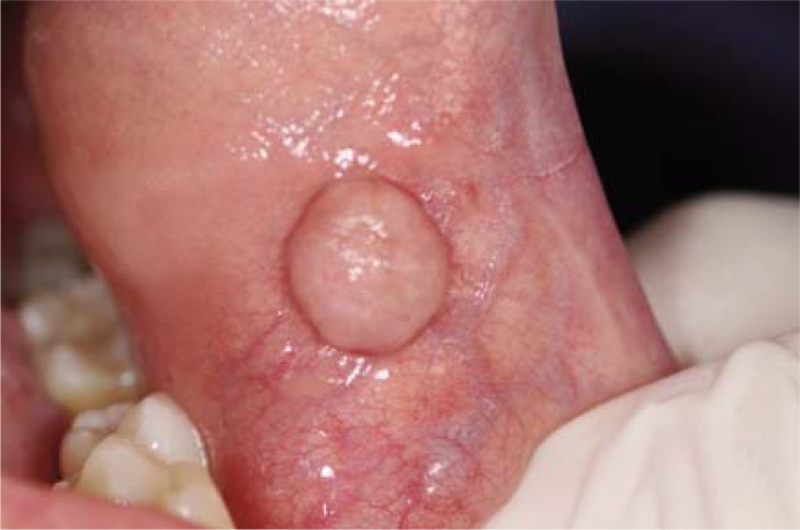
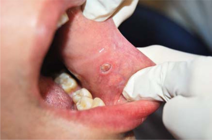
The female patient of 37 years in good general health has a sessile lesion with a diameter of 9 mm on the left cheek mucosa (Fig. 1). The lesion involved the patient not only an aesthetic discomfort but also a problem during functional mastication and phonation.
Figure 1.
Sessile lesion with a diameter of 9 mm on the left cheek mucosa.
The patient has a family history with the absence of neoplastic diseases. Physiological personal history: no allergies to medications, is not pregnant, has a weight of 62 kg and a height of 1.70 cm.
Personal pathological medical history: she claims to be in good general health and not to be smoker. On physical extraoral examination after palpation of submandibular lymph nodes and neck they not appear to be enlarged. On physical intraoral examination a lesion fibromatosis is clearly visible on the left cheek mucosa.
It was decided to use laser diodes to affect the mucous membrane and remove the lesion without the use of local anesthetic infiltration. The protocol used includes a 300-micron fiber and the emission of continuous light of 1.5 Watt with a range of wave of 940 nm.
Before starting surgery, the patient was verbally informed of the risks and complications, and subsequently read and signed informed consent.
After using 10% lidocaine spray flow around the lesion through a small roll of cotton was carried painless and bloodless incision.
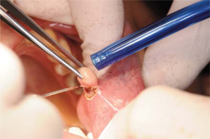
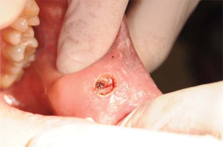
The aspiration was used only for the vapors produced during cutting. The fiber laser has been tilted toward the center of the lesion and in depth to prevent the recurrence of the same (Fig. 2). It was not necessary to suture the wound after excision of the lesion (Fig. 3).
Figure 2.
Removal of the lesion with the laser and suction of fumes produced during cutting.
Figure 3.
Image of the surgical site following excision of the lesion.
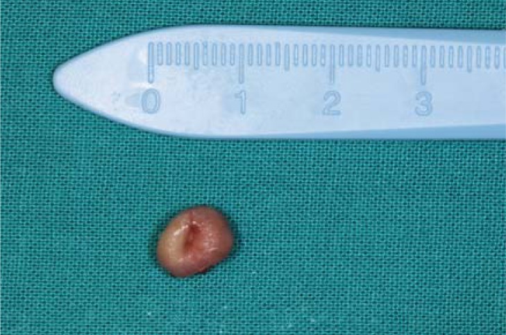
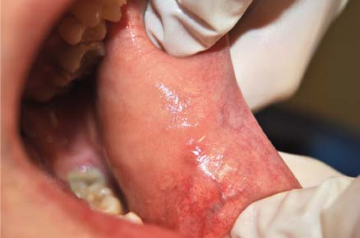
The biopsy sample was analyzed by histopathologic outcome completely benign (Fig. 4). The analysis was done correctly because the removal was extensive and the tip of the laser has been working on healthy tissue without affecting the outcome of healing. The follow-up of the patient was documented to 7 (Fig. 5) and 21 days (Fig. 6) showing a soft tissue recovered fully.
Figure 4.
Fibroid diameter: 9 mm.
Figure 5.
Tissue healing after 7 days.
Figure 6.
Tissue healing after 21 days.
Results
To date, the application of the soft tissues is the first area for the clinical use of lasers in dentistry. Surgical removal of fibroids or other tumors of the oral mucosa through the use of diode laser has become a common procedure for dentists and maxillofacial surgeons (5,7,8).
The effectiveness of laser diodes has been proven to work well as the treatment of intraoral pigmentation, vascular lesions of soft tissues, lingual and labial frenulectomy upper and lower, epulidi and gingival hyperplasia (1–4). The proven benefits of using laser diodes for minor surgery are:
drastic reduction of intraoperative bleeding and in the hours after the surgery (3)
better and faster healing without scarring and better cosmetic result (2)
does not require sutures
reducing the operating time thanks to no need for anesthetic infiltration (9)
in most cases totally absent or less post-operative pain on the surgical site (5,10,11).
Conclusions
The laser diodes give a significant contribution to improving the surgical treatment of tumors of the oral cavity infact during the surgery reduce bleeding and surgical time, while in the process of healing by reduce swelling and post-operative pain and better results appearance without scarring (6–8).
References
- 1.Walinski CJ. Irritation fibroma removal: a comparison of two laser wavelengths. Gen Dent. 2004 May-Jun;52(3):236–8. [PubMed] [Google Scholar]
- 2.Marangoni O, Melato M, Longo L. 808-nm laser with exogenous chromophores for the treatment of benign oral lesions. Photomed Laser Surg. 2005 Jun;23(3):324–7. doi: 10.1089/pho.2005.23.324. [DOI] [PubMed] [Google Scholar]
- 3.Stübinger S, Saldamli B, Jürgens P, Ghazal G, Zeilhofer HF. Soft tissue surgery with the diode laser--theoretical and clinical aspects. Schweiz Monatsschr Zahnmed. 2006;116(8):812–20. [PubMed] [Google Scholar]
- 4.Lomke MA. Clinical applications of dental lasers. Gen Dent. 2009 Jan-Feb;57(1):47–59. [PubMed] [Google Scholar]
- 5.Desiate A, Cantore S, Tullo D, Profeta G, Grassi FR, Ballini A. 980 nm diode lasers in oral and facial practice: current state of the science and art. Int J Med Sci. 2009 Nov 24;6(6):358–64. doi: 10.7150/ijms.6.358. [DOI] [PMC free article] [PubMed] [Google Scholar]
- 6.Goldberg DJ. Laser Treatment of Vascular Lesions. Berlin Heidelberg: Springer; 2005. Laser Dermatology; pp. 13–35. [Google Scholar]
- 7.Adams TC, Pang PK. Lasers in aesthetic dentistry. Dental Clinics of North America. 2004;48(4):833–860. doi: 10.1016/j.cden.2004.05.010. [DOI] [PubMed] [Google Scholar]
- 8.Antenucci EL. Integration of lasers into a soft tissue management program. Dent Clin North Am. 2000 Oct;44(4):811–9. [PubMed] [Google Scholar]
- 9.Kafas P, Angouridakis N, Dabarakis N, Jerjes W. Diode laser lingual frenectomy may be performed without local anaesthesia. Int J Orofac Sci. 2008;1:1. [Google Scholar]
- 10.Reichwage DP, Barjenbruch T, Lemberg K, Janiszewski T, Marr D. Esthetic contemporary dentistry and soft tissue recontouring with the diode laser. J Indiana Dent Assoc. 2004;83(1):13–5. [PubMed] [Google Scholar]
- 11.Sarver DM, Yanosky M. Principles of cosmetic dentistry in orthodontics: part 2. Soft tissue laser technology and cosmetic gingival contouring. Am J Orthod Dentofacial Orthop. 2005;127:85–90. doi: 10.1016/j.ajodo.2004.07.035. [DOI] [PubMed] [Google Scholar]


