Abstract
Background
Neuronal stem cells (NSCs) are promising for neurointestinal disease therapy. While NSCs have been isolated from intestinal musclularis, their presence in mucosa has not been well described. Mucosa-derived NSCs are accessible endoscopically and could be used autologously. Brain-derived Nestin-positive NSCs are important in endogenous repair and plasticity. The aim was to isolate and characterize mucosa-derived NSCs, determine their relationship to Nestin-expressing cells and demonstrate capacity to produce neuroglial networks in vitro and in vivo.
Methods
Neurospheres were generated from periventricular brain, colonic muscularis (Musc), and mucosa-submucosa (MSM) of mice expressing green fluorescent protein (GFP) controlled by the Nestin promoter (Nestin-GFP). NSCs were also grown as adherent colonies from intestinal mucosal organoids. Their differentiation potential was assessed by immunohistochemistry using glial and neuronal markers. Brain and gut derived neurospheres were transplanted into explants of chick embryonic aneural hindgut to determine their fate.
Results
Musc- and MSM-derived neurospheres expressed Nestin and gave rise to cells of neuronal, glial and mesenchymal lineage. While Nestin expression in tissue was mostly limited to glia colabelled with glial fibrillary acid protein (GFAP), neurosphere-derived neurons and glia both expressed Nestin in vitro, suggesting Nestin+/GFAP+ glial cells may give rise to new neurons. Moreover, following transplantation into aneural colon, brain- and gut-derived NSCs were able to differentiate into neurons.
Conclusions
Nestin-expressing intestinal NSCs cells give rise to neurospheres, differentiate into neuronal, glial and mesenchymal lineages in vitro, generate neurons in vivo and can be isolated from mucosa. Further studies are needed exploring their potential for treating neuropathies.
Keywords: Neural Stem Cell, Enteric Nervous System, Hirschsprung’s disease, Neurosphere, Nestin
Introduction
The Enteric Nervous System (ENS) regulates intestinal functions including motility, absorption, secretion, sensory function, blood flow, hormonal secretion and interaction with the immune system [1] [2] [3]. The ENS is known to share many features with the Central Nervous System (CNS), including a similar repertoire of neurotransmitters and the identification of glial cells comparable to mature GFAP+ astrocytes in the CNS [4] [5]. Until recently, enteric neurons were believed to be non-renewable; however, as in the CNS, several groups have identified the presence of post-natal neural stems cells in the gut [6] [7] [8] [9]. These enteric neural stem cells (ENSCs) are believed to contribute to intestinal neurogenesis under specific conditions; however, their exact role in physiological and pathological processes remains elusive. In addition, it has been unclear whether ENSCs reside in and can be isolated from all intestinal segments and layers.
While there has not been a marker identified that exclusively and specifically labels NSCs, Nestin, an intermediate filament protein, has been extensively used to mark these cells in the CNS [10] [11] [12] [13]. During embryogenesis, Nestin regulates proliferation, survival and differentiation of neural stem cells [14]. In adults, Nestin expressing cells are increased during CNS regeneration, in injured muscle, and also have frequently been identified in neuroepithelial tumors [15] [16] [17]. Nestin is expressed in multipotent progenitor cells with regenerative potential [14], but very little is known about Nestin-expressing cells and their function in the adult intestine.
In the gastrointestinal (GI) tract, Nestin is expressed by glial cells, interstitial cells of Cajal (ICC), endothelial cells, pericytes, and stromal tumor cells [18]. It has also been found to be expressed within and surrounding myenteric and submucosal ganglia - in cells smaller and more numerous than neurons [19]. To study the role and identity of intestinal Nestin expressing cells and their possible relationship to ENSCs, we used a transgenic Nestin-GFP mouse model [20], previously used to characterize neural stem and progenitor cells in the central nervous system. We hypothesized that Nestin-expressing cells in the intestine had a similar role with respect to postnatal enteric neurogenesis. To test this hypothesis, we examined the distribution of nestin expression in the enteric nervous system, generated adherent cultures and neurospheres from Nestin+ cells isolated from different areas and layers of the gut wall, determined their neurogenic and gliogenic potential, and compared our results with those of CNS-derived Nestin+ cells. Our findings suggest strong similarities between CNS- and ENS-derived Nestin+ cells in regard to their potential to differentiate into multiple lineages. Furthermore, we were able to demonstrate that Nestin+ cells from either the muscular or mucosal/submucosal layers of the gut can give rise to both neurons and glial cells in culture, and that these cells can be transplanted into aneural hindgut to form neurons ex vivo. Our results support further studies of these cells as a potential source of ENSCs for diagnosis and cell-based therapy in neurointestinal diseases.
Methods
Wild-type and Nestin-GFP mice
NS cultures were generated from wild-type and transgenic mice expressing GFP under the control of the Nestin promoter, as previously described (Mignone 2004). Our local animal care and use committee approved procedures. All neurosphere experiments were performed between post-natal day 21 and 28 (P21–P28) and lactating mice were used for organoid cultures. Mice were sacrificed by CO2 asphyxiation prior to removing tissue for culture.
Adherent organoid cultures
Nestin-GFP mouse small intestine was digested enzymatically with collagenase XI (750ug/mL, Sigma) and dispase 1 (250ug/mL, Roche) for 20 minutes at 37°C, followed by mechanical dissociation of the tissue through progressively smaller pipette tips. Organoids were purified by differential sedimentation through an osmotic gradient with 2% sorbitol (Sigma). Organoids were plated with Dulbecco’s Modified Eagle Medium (DMEM) (Gibco, Invitrogen) containing 10% fetal bovine serum (Gibco, Invitrogen), insulin 0.25U/mL insulin (Sigma) and epidermal growth factor (EGF; Stem Cell Tech, 20ng/ml) and Penicillin/Streptomycin (Gibco, Invitrogen). Once organoids attached to the culture dish and formed colonies, Nestin expression was detected immunohistochemically by GFP fluorescence. On culture days 7–10, colonies were fixed and processed for immunofluorescence.
Non-adherent cultures
Nestin-GFP mouse colon was removed and mechanically separated into two layers, mucosa-submucosa (MSM) and muscularis (Musc). Tissues were digested as described above and passed through a 40μM nylon pore mesh (BD Falcon), then cultured in Neural Stem Cell (NSC) proliferation media containing NeuroCult basal media with 10% proliferation supplement (Stem Cell Technologies), heparin (Stem Cell Technologies (final concentration 0.0002%), EGF (Stem Cell Tech, 20ng/ml) and basic fibroblast growth factor (bFGF) (Stem Cell Tech, 20ng/ml). Floating colonies were grown in ultra-low attachment chambers (Corning), allowed to expand prior to transfer to culture dishes coated with laminin (2mg/cm2; Sigma) and fibronectin (2mg/cm2; Sigma). Cultures were then grown in NSC differentiation media, containing NeuroCult basal media with 10% differentiation supplement (Stem Cell Technologies) and 10ng/ml glial-derived neurotrophic factor (GDNF) (R&D Systems). Once cells attached, 7–10 days after culturing, they were fixed with 4% paraformaldehyde and processed for immunohistochemistry or PCR studies.
Brain neurosphere cultures
Periventricular cerebral tissue was removed and dissociated per the Stemcell Technologies protocol (www.stemcell.com). Single cells were cultured in proliferation media, as described above. At 7–10 days, neurospheres were allowed to attach by adding differentiation media, as above. After 7–14 days, attached cells were fixed and processed for immunofluorescence.
Immunofluorescence and Immunohistochemistry
Cultures were xed in 4% paraformaldehyde, washed, and permeabilized. Primary antibodies used included Tuj1 (1:400; Covance, Princeton, NJ), GFAP (1:500, Dako), S-100 (1:200 NeoMarkers), calretinin (1:200; Invitrogen) and PGP9.5 (1:50, Abcam). Sections were incubated with primary antibody for 45minutes, followed by biotinylated goat anti-mouse IgM or IgG (Vector Labs, Burlingame, CA) and avidin-biotinylated peroxidase complex (Vectastain Elite ABC kit, Vector Labs). Antibody binding sites were visualized with 4-chloro-1-naphthol (Sigma) or diaminobenzidine (DAB). For immuno uorescence, secondary antibodies used included Alexa Fluor 594(1:1000, Invitrogen), and 488 (1:1000, Invitrogen), goat anti-mouse IgG, Alexa Fluor 594 (1:1000, Invitrogen), Alexa Fluor 488 goat anti-mouse IgM (1:1000, Invitrogen), and Alexa Fluor 594 (1:1000, Invitrogen), and Alexa Fluor 488 goat anti-rabbit IgG (Molecular Probes). Cell nuclei were stained with DAPI (Vector Labs).
Neurosphere transplants to aneural chick hindgut
Chick hindgut was explanted on embryonic day #5 (E5), prior to colonization by enteric neural crest cells. A single neurosphere grown for 7–10 days in non-differentiating conditions was implanted into the proximal hindgut using fine forceps under microscopic visualization. The hindgut was cultured in a three-dimensional collagen matrix [21] for 24 hours at 37°C with 5% CO2 in the same proliferation media as described above. The following day, the gut was removed from the plate and placed onto the chorioallantoic membrane (CAM) of E10 host chick embryo. After 7 days on the CAM, the gut was removed and processed for immunohistochemistry.
Quantitative PCR
Total mRNA was extracted from proliferating neurospheres using the RNeasy Mini kit (Qiagen) and cDNA produced with Superscript III Reverse Transcription Kit (Invitrogen). Sox2, cMyc, and Klf4 transcript levels were measured by quantitative PCR (qPCR) with Gapdh as the internal standard. The following primers were used: Sox2 F: GCTGCCTCTTTAAGACTAGGGCTG, R: GCCGCCGCGATTGTTGTGAT; cMyc F: TTCTCTGCCTCTGCCCGCGA, R: GGGCATCGTCGTGGCTGTCT; Klf4 F: GGTCGTGGCCCCGGAAAAGA, R: ACCCACAGCCGTCCCAGTCA. Relative expression was calculated by 2-ΔCt. Statistical comparisons were made by nonparametric one-way analysis of variance (ANOVA) with Tukey’s test.
Results
Colon-derived neurospheres
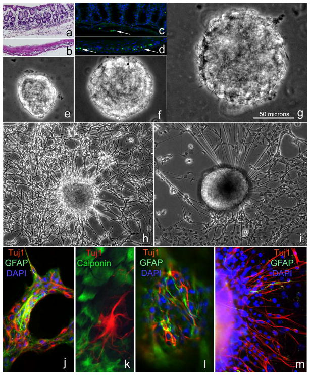
MSM and Musc layers of the colon of wild-type mice were microdissected, and the composition of the layers confirmed by histology (Fig. 1a, b). The submucosal plexus is present in the MSM layer, as shown by the presence of Hu-immunoreactive neuronal cell bodies in the submucosa attached to the MSM (Fig. 1c). The myenteric plexus is present in the Musc layer (Fig. 1d). Single-cell suspensions were prepared from each layer separately to generate neurosphere (NS) cultures. The diameter of NS derived from MSM, Musc, and brain were measured following 5 days in culture and were 48 ± 6 μm, 98 ± 19 μm, and 144 ± 36 μm, respectively. These size differences were all statistically significant (p<0.005, Student’s t-test). Representative images of neurospheres from the three different sources are shown in Figs. 1e–g. Following addition of differentiation medium, NS attached to the plate. From all types of neurospheres we see “bridge-like” structures that form networks of cells with elongated projections. The morphology of the NS and cells emanating from them was very similar to brain-derived cultures, although brain-derived spheres (Fig. 1h) were more densely packed than Musc-derived spheres (Fig. 1i). One week after cell attachment, immunofluorescence showed neurons and glial cells present in Musc-derived NS cultures (Fig. 1j). Cells with a non-neuronal morphology were also observed surrounding the NS and these were positive for smooth muscle markers, calponin (Fig. 1k) and smooth muscle actin (not shown). After attaching and differentiating, these MSM-derived NS also contained glia, neurons (Fig. 1l), and smooth muscle cells (not shown), with many Tuj1+ neurons migrating out of the NS (Fig. 1m).
Figure 1. Brain and gut neurospheres contain neurons and glia cells.
Postnatal mouse colon was separated into MSM (a) and Musc (b) layers, shown by H&E staining. Hu-labeled (green) neuronal cell bodies are present in both layers (c, d, arrows). NS were derived from MSM (e), Musc(f), and brain (g). Scale bar in (g) refers to panels e–g. Following 14 days in culture, adherent brain-derived (h) and Musc-derived (i) neurospheres produce elaborate neuroglial networks. Neuronal (Tuj1) and glial cells (GFAP) are present in Musc-derived NS cultures (j), as are smooth muscle cells (k; calponin). MSM-derived NS (l) similarly contain glial cells (GFAP) and neurons (Tuj1), with neurons extending out from the NS (m).
Transplant experiments
To determine the capacity of gut- and brain-derived NS to survive and generate neurons following transplantation, floating NS were placed into aneural hindgut from embryonic day #5 (E5) chick embryos. At this stage, the hindgut has not yet received enteric neural crest-derived cells. Following transplantation, hindguts were cultured in a collagen gel for 24 hours, and then placed onto the chorioallantoic membrane (CAM) of an E10 host chick embryo (Fig. 2a). After 7 days, guts were harvested and processed for immunohistochemistry. Tuj1-immunoreactivity was present in the transplanted hindgut (Fig. 2b). No staining was observed with CN antibody, a chick-specific neuronal antibody, confirming that the Tuj1+ neurons were not chick-derived (Fig. 2b, inset). Tuj1+ cells with neuronal-like morphology were present following transplantation of both brain- and gut-derived neurospheres, with small groups of Tuj1+ cells forming structures resembling ganglia and extending fine neurites (Fig. 2c, d).
Figure 2. Transplanted gut-derived neurospheres create neuronal networks in aneural colon.
A gut-derived NS (a; inset) was transplanted into aneural E5 chick hindgut (a) and cultured on a CAM for 7 days. Tuj1-immunoreactivity (arrow) was present in the transplanted hindgut (b). No labeling was observed using CN, a chick-specific neuronal antibody, confirming that neurons were mouse-derived (b; inset). Clusters of Tuj1+ cells formed ganglion-like structures extending fine neurites (c, d). ep, epithelium; sm, submucosa; mp, muscularis propria
Nestin is expressed by enteric glial cells
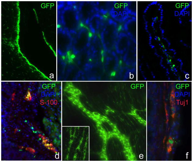
Based on the similarities between brain- and gut-derived NS, we hypothesized that ENSCs express Nestin, a marker of brain NSCs. Nestin expression was examined in Nestin-GFP transgenic mice. As expected, the periventricular region of the brain was strongly GFP+ (Fig. 3a). Nestin was also strongly expressed throughout the gut. In the small intestinal mucosa, Nestin+ cells surrounded each crypt (Fig. 3b) and were present in the villus core (Fig. 3c). Nestin was also expressed by submucosal and myenteric ganglia (Fig, 3d), and by interganglionic fibers (Fig. 3e). Nestin-immunoreactive cells in the mucosa and muscularis rarely co-expressed Tuj1, with only occasional double-labeled cells seen in the myenteric ganglia (Fig. 3f). In contrast, Nestin expression strongly overlapped with glial markers, GFAP and S-100 (Fig. 3d), in both plexuses.
Figure 3. Nestin is expressed by glial cells in the enteric ganglia.
Nestin is strongly expressed in the periventricular region of Nestin-GFP mouse brain (a). In the small intestinal mucosa, GFP+ cells are present around crypts (b) and within the villus core (c). GFP is expressed by cells in submucosal (d) and myenteric (d–f) ganglia. Nestin- immunoreactive cells (GFP+) in the enteric ganglia co-express S-100 (d) but not Tuj1 (f).
Nestin-expressing cells generate NS
Brain- and gut-derived NS were prepared from Nestin-GFP mice. Following 7 days in culture, all NS were GFP+. All gut-derived NS, whether MSM- or Musc-derived, were intensely fluorescent, with the vast majority of cells expressing GFP (Fig. 4a). When allowed to attach and differentiate, these NS generated extensive neuro-glial networks (Fig. 4b), containing both Tuj1+ neurons (Fig. 4c) and S-100+ glial cells (Fig. 4d). Both neurons and glia co-expressed GFP (Fig. 4c, d).
Figure 4. Nestin-expressing gut-derived NS co-express neuronal and glial markers.
All gut-derived NS are strongly GFP+ (a), and extend intricate neuroglial networks (b–d). Many GFP+ cells also co-express neuronal (c) and glial (d) markers.
Nestin is highly expressed in adherent gut organoid-derived colonies
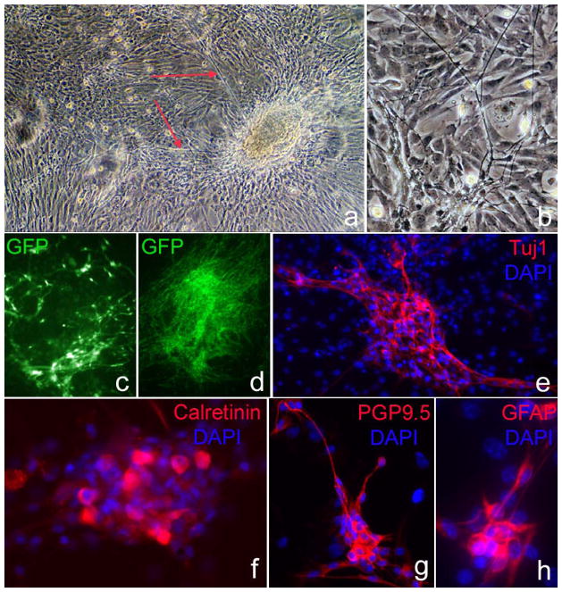
To confirm that Nestin-expressing cells in the MSM layer include ENSCs, we used gut organoid cultures as previously described [6]. These organoids prepared from the small intestine contain crypt-villus units, which attach to plastic surface within 24 hours. Cells proliferate and form large colonies over 7–10 days. In approximately 10–20% of colonies, elongated cell processes emerge from the core of the organoid (Fig. 5a) and form intricate networks with a neuronal morphology (Fig. 5b). Cells with this neuronal-like morphology were GFP+ (Fig. 5c, d). Over the following 14 days, GFP+ cells proliferated, expanded, and formed networks of elongated cells (Fig. 5e) that stained for neural and glial markers (Fig. 5f–i).
Figure 5. Intestinal organoid cultures produce neuroglial colonies.
Gut organoid cultures generate colonies that extend long cell processes emerging from their core (a) and form networks with a neural morphology (b). These neuron-like cells were GFP+ (c, d) and, over the following 14 days, proliferate and expand to form larger networks of elongated cells expressing neural (e–g) and glial markers (h).
Gut and Brain Neurospheres express similar pluripotency markers
Transcript levels for the pluripotency markers Sox2, cMyc, and Klf4 were quantified by qPCR, and the results normalized to Gapdh. All three factors were expressed in Musc, MSM, and brain-derived proliferating neurospheres (Fig. 6). Expression of cMyc and Klf4 was not statistically different among the three sources of neurospheres. Sox2 expression was significantly greater in Musc-derived than MSM-derived neurospheres (p=0.016), but not different from brain.
Figure 6. Markers of pluripotency are expressed in Musc, MSM, and brain neurospheres.
Quantitative PCR on proliferating neurospheres demonstrates Sox2 (A), cMyc (B), and Klf4 (C) expression in neurospheres derived from Musc, MSM, and brain. Results represent averages following three separate experiments, each performed in triplicate. The only statistical difference observed was in Sox2 expression between Musc- and MSM-derived neurospheres.
Discussion
NSCs can be isolated from specific areas of the post-natal CNS including the subventricular zone [22], hypothalamus [23], hippocampus [24] and spinal cord [25]. In response to injury, these cells proliferate, home to the site of tissue damage, and differentiate into neurons and glia that are engaged in repair processes [26] [27]. CNS-derived NSCs also provide trophic support to damaged tissue, enhance vasculogenesis, and promote survival, migration, and differentiation of the endogenous precursor cells following stroke [27]. We and others [6] [7] [28] [29] [30], have found that the intestine also possesses NSCs, but these cells are not nearly as well characterized as those in the CNS and their role in health and disease remains unknown. Intestinal NSCs are most commonly isolated from the muscularis layer of the gut [7] [31], although some have reported successful isolation from mucosa [6] [32]. Kruger isolated NSCs from the intestinal muscle layer based on p75 and α4 integrin expression, but did not find these markers expressed in the mucosa [7]. More recently Metzger et al. [32] seeded cells from human intestinal mucosal-submucosal biopsies. They were able to produce floating colonies that expanded, yielded neurons and glial cells, and could be transplanted into embryonic aganglionic mouse hindgut. Our results are consistent with those of Metzger et al., as cells isolated from rodent MSM also yielded NS that contained neurons and glia. We were able to confirm this result using organoid cultures from small intestine, an established model for culturing only mucosa [33], although some submucosal cells may also be present. These organoid cultures produced colonies that contained both neurons and glia. The source of these cells, whether mucosal or submucosal, is unclear. However, both layers are readily accessible by endoscopic biopsy, making these cells a potentially important source of multipotent progenitor cells able to generate neurons and glia for autologous cell replacement therapies and diagnostic cell-based assays.
Sohal et al. [34] followed a population of cells emigrating ventrally from the cranial neural tube in the developing chick and into the gastrointestinal tract. These cells colonized only the foregut and were located in enteric ganglia, smooth muscle, and mucosa. These cells differentiated into neurons, glial cells, interstitial cells of Cajal, and epithelial cells, possibly indicating a common progenitor cell for both epithelial and neuroglial lineages. Whether a subset of these cells retain multipotentiality in the adult mammalian organism remains to be determined.
To determine whether NSC can be isolated from different locations along the gastrointestinal tract, we used adherent and non-adherent culture techniques and successfully isolated NSCs from both small and large intestine. Other authors have similarly found them both in large and small intestine [35]. While NSCs appear to be present along the entire length of the gut, it remains to be shown whether the density of NSCs differs along the proximo-distal axis. The possibility of using autologous NSCs from one area of the GI tract for transplantation into another area, such as transplanting small intestine-derived NSCs into aganglionic colon, may have important clinical value. Previous animal models of NSC transplantation suggest this to be a feasible approach [36], although the practicability of autologous transplantation in humans has yet to be evaluated.
Adherent and floating colonies prepared from Nestin-GFP mice were strongly GFP+ early in their formation. We have previously shown that small intestine-derived NSCs express Nestin [6], and others have also found Nestin expression by intestinal NSCs [37] [38]. These results suggest that gut-derived colonies originate from a Nestin-expressing stem/progenitor cell. This conclusion is supported by recent evidence showing that targeted deletion of Hand2 in nestin-expressing cells leads to substantial loss of enteric neurons, confirming that a population of neural precursors expresses nestin [39]. In both MSM and Musc-derived NS, we find expression of Sox-2, c-Myc, and Klf4. These genes are associated with stem cells and multipotentiality in other tissues, including skin [40] [41], pancreas [42], umbilical cord [43], and brain [44]. These genes are also capable of generating induced pluripotent stem cells [45], confirming their important role in stem cell development and differentiation. Heanue et al. also found Sox-2 expressed in intestinal NSCs and targeted this transcription factor to isolate NSCs [46]. Their studies yielded a cellular morphology very similar to that observed in our Nestin+ NSCs.
Differentiating neurons and glia that develop from Nestin+ colonies continue to express Nestin strongly as they grow and differentiate. Interestingly, while Nestin expression is initially limited to the glial population in the normal intestine, it is expressed by both newly differentiated neurons and glia in vitro. This raises the intriguing possibility that new neurons may arise from glial precursors. Recent evidence is consistent with this hypothesis, suggesting that enteric glial cells may represent the NSC in the gut [47] [48]. In the CNS, radial glia serve not only as a source for neurogenesis but also as a scaffold for remodeling newly nascent neurons [49]. In both brain- and gut-derived cultures, we found that cells emerging from adherent NS create long, bridging extensions used as migratory pathways by later generations of cells emerging from the NS. We hypothesize that in the gut a subset of glial cells may have a similar neurogenic and scaffolding potential. Finally, this work reveals fundamental similarities between intestinal-derived and brain-derived NSCs. Both are capable of generating NS that share similar morphologic and immunophenotypic features, with glial, neuronal, and smooth muscle cells. Importantly, both appear to arise from Nestin-expressing cells.
Acknowledgments
The authors gratefully acknowledge support from the American Academy of Neurology (JD) and the Stephen E. and Catherine Pappas Foundation (JD). JD is a recipient of a Paul Calabresi Career Development Award for Clinical Oncology. NN is supported be a Bolyai Fellowship of the Hungarian Academy of Sciences. AMG is supported by NIH grant DK080914.
Funding:
No funding declared
Footnotes
Author contribution:
JBG designed the research study and performed the cell culture experiments and wrote the paper
CS Performed the tissue processing, immunostains and photographs and assisted the transplants
AP Performed the gut micro dissection and assisted the transplants
NN Performed the transplants and some of the immunostains
LAB Performed the RT-PCR experiments
JD Contributed essential reagents, the mouse model, expertise and helped design the research
AC Assisted and performed some of the cell culture experiments
AMG Helped design the research, analyzed results, contributed expertise and helped write the paper
Conflicts of Interest:
No competing interests declared
References
- 1.Kiba T. Relationships between the autonomic nervous system, humoral factors and immune functions in the intestine. Digestion. 2006;74(3–4):215–27. doi: 10.1159/000100512. [DOI] [PubMed] [Google Scholar]
- 2.Margolis KG, Stevanovic K, Karamooz N, et al. Enteric neuronal density contributes to the severity of intestinal inflammation. Gastroenterology. 2011;141(2):588–98. 598 e1–2. doi: 10.1053/j.gastro.2011.04.047. [DOI] [PMC free article] [PubMed] [Google Scholar]
- 3.Bischoff SC, Mailer R, Pabst O, et al. Role of serotonin in intestinal inflammation: knockout of serotonin reuptake transporter exacerbates 2,4,6-trinitrobenzene sulfonic acid colitis in mice. Am J Physiol Gastrointest Liver Physiol. 2009;296(3):G685–95. doi: 10.1152/ajpgi.90685.2008. [DOI] [PubMed] [Google Scholar]
- 4.Jessen KR, Mirsky R. Glial cells in the enteric nervous system contain glial fibrillary acidic protein. Nature. 1980;286(5774):736–7. doi: 10.1038/286736a0. [DOI] [PubMed] [Google Scholar]
- 5.Jessen KR, Mirsky R. Astrocyte-like glia in the peripheral nervous system: an immunohistochemical study of enteric glia. J Neurosci. 1983;3(11):2206–18. doi: 10.1523/JNEUROSCI.03-11-02206.1983. [DOI] [PMC free article] [PubMed] [Google Scholar]
- 6.Suarez-Rodriguez R, Belkind-Gerson J. Cultured nestin-positive cells from postnatal mouse small bowel differentiate ex vivo into neurons, glia, and smooth muscle. Stem Cells. 2004;22(7):1373–85. doi: 10.1634/stemcells.2003-0049. [DOI] [PubMed] [Google Scholar]
- 7.Kruger GM, Mosher JT, Bixby S, Joseph N, Iwashita T, Morrison SJ. Neural crest stem cells persist in the adult gut but undergo changes in self-renewal, neuronal subtype potential, and factor responsiveness. Neuron. 2002;35(4):657–69. doi: 10.1016/s0896-6273(02)00827-9. [DOI] [PMC free article] [PubMed] [Google Scholar]
- 8.Mosher JT, Yeager KJ, Kruger GM, et al. Intrinsic differences among spatially distinct neural crest stem cells in terms of migratory properties, fate determination, and ability to colonize the enteric nervous system. Dev Biol. 2007;303(1):1–15. doi: 10.1016/j.ydbio.2006.10.026. [DOI] [PMC free article] [PubMed] [Google Scholar]
- 9.Bondurand N, Natarajan D, Thapar N, Atkins C, Pachnis V. Neuron and glia generating progenitors of the mammalian enteric nervous system isolated from foetal and postnatal gut cultures. Development. 2003;130(25):6387–400. doi: 10.1242/dev.00857. [DOI] [PubMed] [Google Scholar]
- 10.Chouaf-Lakhdar L, Fevre-Montange M, Brisson C, Strazielle N, Gamrani H, Didier-Bazes M. Proliferative activity and nestin expression in periventricular cells of the adult rat brain. Neuroreport. 2003;14(4):633–6. doi: 10.1097/00001756-200303240-00022. [DOI] [PubMed] [Google Scholar]
- 11.Kukekov VG, Laywell ED, Thomas LB, Steindler DA. A nestin-negative precursor cell from the adult mouse brain gives rise to neurons and glia. Glia. 1997;21(4):399–407. doi: 10.1002/(sici)1098-1136(199712)21:4<399::aid-glia7>3.0.co;2-z. [DOI] [PubMed] [Google Scholar]
- 12.Zhou FC, Chiang YH. Long-term nonpassaged EGF-responsive neural precursor cells are stem cells. Wound Repair Regen. 1998;6(4):337–48. doi: 10.1046/j.1524-475x.1998.60409.x. [DOI] [PubMed] [Google Scholar]
- 13.Kawaguchi A, Miyata T, Sawamoto K, et al. Nestin-EGFP transgenic mice: visualization of the self-renewal and multipotency of CNS stem cells. Mol Cell Neurosci. 2001;17(2):259–73. doi: 10.1006/mcne.2000.0925. [DOI] [PubMed] [Google Scholar]
- 14.Wiese C, Rolletschek A, Kania G, et al. Nestin expression--a property of multi-lineage progenitor cells? Cell Mol Life Sci. 2004;61(19–20):2510–22. doi: 10.1007/s00018-004-4144-6. [DOI] [PubMed] [Google Scholar]
- 15.Frisen J, Johansson CB, Torok C, Risling M, Lendahl U. Rapid, widespread, and longlasting induction of nestin contributes to the generation of glial scar tissue after CNS injury. J Cell Biol. 1995;131(2):453–64. doi: 10.1083/jcb.131.2.453. [DOI] [PMC free article] [PubMed] [Google Scholar]
- 16.Vaittinen S, Lukka R, Sahlgren C, et al. Specific and innervation-regulated expression of the intermediate filament protein nestin at neuromuscular and myotendinous junctions in skeletal muscle. Am J Pathol. 1999;154(2):591–600. doi: 10.1016/S0002-9440(10)65304-7. [DOI] [PMC free article] [PubMed] [Google Scholar]
- 17.Dahlstrand J, V, Collins P, Lendahl U. Expression of the class VI intermediate filament nestin in human central nervous system tumors. Cancer Res. 1992;52(19):5334–41. [PubMed] [Google Scholar]
- 18.Vanderwinden JM, Gillard K, De Laet MH, Messam CA, Schiffmann SN. Distribution of the intermediate filament nestin in the muscularis propria of the human gastrointestinal tract. Cell Tissue Res. 2002;309(2):261–8. doi: 10.1007/s00441-002-0590-3. [DOI] [PubMed] [Google Scholar]
- 19.Cantarero Carmona I, Luesma Bartolome MJ, Lavoie-Gagnon C, Junquera Escribano C. Distribution of nestin protein: immunohistochemical study in enteric plexus of rat duodenum. Microsc Res Tech. 2011;74(2):148–52. doi: 10.1002/jemt.20884. [DOI] [PubMed] [Google Scholar]
- 20.Mignone JL, Kukekov V, Chiang AS, Steindler D, Enikolopov G. Neural stem and progenitor cells in nestin-GFP transgenic mice. J Comp Neurol. 2004;469(3):311–24. doi: 10.1002/cne.10964. [DOI] [PubMed] [Google Scholar]
- 21.Nagy N, Goldstein AM. Endothelin-3 regulates neural crest cell proliferation and differentiation in the hindgut enteric nervous system. Dev Biol. 2006;293(1):203–17. doi: 10.1016/j.ydbio.2006.01.032. [DOI] [PubMed] [Google Scholar]
- 22.Sanai N, Nguyen T, Ihrie RA, et al. Corridors of migrating neurons in the human brain and their decline during infancy. Nature. 2011;478(7369):382–6. doi: 10.1038/nature10487. [DOI] [PMC free article] [PubMed] [Google Scholar]
- 23.Yuan TF, Arias-Carrion O. Adult neurogenesis in the hypothalamus: evidence, functions, and implications. CNS Neurol Disord Drug Targets. 2011;10(4):433–9. doi: 10.2174/187152711795563985. [DOI] [PubMed] [Google Scholar]
- 24.Amrein I, Isler K, Lipp HP. Comparing adult hippocampal neurogenesis in mammalian species and orders: influence of chronological age and life history stage. Eur J Neurosci. 2011;34(6):978–87. doi: 10.1111/j.1460-9568.2011.07804.x. [DOI] [PubMed] [Google Scholar]
- 25.Leung C, Chan SC, Tsang SL, Wu W, Sham MH. Cyp26b1 mediates differential neurogenicity in axial-specific populations of adult spinal cord progenitor cells. Stem Cells Dev. 2012 doi: 10.1089/scd.2011.0613. [DOI] [PubMed] [Google Scholar]
- 26.Clausen M, Nakagomi T, Nakano-Doi A, et al. Ischemia-induced neural stem/progenitor cells express pyramidal cell markers. Neuroreport. 2011;22(16):789–94. doi: 10.1097/WNR.0b013e32834acb54. [DOI] [PubMed] [Google Scholar]
- 27.Chang YC, Shyu WC, Lin SZ, Li H. Regenerative therapy for stroke. Cell Transplant. 2007;16(2):171–81. [PubMed] [Google Scholar]
- 28.Bixby S, Kruger GM, Mosher JT, Joseph NM, Morrison SJ. Cell-intrinsic differences between stem cells from different regions of the peripheral nervous system regulate the generation of neural diversity. Neuron. 2002;35(4):643–56. doi: 10.1016/s0896-6273(02)00825-5. [DOI] [PubMed] [Google Scholar]
- 29.Schafer KH, Van Ginneken C, Copray S. Plasticity and neural stem cells in the enteric nervous system. Anat Rec (Hoboken) 2009;292(12):1940–52. doi: 10.1002/ar.21033. [DOI] [PubMed] [Google Scholar]
- 30.Almond S, Lindley RM, Kenny SE, Connell MG, Edgar DH. Characterisation and transplantation of enteric nervous system progenitor cells. Gut. 2007;56(4):489–96. doi: 10.1136/gut.2006.094565. [DOI] [PMC free article] [PubMed] [Google Scholar]
- 31.Silva AT, Wardhaugh T, Dolatshad NF, Jones S, Saffrey MJ. Neural progenitors from isolated postnatal rat myenteric ganglia: expansion as neurospheres and differentiation in vitro. Brain Res. 2008;1218:47–53. doi: 10.1016/j.brainres.2008.04.051. [DOI] [PubMed] [Google Scholar]
- 32.Metzger M, Caldwell C, Barlow AJ, Burns AJ, Thapar N. Enteric nervous system stem cells derived from human gut mucosa for the treatment of aganglionic gut disorders. Gastroenterology. 2009;136(7):2214–25. e1–3. doi: 10.1053/j.gastro.2009.02.048. [DOI] [PubMed] [Google Scholar]
- 33.Evans GS, Flint N, Somers AS, Eyden B, Potten CS. The development of a method for the preparation of rat intestinal epithelial cell primary cultures. J Cell Sci. 1992;101(Pt 1):219–31. doi: 10.1242/jcs.101.1.219. [DOI] [PubMed] [Google Scholar]
- 34.Sohal GS, Ali MM, Farooqui FA. A second source of precursor cells for the developing enteric nervous system and interstitial cells of Cajal. Int J Dev Neurosci. 2002;20(8):619–26. doi: 10.1016/s0736-5748(02)00103-x. [DOI] [PubMed] [Google Scholar]
- 35.Metzger M, Bareiss PM, Danker T, et al. Expansion and differentiation of neural progenitors derived from the human adult enteric nervous system. Gastroenterology. 2009;137(6):2063–2073. e4. doi: 10.1053/j.gastro.2009.06.038. [DOI] [PubMed] [Google Scholar]
- 36.Lindley RM, Hawcutt DB, Connell MG, et al. Human and mouse enteric nervous system neurosphere transplants regulate the function of aganglionic embryonic distal colon. Gastroenterology. 2008;135(1):205–216. e6. doi: 10.1053/j.gastro.2008.03.035. [DOI] [PubMed] [Google Scholar]
- 37.Rauch U, Klotz M, Maas-Omlor S, et al. Expression of intermediate filament proteins and neuronal markers in the human fetal gut. J Histochem Cytochem. 2006;54(1):39–46. doi: 10.1369/jhc.4A6495.2005. [DOI] [PubMed] [Google Scholar]
- 38.Schafer KH, Hagl CI, Rauch U. Differentiation of neurospheres from the enteric nervous system. Pediatr Surg Int. 2003;19(5):340–4. doi: 10.1007/s00383-003-1007-4. [DOI] [PubMed] [Google Scholar]
- 39.Lei J, Howard MJ. Targeted deletion of Hand2 in enteric neural precursor cells affects its functions in neurogenesis, neurotransmitter specification and gangliogenesis, causing functional aganglionosis. Development. 2011;138(21):4789–800. doi: 10.1242/dev.060053. [DOI] [PMC free article] [PubMed] [Google Scholar]
- 40.Clewes O, Narytnyk A, Gillinder KR, Loughney AD, Murdoch AP, Sieber-Blum M. Human epidermal neural crest stem cells (hEPI-NCSC)--characterization and directed differentiation into osteocytes and melanocytes. Stem Cell Rev. 2011;7(4):799–814. doi: 10.1007/s12015-011-9255-5. [DOI] [PMC free article] [PubMed] [Google Scholar]
- 41.Sieber-Blum M, Hu Y. Epidermal neural crest stem cells (EPI-NCSC) and pluripotency. Stem Cell Rev. 2008;4(4):256–60. doi: 10.1007/s12015-008-9042-0. [DOI] [PubMed] [Google Scholar]
- 42.Shankar S, Nall D, Tang SN, et al. Resveratrol inhibits pancreatic cancer stem cell characteristics in human and KrasG12D transgenic mice by inhibiting pluripotency maintaining factors and epithelial-mesenchymal transition. PLoS One. 2011;6(1):e16530. doi: 10.1371/journal.pone.0016530. [DOI] [PMC free article] [PubMed] [Google Scholar]
- 43.Pessina A, Bonomi A, Sisto F, et al. CD45+/CD133+ positive cells expanded from umbilical cord blood expressing PDX-1 and markers of pluripotency. Cell Biol Int. 2010;34(8):783–90. doi: 10.1042/CBI20090236. [DOI] [PubMed] [Google Scholar]
- 44.Ma HI, Kao CL, Lee YY, et al. Differential expression profiling between atypical teratoid/rhabdoid and medulloblastoma tumor in vitro and in vivo using microarray analysis. Childs Nerv Syst. 2010;26(3):293–303. doi: 10.1007/s00381-009-1016-2. [DOI] [PubMed] [Google Scholar]
- 45.Eminli S, Utikal J, Arnold K, Jaenisch R, Hochedlinger K. Reprogramming of neural progenitor cells into induced pluripotent stem cells in the absence of exogenous Sox2 expression. Stem Cells. 2008;26(10):2467–74. doi: 10.1634/stemcells.2008-0317. [DOI] [PubMed] [Google Scholar]
- 46.Heanue TA, Pachnis V. Prospective identification and isolation of enteric nervous system progenitors using Sox2. Stem Cells. 2011;29(1):128–40. doi: 10.1002/stem.557. [DOI] [PMC free article] [PubMed] [Google Scholar]
- 47.Laranjeira C, Sandgren K, Kessaris N, et al. Glial cells in the mouse enteric nervous system can undergo neurogenesis in response to injury. J Clin Invest. 2011;121(9):3412–24. doi: 10.1172/JCI58200. [DOI] [PMC free article] [PubMed] [Google Scholar]
- 48.Joseph NM, He S, Quintana E, Kim YG, Nunez G, Morrison SJ. Enteric glia are multipotent in culture but primarily form glia in the adult rodent gut. J Clin Invest. 2011;121(9):3398–411. doi: 10.1172/JCI58186. [DOI] [PMC free article] [PubMed] [Google Scholar]
- 49.Sild M, Ruthazer ES. Radial glia: progenitor, pathway, and partner. Neuroscientist. 2011;17(3):288–302. doi: 10.1177/1073858410385870. [DOI] [PubMed] [Google Scholar]


