Abstract
Aim:
The purpose of this study was to evaluate and compare the tensile bond strength and microleakage of Fuji IX GP, Fuji II LC, and compoglass and to compare bond strength with degree of microleakage exhibited by the same materials.
Materials and Methods:
Occlusal surfaces of 96 noncarious primary teeth were ground perpendicular to long axis of the tooth. Preparations were distributed into three groups consisting of Fuji IX GP, Fuji II LC and Compoglass. Specimens were tested for tensile bond strength by mounting them on Instron Universal Testing Machine. Ninety-six primary molars were treated with Fuji IX GP, Fuji II LC, and compoglass on box-only prepared proximal surface. Samples were thermocycled, stained with dye, sectioned, and scored for microleakage under stereomicroscope. ANOVA and Bonferrani correction test were done for comparisons. Pearson Chi-square test and regression analysis were done to assess the association between the parameters.
Results:
Compoglass showed highest tensile strength and Fuji II LC showed least microleakage. There was a significant difference between the three groups in tensile strength and microleakage levels. The correlation between tensile strength and microleakage level in each group showed that there was a significant negative correlation only in Group 3.
Conclusion:
Fuji II LC and compoglass can be advocated in primary teeth because of their superior physical properties when compared with Fuji IX GP.
Keywords: Compomer, glass ionomer, microleakage, tensile bond strength
Introduction
A major advancement in the current practice of dentistry is the restoration of teeth with tooth colored, adhesive materials. The success and longevity of a dental restoration depends on the sealing of the cavity walls as well as the retention to the tooth surface.
When glass ionomer cements were developed by Wilson and Kent in 1972, it caught the attention of researchers and practicing dentists, because it was reported to form chemically adhesive bonds to the tooth structure. The materials are tooth colored, unite with tooth structure, have tissue compatibility, are radiopaque, release fluoride over time, inhibit demineralization, and contribute to remineralization of adjacent dentin. The use of glass ionomer cements has grown since their introduction in the 1970s because of their advantages.
However, the conventional glass ionomer cement has been plagued by several negative characteristics: Prolonged setting time that restricts finishing and polishing for approximately 24 hours, sensitivity to moisture during initial hardening, dehydration, rough surface texture, opaqueness, low fracture toughness, and poor wear resistance. Several authors assume that gap size is positively correlated to microleakage values and attempts have been made to correlate bond strength values with marginal gap size.
The purpose of this study was to evaluate the tensile bond strength and microleakage of a conventional glass ionomer (Fuji IX GP, GC products), resin modified glass ionomer (Fuji II LC, GC products) and a compomer, vivadent products (Compoglass) of primary molars and also to compare the bond strengths with the degree of microleakage exhibited by the same materials.
Materials and Methods
A total of 192 extracted noncarious primary molars were used as test specimens. Out of this, 96 teeth were used as test specimens for tensile bond strength and the other 96 teeth for microleakage. For the assessment of tensile bond strength, teeth were stored in ringer's solution, at room temperature. The occlusal surfaces of noncarious teeth were ground perpendicular to the long axis of the tooth using a water cooled diamond disc mounted on an air motor hand-piece.
Horizontal indentations were placed on the radicular portion of the specimens. The teeth were then embedded into self-curing acrylic resin.
96 teeth were randomly divided into 3 groups of 32 teeth each.
Group 1: Fuji IX GP (conventional glass ionomer cement)
Group 2: Fuji II LC (cesin modified glass ionomer cement)
Group 3: Compoglass (compomer).
Group 1
In this group, Fuji IX GP was used. The dentin surface was conditioned for 20 seconds with dentin conditioner and then rinsed off with water and dried by gently blowing with an air syringe. A hollow polyvinyl mould with an inner diameter of 4 mm and height 6 mm was placed on the treated dentin surface. Fuji IX GP was inserted into the polyvinyl mould. A 26 gauge ligature wire was twisted to form a loop at one end and was placed inside the cement. Following complete setting, the polyvinyl cylindrical moulds were removed and the specimens were returned to ringer's solution.
Group 2
The same methodology as group 1 was followed except for the restorative materials, which were used. The material used for restoration was Fuji II LC.
Group 3
In group 3, compoglass was used. Acid etching was done on the dentin surface with 37% phosphoric acid gel for 15 seconds, which was then rinsed off with water and gently air-dried using an air syringe. Syntac single component was coated on the prepared surface and cured for 20 seconds. A second layer of Syntac single-component was applied and then light-cured for 20 seconds. A hollow polyvinyl cylindrical mould with inner diameter of 4 mm and height 6 mm was placed on the treated dentin surface and compoglass was inserted. A 26 gauge ligature wire was twisted to form a loop at one end and was placed inside the restoration and then light cured for 40 seconds. Following complete curing, the polyvinyl cylindrical moulds were removed. The specimens were then returned to ringer's solution.
The specimens were tested for tensile bond strength by mounting them onto an Instron Universal Testing Machine running at a crosshead speed of 5 mm/minute.
The data base was analyzed in Statistical Package for Social Sciences (SPSS). version 10.0.5. One way analysis of variance (ANOVA) was done to find out the statistically significant difference in the tensile bond strength between the three groups. Bonferrani correction test was done for multiple comparisons.
For the assessment of microleakage, teeth were stored at room temperature, in 1% sodium hypochlorite solution for disinfection purposes and used within a month. In all the teeth, box-only preparations were done on the proximal surface. The preparations were standardized to a depth of 3 mm and width of 3 mm.
All the three groups were restored with the respective restorative materials according to the manufacturer's instructions. The teeth were finished to contour with a composite finishing bur 7901 and polished using soflex discs (3M ESPE products) in a low speed contra-angled hand piece on a micromotor. The teeth were impermeabilized by coating the apices with sticky wax. One coat of nail varnish was applied on the entire tooth except up to 1 mm from the restoration margin.
All the specimens were subjected to 1000 thermocycles. Each cycle consisted of 30 seconds at 6°C±2°C and 30 seconds at 60°C±2°C. After thermocycling, the teeth were placed in 2% basic fuchsin dye for 24 hours at room temperature. After removal of the specimens from the dye solution, the superficial dye was removed with a pumice slurry and rubber cup. The specimens were then sectioned longitudinally with double-sided diamond discs. Microleakage was studied under the stereomicroscope at ×16 magnification.
Staining along the tooth restoration interface was recorded according to the following scores:
0=No dye penetration
1=Partial dye penetration
2=Dye penetration along the occlusal or gingival wall, but not including the axial wall.
3=Dye penetration to and along the axial wall.
Pearson Chi-square test was used to find out the association between the three groups versus microleakage. Regression analysis was done to assess the association between both the parameters, that is, tensile bond strength and microleakage. Pearson correlation was used to find out the correlation between the two parameters, that is, tensile bond strength and microleakage.
Results
The mean tensile bond strength, standard deviation values and 95% confidence interval (CI) values are presented in Table 1. One way ANOVA was done to find out the statistically significant difference in the tensile bond strength between the three groups. P<0.001 indicates that calculated values are highly significant. Group 1 has a mean of 1.02±0.39 (95% CI: 0.88–1.16). Group 2 has a mean of 1.52±0.46 (95% CI: 1.36–1.69). Group 3 has a mean of 2.44±0.52 (95% CI: 2.25–2.62). One way ANOVA shows that there is a significant difference between the three groups in tensile bond strength (P<0.01) [Figure 1].
Table 1.
Mean Tensile bond strengths

Figure 1.
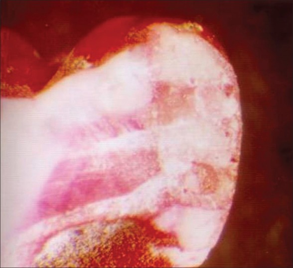
Group 1-Fuji IX GP at ×16 magnification
Table 2 shows the statistical analysis using Pearson Chi-Square test which shows the count and the percentage of the microleakage level in the overall, occlusal, gingival, and axial surface and the P-value. P<0.01 indicates that calculated values are highly significant. Pearson Chi-square test shows that there was a significant difference between the microleakage levels (overall, occlusal, gingival, and axial) in the three groups (P<0.01).
Table 2.
Pearson Chi-Square test
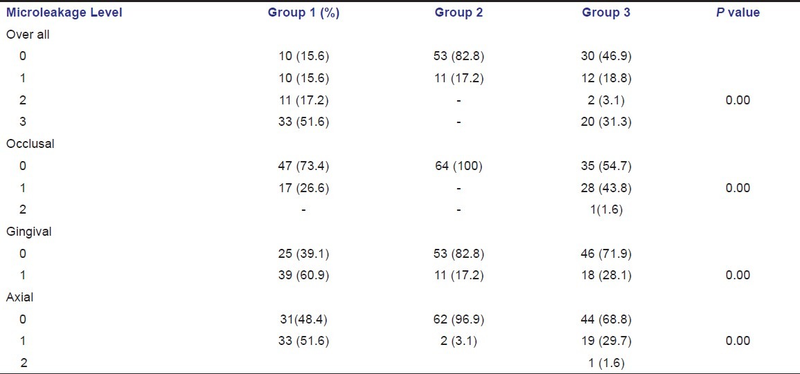
The microleakage level was more in group 1 [Figure 2] and group 2 [Figure 3] showed the least microleakage, group 3 [Figure 4] also showed more microleakage when compared with group 2, that is, no microleakage in 82.8% of the samples in group 2, whereas groups 1 and 3 showed that only 15.6% and 46.9% had no microleakage, respectively [Figure 5].
Figure 2.
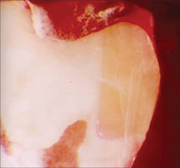
Group 2-Fuji II LC at ×16 magnification
Figure 3.
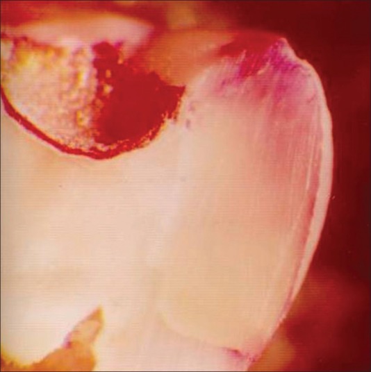
Group 3-Compoglass at ×16 magnification
Figure 4.
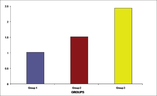
Mean tensile bond strengths for the three groups
Figure 5.
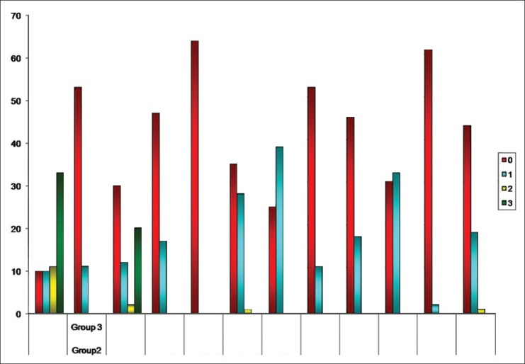
Count of the microleakage level
Table 3 shows the Pearson correlation coefficient exhibiting a significant negative correlation in group 3 between tensile bond strength and microleakage level. Even though group 2 also showed significant negative correlation between tensile bond strength and microleakage level, but the significance was not much. Group 1 did not have any significant correlation between tensile bond strength and microleakage level.
Table 3.
Pearson Correlation Coefficient

Discussion
Interest in adhesion of restorative materials to enamel and dentin has gained considerable interest during recent years.
The old and traditional methods of cavity preparation were material driven and tooth destructive. Based on the possibilities of adhering the restorations to tooth structure, a new cavity preparation philosophy emerged: Cavity size and shape is strictly defect oriented and minimally invasive. The maximum amount of healthy tissue is preserved and the cavity extension is limited to technical parameters.
During the past decades, glass ionomers have become important dental restorative and luting materials for use in children. The advantages of glass ionomer cements are fluoride ion release and uptake, biocompatibility and chemical bonding to both enamel and dentin and its co-efficient of thermal expansion is almost similar to the tooth structure. In spite of the advantages, dentists were reluctant to use the material in clinical situations as they were difficult to handle, had low wear resistance and strength, were brittle, and proved unreliable in the long-term because of poor reliability.
An important advancement in glass ionomer technology that has influenced dentistry for children is development of the Resin Modified Glass Ionomer Cements. The resin modified glass ionomer cements consist primarily of glass ionomer and a minor amount of resin. The resin modified glass ionomer cements harden initially by free-radical photopolymerization of the resin component in the formulation. A chemical resin polymerization reaction and the glass ionomer setting reaction subsequently progress. Addition of the resin component not only decreases initial hardening time and handling difficulties, but also substantially increases wear resistance and physical strengths of the cement.
Compomers have become available more recently and are recommended for use as a pediatric restorative material. Compomers are actually a cross between composite resin and glass ionomer cement and are officially termed polyacid-modified, resin-based composites. The fluoride release from compomers is less than that of glass ionomer cements. The mechanical properties of tensile and flexural strength as well as wear resistance of compomers are superior to that of glass ionomers.
The effectiveness of an adhesive dental restorative material has been evaluated by one or a combination of the following methods: Direct bond strength measurements and study of marginal leakage.
From the results of the present in vitro study, the highest tensile bond strength was observed with compomers and the least tensile bond strength for chemically cured glass ionomer cement.
The increased bond strength value in group 3, where compomer was used may be due to the fact that they are composed primarily of the resin component in a larger fraction by weight. Moreover, pretreatment of the dentin surface with orthophosphoric acid gives higher bond strength, because bonding of compomers to tooth structure is primarily mediated by micromechanical retention.
The inherent morphology of the dentin is complicated by the formation of a smear layer. The smear layer acts as a diffusion barrier that decreases dentinal permeability and is also considered as an obstruction that prevents resin from reaching the underlying dentin substrate. The phosphoric-acid etching removes the smear layer, opens the dentinal tubules and allows deeper penetration of the resin matrix.
An adhesive may be applied over the entire cavity preparation as it improves the retention of the restoration. The bonding agent penetrates into the dentin. The bonding agent co-polymerizes with the primer to form an intermingled layer of collagen fibers and resin called the hybrid layer. This hybrid layer has been considered the most important factor for ensuring a good bond between resin and dentin.
The higher tensile bond strength of resin modified glass ionomer cement may be due to the presence of 20% resin component in group 2 and none in group 1. Oilo et al.[1] and Lin et al.[2] also observed a tensile bond strength for resin modified glass ionomer cement, which was greater than chemically cured glass ionomer cement.
The results obtained in this study shows that the resin modified glass ionomer cement-Fuji II LC exhibited the least microleakage. A total of 82.8% of Fuji II LC samples did not exhibit any microleakage. This suggests the superior adhesion of Fuji II LC. This may be attributed to the fact of the similarity of coefficient of thermal expansion of tooth and restorative material contributing to superior adaptation of resin modified glass ionomer cement to the tooth structure (11.4×10⁻⁶/°C and 11×10-6/°C). This correlates with the findings of references.[3,4,5,6,7] The microleakage of resin modified glass ionomer cements was lesser than compomers. Only 46.9% of the compomer samples showed no microleakage. The resin modified glass ionomer cements have a significant auto-cure resin feature. This accomplishes a complete cure even in those areas of the preparation where the light has not reached. The glass ionomers are known to form an ion-exchange layer. This unites the cement to the tooth structure and completely prevents microleakage. The factor which might contribute to the compomers having more microleakage than resin modified glass ionomers is that the polymerization shrinkage of the light-cured resin pulls the material away from the cavity walls, forming a gap. This gap at the restoration margins may allow microleakage. This is in agreement with the study of Brackett et al.[8]
In our study, the association between tensile bond strength and microleakage was also studied. Pearson correlation coefficient [Table 3] was used to know the correlation between tensile bond strength and microleakage in all the three groups.
Pearson correlation coefficient showed that, there was a negative correlation in compomers, between tensile bond strength and microleakage level (P>0.05).
Regression analysis predicted that compomers would show a 0.83 unit decrease in microleakage level, when there was an increase in one unit of tensile bond strength.
Hence, we conclude that the tensile bond strength of compoglass is significantly greater than Fuji IX GP and Fuji II LC.
Fuji IX GP and compoglass showed moderate to severe leakage in contrast to minimal leakage with Fuji II LC.
The significant negative correlations between tensile bond strength and microleakage obtained with compomer indicate that high bond strengths are associated with low microleakage and vice versa.
Resin modified glass ionomer cements and compomers should be considered for restorations in primary teeth because of their biocompatibility, antibacterial properties, ability to leach fluorides, and because of their better physical properties.
Footnotes
Source of Support: Nil,
Conflict of Interest: None declared
References
- 1.Oilo G, Um CM. Bond strength of glass-ionomer cement and composite resin combinations. Quintessence Int. 1992;23:633–9. [PubMed] [Google Scholar]
- 2.Lin A, McIntyre NS, Davidson RD. Studies on the adhesion of glass-ionomer cements to dentin. J Dent Res. 1992;71:1836–41. doi: 10.1177/00220345920710111401. [DOI] [PubMed] [Google Scholar]
- 3.Toledano M, Osorio E, Osorio R, García-Godoy F. Microleakage of class V resin-modified glass ionomer and compomer restorations. J Prosthet Dent. 1999;81:610–5. doi: 10.1016/s0022-3913(99)70217-9. [DOI] [PubMed] [Google Scholar]
- 4.Castro A, Feigal RE. Microleakage of a new improved glass ionomer restorative material in primary and permanent teeth. Pediatr Dent. 2002;24:23–8. [PubMed] [Google Scholar]
- 5.Zyskind D, Frenkel A, Fuks A, Hirschfeld Z. Marginal leakage around V-shaped cavities restored with glass-ionomer cements: An in vitro study. Quintessence Int. 1991;22:41–5. [PubMed] [Google Scholar]
- 6.Gladys S, Van Meerbeek B, Lambrechts P, Vanherle G. Marginal adaptation and retention of a glass-ionomer, resin-modified glass-ionomers and a polyacid-modified resin composite in cervical class-V lesions. Dent Mater. 1998;14:294–306. doi: 10.1016/s0109-5641(98)00043-8. [DOI] [PubMed] [Google Scholar]
- 7.Rodrigues JA, De Magalhães CS, Serra MC, Rodrigues Júnior AL. In vitro microleakage of glass-ionomer composite resin hybrid materials. Oper Dent. 1999;24:89–95. [PubMed] [Google Scholar]
- 8.Brackett WW, Gunnin TD, Gilpatrick RO, Browning WD. Microleakage of class V compomer and light-cured glass ionomer restorations. J Prosthet Dent. 1998;79:261–3. doi: 10.1016/s0022-3913(98)70234-3. [DOI] [PubMed] [Google Scholar]


