Abstract
Patients with acute and chronic liver disease have long been assumed to have a bleeding tendency on the basis of abnormal results for standard tests of hemostasis. However, recent studies have suggested that hemostasis in patients with liver disease exists in a state of rebalance, in which defects in prohemostatic drivers are compensated for by commensurate changes in antihemostatic drivers. Standard assays of hemostasis cannot evaluate this potential state of rebalance because they only assess components of clot formation and, therefore, may provide misleading information regarding the risk of bleeding, possibly leading clinicians to administer unneeded or even harmful prohemostatic factors. Thromboelastography (TEG) is a commercially available, rapid, point-of-care assay that assesses clot formation in whole blood, including plasmatic and cellular components. Studies using TEG in patients with cirrhosis and acute liver failure have suggested that rebalanced hemostasis exists in many patients, even in the presence of thrombocytopenia and an elevated prothrombin time/international normalized ratio. TEG has also been used to study mechanisms of rebalanced hemostasis and the pathogenesis of specific complications of liver disease, such as variceal rebleeding and infection. Finally, TEG has become widely used to guide factor repletion and fibrinolytic therapy during liver transplantation. The present clinical review will summarize these potential applications of TEG in patients with liver disease.
Keywords: Thromboelastography, coagulopathy, cirrhosis, liver failure, hemostasis
Thromboelastography (TEG) records the assembly of a clot in whole blood and provides an assessment of overall hemostasis, including contributions from plasmatic and cellular components. In a single, point-of-care assay, TEG dissects the dynamics of clot formation, ultimate clot strength, and the stability of the clot.1 In contrast to TEG, standard tests of coagulation, such as prothrombin time (PT)/international normalized ratio of PT (INR) or activated partial thromboplastin time (aPTT), assess only plasmatic events in hemostasis; these tests omit the contribution of the platelet scaffold as well as interactions with other cellular components of whole blood. INR and aPTT may, therefore, provide an inadequate and possibly misleading assessment of bleeding risk in patients with liver disease.
Patients with severe liver disease such as cirrhosis or acute liver failure (ALF) are assumed to have a bleeding diathesis based on elevated INR, thrombocytopenia, and the presence of portal hypertension, but the magnitude of these contributors difers between acute and chronic conditions (Table 1). In patients with cirrhosis, INR increases modestly with decompensation of hepatic synthetic function and accurately reflects short-term patient mortality but does not predict bleeding risk.2,3 Instead, distortion of the hepatic architecture due to fibrosis leads to progressive portal hypertension, hypersplenism, moderate-to-severe thrombocytopenia, and gastroesophageal varices; these factors determine bleeding risk in patients with cirrhosis more accurately than INR. In contrast, patients with ALF develop a more dramatically elevated INR, which again predicts short-term mortality; however, these patients have relatively mild portal hypertension as a consequence of the collapse of the liver parenchyma.4,5 Patients with ALF frequently have a modest degree of thrombocyto-penia.6 Despite marked elevation of INR and variable thrombocytopenia, patients with ALF seldom bleed spontaneously, and clinically significant bleeding after invasive procedures is uncommon.7,8
Table 1.
Contributors to the Perception of a Bleeding Tendency in Patients with Acute and Chronic Liver Disease
| Contributor | Cirrhosis | Acute liver failure |
|---|---|---|
| Elevated international normalized ratio | Mild-moderate | Moderate-severe |
| Thrombocytopenia | Moderate-severe | Mild-moderate |
| Portal hypertension | Moderate-severe | None-mild |
This review will highlight the possible clinical utility of TEG in assessing the magnitude of potential bleeding risk in patients with ALF and cirrhosis. The use of TEG in exploring the state of overall hemostasis and the pathogenesis of specific complications of liver disease will also be highlighted.
Overview of Hemostasis in Liver Disease
Each of the 3 phases of hemostasis-platelet-endothelial interaction, coagulation, and fibrinolysis-is affected by liver disease, but the effects of laboratory abnormalities on overall hemostasis have only recently been assessed.
Primary hemostasis, the formation of the platelet plug on an endothelial defect, is considered to be defective in patients with cirrhosis due to qualitative platelet dysfunction and thrombocytopenia.9 However, compensatory mechanisms exist to restore platelet-endothelial interactions toward normal in patients with cirrhosis. Plasma concentrations of von Willebrand factor (vWF), the endothelial-derived protein responsible for adhesion of platelets to endothelial cells, are increased in patients with liver disease, which increases the stickiness of platelets. In addition, plasma concentrations of ADAMTS-13, a liver-derived protease that cleaves vWF into smaller and less sticky multimers, are decreased in patients with liver disease, again resulting in a relative increase in platelets' adhesion to the endothelium.3
The coagulation phase of hemostasis is considered to be defective in patients with liver disease due to their elevated INR. However, Tripodi and colleagues showed that patients with cirrhosis can generate as much thrombin as healthy controls, as long as the assay mixture includes thombomodulin, the endogenous activator of the anticoagulant protein C.10,11 In fact, patients with cirrhosis appear to have more of a hypercoagulable state than a bleeding tendency, as a result of elevated factor VIII levels from endothelial activation and/or injury and depressed protein C levels from decreased hepatic biosynthesis and possibly increased consumption.12
Finally, the clot dissolution phase of hemostasis is considered to be defective in patients with cirrhosis due to diminished levels of profibrinolytic proteins. However, deficits in profibrinolytic proteins such as plasminogen appear to be counterbalanced by low levels of anti-fibrinolytic proteins such as α2-antiplasmin and high levels of tissue plasminogen activator.
The above discussion forms the rationale for the modern concept of rebalanced hemostasis in patients with liver disease; in this state, abnormalities in prohemostatic drivers are compensated for by commensurate changes in antihemostatic drivers.3 However, patients with liver disease maintain a more fragile balance of hemostasis than normal subjects due to deficiencies in both prohemostatic and antihemostatic drivers, as well as the presence of other conditions that commonly accompany liver failure, such as renal failure, infection, and endothelial dysfunction. This discussion clearly suggests that standard laboratory tests of hemostasis such as INR do not adequately assess this state of rebalance, and more global assays such as TEG may be very informative.
Thromboelastography Methodology
Basic Thromboelastography Theory and Hardware
TEG was first introduced in the 1940s, and technical refinements have since led to standardization of the technique. Two TEG devices are commercially available: the Thrombelastograph Haemostasis Analyzer 5000 (Hae-monetics Corp., Haemoscope Division) and the Rotem (Pentapharm GmbH).13 In the Thromboelastograph Haemostasis Analyzer 5000, whole blood in a cuvette is rotated around a suspended pin, which registers resistance to rotation as clotting ensues. In the Rotem device, whole blood is placed into a cuvette into which a pin is immersed and rotated.
Originally, TEG was performed on freshly drawn, noncitrated blood, and the test required up to 60 minutes to complete. Currently, citrated blood is used and recalcified at the time of TEG, which has increased the allowable time from blood draw to assay to 2 hours, and the time required for the assay (excluding the clot dissolution phase) has decreased to approximately 30 minutes. In order to further decrease the time required for the TEG assay, kaolin is often added as an initiator of clotting. Although the addition of kaolin changes non-kaolin-activated TEG parameters in normal volunteers and patients with cirrhosis, the effects of such changes on bleeding risk have not been studied.14
Thromboelastography Parameters
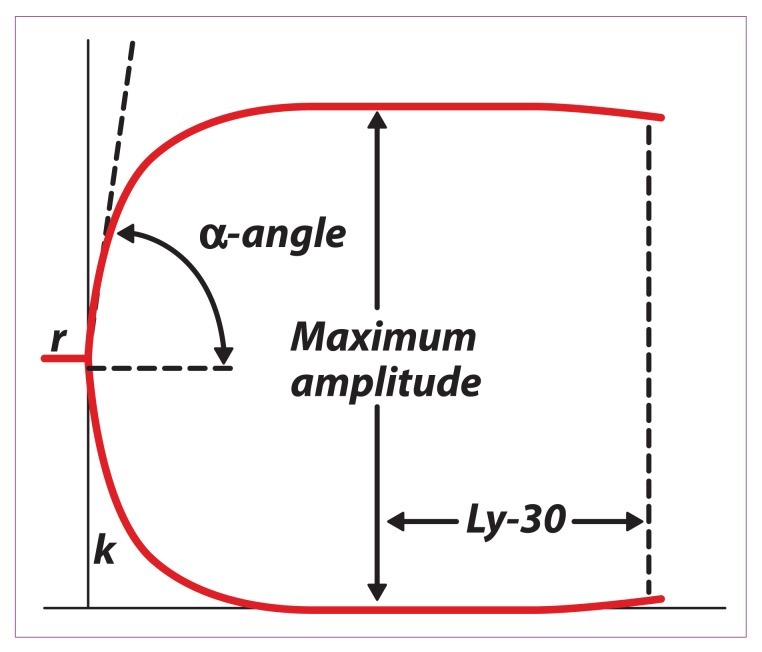
Five parameters are recorded in a standard TEG (Table 2 and Figures 1). The reaction (r)-time (in minutes) represents the latency of clot formation from the beginning of the clotting reaction to the initial formation of fibrin (defined as a clot amplitude of 2 mm). The r-time generally corresponds to INR and aPTT but may depend on details of TEG methodology.15 The kinetic (k)-time (in minutes) describes the time required for the initial fibrin formation (2 mm) to reach a specific clot firmness (defined as a clot amplitude of 20 mm). The α-angle (in degrees) corresponds to the kinetics of clot formation; this value is extrapolated from a line drawn from the baseline to the tangent of the curve at the end of the r-time. The α-angle reflects the rate of fibrin formation and crosslinking of platelets and is mostly affected by fibrinogen concentration and platelet count. The maximum amplitude (in mm) measures the maximum clot strength; it is also most dependent on platelet count/function and fibrinogen concentration. Finally, clot lysis at 30 minutes (Lysis-30; in percent) reflects clot dissolution within 30 minutes of reaching maximum amplitude; this value is a measure of fibrinolysis.
Table 2.
Correlation of Individual Thromboelastography Parameters with Phases of Hemostasis and Standard Hemostatic Laboratory Tests
| Thromboelastography parameter | Correlations with physiologic phase of hemostasis | Correlations with standard hemostatic laboratory tests |
|---|---|---|
| Reaction time (min) | Time between initiation of coagulation cascade to initial formation of fibrin | INR, aPTT, procoagulant factor levels |
| Kinetic time (min) | Time between initial formation of fibrin to specific clot firmness (20 mm) | Fibrinogen, platelet count |
| α-angle (degrees) | Rate of fibrin formation and crosslinking | Fibrinogen, platelet count |
| Maximum amplitude (mm) | Maximum clot strength | Fibrinogen, platelet count |
| Lysis-30 (%) | Fibrinolysis 30 minutes after maximum amplitude | Fibrin degradation products |
aPTT=activated partial thromboplastin time; INR=international normalized ratio.
Adapted from Zuckerman L, Cohen E, Vagher JP, Woodward E, Caprini JA.15
Figure 1.
A normal thromboelastogram displaying individual parameters. See the text for details of measurement intervals.
k=kinetic time; Ly-30=clot lysis in 30 minutes; r=reaction time.
In addition to these standard parameters, TEG can also detect the effects of endogenous and exogenous hepa-rinoids, vitamin K antagonists (warfarin), and antiplatelet agents, depending on specific details in technique—for example, whether blood samples are citrated and/or whether activators of coagulation such as kaolin are included in the reaction mixture.14,16–18
Use of Thromboelastography to Guide Hemostatic Factor Repletion in Patients Undergoing Liver Transplantation
The first use of TEG in patients with liver disease was to guide prohemostatic factor repletion in patients undergoing liver transplantation (LT).19 During the preanhepatic and anhepatic phases, TEG revealed impaired hemostasis with a much shorter turnaround time than that of traditional coagulation tests, and TEG-guided factor repletion led to a decrease in red blood cell and plasma infusion volumes.19,20 A systematic quantification of TEG parameter changes after blood component transfusion suggested that 1 unit of platelets decreased the r-time by 0.43 minutes, raised the a-angle by 1.5°, and raised the maximum amplitude of clot formation by 1.4 mm.21 However, transfusion of plasma or cryoprecipitate had little effect on any of the TEG parameters, and transfusion of combinations of blood components did not yield synergistic effects on TEG parameters. Although not standardized across transplantation centers, recommendations for correction of TEG parameters are 2 units of plasma for an r-time greater than 15 minutes, 10 units of platelets for a maximum amplitude less than 40 mm, and 6 units of cryoprecipitate for an a-angle less than 40–45°.19,22 Subsequently, TEG has been used during LT to monitor and treat hyperfibrinolysis with e-aminocaproic acid and aprotinin, also leading to a decrease in transfusion requirements.22–24 Intraoperative TEG has also been used to guide repletion of factor IX in a patient with hemophilia B undergoing LT for cirrhosis due to hepatitis C virus infection.25
Use of Thromboelastography in Patients with Cirrhosis
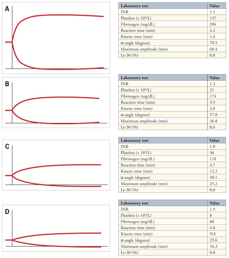
In general agreement with the concept of rebalanced hemostasis, patients with cirrhosis often maintain normal global hemostasis as assessed by TEG (Figures 2A and Table 3). In a cohort of 273 patients with stable cirrhosis, our group found that mean and median TEG parameters were within normal limits, although the maximum amplitude was decreased in proportion to the severity of thrombocytopenia due to hypersplenism (Figures 2B). However, in a subset of 48 patients with more decompensated but stable cirrhosis (INR ≥1.5), we found that the mean maximum amplitude of clot formation was below normal limits, probably due to lower platelet counts in this sicker population (Table 3); the a-angle was also depressed in patients with decompensated cirrhosis and hypofibrinogenemia (Figures 2C).
Figure 2.
Representative thromboelastograms (TEGs) from patients with cirrhosis, with associated standard hemostatic laboratory values measured at the same time point. Panel A shows a normal TEG from a patient with well-compensated (Child-Pugh class A) alcoholic cirrhosis.
Panel B shows an abnormal TEG with low maximum amplitude due to thrombocytopenia in a patient with stable but mildly decompensated (Child-Pugh class B) cirrhosis due to hepatitis C virus infection. Panel C shows an abnormal TEG with a high kinetic time, a low α-angle, and low maximum amplitude due to thrombocytopenia and hypofibrinogenemia in a patient with severely decompensated (Child-Pugh class C) alcoholic cirrhosis. Panel D shows an abnormal TEG with unmeasurably high (NA) kinetic time, a very low α-angle, and very low maximum amplitude due to sepsis; this TEG is from the same patient shown in panel B but was taken 3 weeks later, during presentation for a fatal acute infection. Normal ranges for TEG parameters and standard laboratory tests are noted in Table 3 (RT Stravitz, unpublished observations).
INR=international normalized ratio; Ly-30=clot lysis in 30 minutes; NA=not available.
Table 3.
Comparison of Standard Hemostatic Laboratory Tests and Thromboelastography (TEG) Parameters Between Patients with Acute Liver Injury (ALI)/Acute Liver Failure (ALF) and Cirrhosis
| Parameter | Normal range | ALI/ALF (N=51) | Cirrhosis (N=273) | Cirrhosis INR ≥1.5 (N=48) |
|---|---|---|---|---|
| Standard hemostatic laboratory tests | ||||
| INR | 0.9–1.1 | 3.4±1.7 | 1.3±0.3† | 1.7±0.4† |
| Fibrinogen (mg/dL) | 200–450 | 195±84 | 263±108** | 179±89 |
| Platelets (X 109/L) | 172–440 | 186±95 | 112±79† | 84±46† |
| TEG parameters‡ | ||||
| Reaction time (min) | 2.5–7.5 | 4.7±1.9 | 4.4±1.2 | 4.2±1.5 |
| Kinetic time (min) | 0.8–2.8 | 1.7 [0.8–20.0] | 2.2 [0.8–16.6] | 2.8 [1.2–16.6]** |
| α–angle (degrees) | 55.2–78.4 | 63.7±12.2 | 62.6±9.3 | 58.1±10.8* |
| Maximum amplitude (mm) | 50.6–69.4 | 55.0±10.9 | 51.5±10.4* | 45.0±9.9† |
| Lysis–30 (%) | 0.0–7.5 | 0.0 [0.0–2.1] | 0.5 [0.0–5.2]† | 0.25 [0.0–3.2]* |
Patients with ALI/ALF have been described in detail.32 Patients with cirrhosis and an international normalized ratio (INR) of 1.5 or greater were selected from the overall cirrhosis cohort. Normal range is for the local laboratory. Values are given as mean±standard deviation or median [range] (RT Stravitz, unpublished data).
P<.05.
P<.001.
P<.0001. All comparisons are versus ALI/ALF.
TEG was performed on a Thrombelastograph Haemostasis Analyzer 5000 (Haemonetics Corp., Haemoscope Division). Clotting was initiated at 37°C by the addition of kaolin to 0.34 mL of recalcified blood.
Burroughs and colleagues also observed that patients with compensated cirrhosis often have normal TEG parameters. In fact, patients with cirrhosis secondary to cholestatic liver disease (primary biliary cirrhosis [PBC] or primary sclerosing cholangitis [PSC]) were found to be relatively hypercoagulable by TEG compared to patients with noncholestatic liver diseases or healthy controls; the proportion of patients with a hypercoagulable TEG was 28%, 43%, 5%, and 0% in patients with PBC, PSC, noncholestatic liver disease, and normal controls, respectively.14,26 In contrast, INR and other standard laboratory tests of hemostasis did not differ between patients with cholestatic versus noncholestatic liver diseases.26 These observations may explain why patients with PBC and PSC have fewer bleeding complications and lower intraoperative transfusion requirements during LT compared to noncholestatic cirrhosis patients with similar degrees of portal hypertension. A subsequent study confirmed the relative hypercoagulability, as measured by TEG, of patients with cholestatic liver disease compared to those with noncholestatic liver disease; this study ascribed this difference to increased fibrinogen concentrations and better platelet function in the former population.27 Thus, investigations using TEG in patients with cirrhosis support observations made using thrombin generation assays, which showed that overall hemostasis is relatively well preserved by compensatory mechanisms that maintain a rebalanced state.10–12
In addition to supporting the concept of rebalanced hemostasis in the setting of cirrhosis, TEG has also demonstrated clinical utility in predicting complications of liver disease, such as gastrointestinal bleeding and infection. These 2 complications are pathogenetically linked in patients with cirrhosis, as variceal bleeding promotes infection, and infection promotes bleeding.28,29 TEG has been shown to be superior to INR or platelet count for estimating the risk of rebleeding from esophageal varices.30 In a small number of patients with cirrhosis who presented with acute variceal bleeding, specific parameters of TEG (r-time, k-time, and α-angle) indicated increased hypocoagulability in those who experienced early rebleeding compared to those who did not (P <.001 for all parameters). In contrast, none of the standard laboratory tests of hemostasis (INR, aPTT, or platelet count) differed between those who rebled and those who did not.
TEG can also be useful for detecting infection in patients with cirrhosis. In a prospectively studied cohort of decompensated cirrhotics admitted to the hospital, 36% of patients developed an infection; 4 TEG parameters became more hypocoagulable in these patients compared to patients who did not develop an infection (eg, Figures 2D).31 In contrast, TEG parameters remained unchanged in those who did not develop an infection. Furthermore, resolution of infection was associated with improvement in TEG parameters to preinfection levels, and persistence of infection was associated with continued deterioration of hemostasis as measured by TEG but not by standard laboratory tests of hemostasis. Similar to the prediction of variceal rebleeding, these observations suggest that TEG is a more sensitive test of the balance of hemostasis in patients with chronic liver disease compared to standard coagulation tests.
The mechanism by which infection causes a deterioration of hemostasis has been explored by modifying the TEG assay, which highlights another potential use of this technique as an investigative tool. Patients with cirrhosis express increased plasma concentrations of endotheliumderived endogenous heparinoids due to increased production and decreased hepatic clearance of these molecules; this observation was first made by heparinase-modified TEG in patients undergoing LT.16 In a prospective analysis of patients with cirrhosis who were admitted to the hospital for nonbleeding indications, half developed an infection; r-time, α-angle, and maximum amplitude were hypocoagulable in these patients compared to those who did not develop an infection.32 Of the 30 infected patients, 28 were determined to have significantly improved TEG parameters in heparinase-modified TEG compared to native (nonheparinase) TEG, indicating a significant heparin effect; no difference in TEG parameters was observed in the presence and absence of heparinase in uninfected subjects. Furthermore, the heparin effect disappeared in all patients after resolution of infection. Again, there were no differences in standard laboratory tests of coagulation before and after infection.
Use of Thromboelastography in Patients with Acute Liver Failure
Fewer studies have used TEG to investigate hemostasis in patients with ALF. Similar to patients with cirrhosis, patients with ALF generally have TEG parameters within normal limits.33 As shown in Table 3, the means and medians of all 5 TEG parameters were within normal limits in 51 patients with acute liver injury (ALI)/ALF, and the maximum amplitude of clot formation was significantly higher in this group than in the previously discussed group of stable cirrhotics, despite a much higher INR in the ALI/ALF group (3.4 in the ALI/ALF cohort vs 1.3 in the entire cirrhotic cohort and 1.7 in the more decompensated cirrhotic cohort). This observation may be ascribed to the fact that patients with ALF had milder degrees of thrombocytopenia than patients with cirrhosis (mean platelet counts of 186 × 109/L, 112 × 109/L, and 84 × 109/L in ALI/ALF patients, cirrhotics, and decompensated cirrhotics with INR ≤1.5, respectively).
Similar to patients with cirrhosis, patients with ALI/ALF also show evidence of compensation for hemostatic abnormalities when assessed by TEG. Indeed, the maximum amplitude of clot formation increases in parallel with the number of positive elements of systemic inflammatory response syndrome (SIRS) and correlates directly with venous ammonia concentration and the severity of hepatic encephalopathy.33 Since the degree of hyperammonemia, the severity of hepatic encephalopathy, and the number of SIRS components are closely related in patients with ALF, these data suggest that the severity of SIRS reflects the degree of increase in prohemostatic acute-phase reactants—including factor VIII, vWF, and fibrinogen—which leads to increased clot strength.34,35
The relationship of SIRS to abnormal hemostasis in patients with ALF has also been explored using heparinase-modified TEG.36 Endothelial activation and/or injury is an integral component of ALF, leading not only to increased levels of procoagulant endothelial factors (factor VIII and vWF) but also to increased levels of endogenous heparinoids. Similar to observations in patients with cirrhosis, the estimated total thrombin generation in patients with ALF was not significantly different than that observed in patients with cirrhosis who were undergoing LT or healthy controls, and a significant heparin effect was observed. Also, the maximum amplitude of clot formation as measured by heparinase-modified TEG in patients with ALF was similar to that of healthy controls and patients with cirrhosis.36
Conclusions
In summary, the use of TEG in patients with acute and chronic liver disease has corroborated the concept of rebalanced global hemostasis that was proposed by Tripodi and colleagues and Lisman and colleagues using other laboratory methods.10,11,37,38 In addition, TEG has become widely used during LT to guide prohemostatic factor repletion and antifibrinolytic therapy and to estimate the degree to which endogenous heparinoids may contribute to bleeding during LT. Finally, the use of TEG has elucidated new mechanisms involved in rebalanced hemostasis in patients with liver disease (including the role of the acute-phase reaction in augmenting clot strength) and has identified endogenous heparinoids as an important link between infection and impaired hemostasis.
Although most studies imply that TEG provides a better assessment of bleeding risk than standard tests of hemostasis in patients with liver disease, it should be emphasized that no studies have directly tested this possibility. However, the absence of guidelines for prohemostatic factor repletion in specific settings, such as before invasive procedures, remains a major obstacle in the management of patients with liver disease, especially since factor repletion has serious potential adverse effects.39 Considering the last decade of research, it seems certain that clinicians have overestimated the bleeding diathesis associated with liver disease based upon standard laboratory tests of hemostasis. A prospective randomized trial using a global test of hemostasis, such as TEG, is now urgently needed to assess bleeding risk after invasive procedures and to more judiciously guide factor repletion in patients with acute and chronic liver disease.
References
- 1.Reikvam H, Steien E, Hauge B, et al. Thrombelastography. Transfus Apher Sci. 2009;40:119–123. doi: 10.1016/j.transci.2009.01.019. [DOI] [PubMed] [Google Scholar]
- 2.Malinchoc M, Kamath PS, Gordon FD, Peine CJ, Rank J, ter Borg PC. A model to predict poor survival in patients undergoing transjugular intrahepatic portosystemic shunts. Hepatology. 2000;31:864–871. doi: 10.1053/he.2000.5852. [DOI] [PubMed] [Google Scholar]
- 3.Tripodi A, Mannucci PM. The coagulopathy of chronic liver disease. N Engl J Med. 2011;365:147–156. doi: 10.1056/NEJMra1011170. [DOI] [PubMed] [Google Scholar]
- 4.O'Grady JG, Alexander GJ, Hayllar KM, Williams R. Early indicators of prognosis in fulminant hepatic failure. Gastroenterology. 1989;97:439–445. doi: 10.1016/0016-5085(89)90081-4. [DOI] [PubMed] [Google Scholar]
- 5.Valla D, Flejou JF, Lebrec D, et al. Portal hypertension and ascites in acute hepatitis: clinical, hemodynamic and histological correlations. Hepatology. 1989;10:482–487. doi: 10.1002/hep.1840100414. [DOI] [PubMed] [Google Scholar]
- 6.Schiodt FV, Balko J, Schilsky M, Harrison ME, Thornton A, Lee WM. Thrombopoietin in acute liver failure. Hepatology. 2003;37:558–561. doi: 10.1053/jhep.2003.50113. [DOI] [PubMed] [Google Scholar]
- 7.Vaquero J, Fontana RJ, Larson AM, et al. Complications and use of intracranial pressure monitoring in patients with acute liver failure and severe encephalopathy. Liver Transpl. 2005;11:1581–1589. doi: 10.1002/lt.20625. [DOI] [PubMed] [Google Scholar]
- 8.Munoz SJ, Stravitz RT, Gabriel DA. Coagulopathy of acute liver failure. Clin Liver Dis. 2009;13:95–107. doi: 10.1016/j.cld.2008.10.001. [DOI] [PubMed] [Google Scholar]
- 9.Ordinas A, Escolar G, Cirera I, et al. Existence of a platelet-adhesion defect in patients with cirrhosis independent of hematocrit: studies under flow conditions. Hepatology. 1996;24:1137–1142. doi: 10.1053/jhep.1996.v24.pm0008903388. [DOI] [PubMed] [Google Scholar]
- 10.Tripodi A, Salerno F, Chantarangkul V, et al. Evidence of normal thrombin generation in cirrhosis despite abnormal conventional coagulation tests. Hepatology. 2005;41:553–558. doi: 10.1002/hep.20569. [DOI] [PubMed] [Google Scholar]
- 11.Tripodi A, Primignani M, Chantarangkul V, et al. Thrombin generation in patients with cirrhosis: the role of platelets. Hepatology. 2006;44:440–445. doi: 10.1002/hep.21266. [DOI] [PubMed] [Google Scholar]
- 12.Tripodi A, Primignani M, Lemma L, et al. Detection of the imbalance of pro-coagulant versus anticoagulant factors in cirrhosis by a simple laboratory method. Hepatology. 2010;52:249–255. doi: 10.1002/hep.23653. [DOI] [PubMed] [Google Scholar]
- 13.Gabriel DA, Carr M, Roberts HR. Monitoring coagulation and the clinical effects of recombinant factor VIIa. Semin Hematol. 2004;41(suppl 1):20–24. doi: 10.1053/j.seminhematol.2003.11.005. [DOI] [PubMed] [Google Scholar]
- 14.Thalheimer U, Triantos CK, Samonakis DN, et al. A comparison of kaolin-activated versus nonkaolin-activated thromboelastography in native and citrated blood. Blood Coagul Fibrinolysis. 2008;19:495–501. doi: 10.1097/MBC.0b013e3282f9adf9. [DOI] [PubMed] [Google Scholar]
- 15.Zuckerman L, Cohen E, Vagher JP, Woodward E, Caprini JA. Comparison of thrombelastography with common coagulation tests. Thromb Haemost. 1981;46:752–756. [PubMed] [Google Scholar]
- 16.Kettner SC, Gonano C, Seebach F, et al. Endogenous heparin-like substances significantly impair coagulation in patients undergoing orthotopic liver transplantation. Anesth Analg. 1998;86:691–695. doi: 10.1097/00000539-199804000-00002. [DOI] [PubMed] [Google Scholar]
- 17.Zmuda K, Neofotistos D, Ts'ao CH. Effects of unfractionated heparin, lowmolecular-weight heparin, and heparinoid on thromboelastographic assay of blood coagulation. Am J Clin Pathol. 2000;113:725–731. doi: 10.1309/Q4AE-BMCW-CQ7J-NUVT. [DOI] [PubMed] [Google Scholar]
- 18.Skolnick BE, Mathews DR, Khutoryamsky NM, Pusateri AE, Carr ME. Exploratory study on the reversal of warfarin with rFVIIa in healthy subjects. Blood. 2010;116:693–701. doi: 10.1182/blood-2009-11-253492. [DOI] [PubMed] [Google Scholar]
- 19.Kang YG, Martin DJ, Marquez J, et al. Intraoperative changes in blood coagulation and thrombelastographic monitoring in liver transplantation. Anesth Analg. 1985;64:888–896. [PMC free article] [PubMed] [Google Scholar]
- 20.Owen CA, Jr, Rettke SR, Bowie EJ, et al. Hemostatic evaluation of patients undergoing liver transplantation. Mayo Clin Proc. 1987;62:761–772. doi: 10.1016/s0025-6196(12)62328-3. [DOI] [PubMed] [Google Scholar]
- 21.Clayton DG, Miro AM, Kramer DJ, Rodman N, Wearden S. Quantification of thrombelastographic changes after blood component transfusion in patients with liver disease in the intensive care unit. Anesth Analg. 1995;81:272–278. doi: 10.1097/00000539-199508000-00011. [DOI] [PubMed] [Google Scholar]
- 22.Porte RJ, Bontempo FA, Knot EA, Lewis JH, Kang YG, Starzl TE. Systemic effects of tissue plasminogen activator-associated fibrinolysis and its relation to thrombin generation in orthotopic liver transplantation. Transplantation. 1989;47:978–984. doi: 10.1097/00007890-198906000-00012. [DOI] [PMC free article] [PubMed] [Google Scholar]
- 23.Kang Y, Lewis JH, Navalgund A, et al. Epsilon-aminocaproic acid for treatment of fibrinolysis during liver transplantation. Anesthesiology. 1987;66:766–773. [PMC free article] [PubMed] [Google Scholar]
- 24.Mallett SV, Cox D, Burroughs AK, Rolles K. The intra-operative use of trasylol (aprotinin) in liver transplantation. Transpl Int. 1991;4:227–230. doi: 10.1007/BF00649108. [DOI] [PubMed] [Google Scholar]
- 25.De Pietri L, Masetti M, Montalti R, et al. Use of recombinant factor IX and thromboelastography in a patient with hemophilia B undergoing liver transplantation: a case report. Transplant Proc. 2008;40:2077–2079. doi: 10.1016/j.transproceed.2008.05.054. [DOI] [PubMed] [Google Scholar]
- 26.Ben-Ari Z, Panagou M, Patch D, et al. Hypercoagulability in patients with primary biliary cirrhosis and primary sclerosing cholangitis evaluated by thrombelastography. J Hepatol. 1997;26:554–559. doi: 10.1016/s0168-8278(97)80420-5. [DOI] [PubMed] [Google Scholar]
- 27.Pihusch R, Rank A, Gohring P, Pihusch M, Hiller E, Beuers U. Platelet function rather than plasmatic coagulation explains hypercoagulable state in cholestatic liver disease. J Hepatol. 2002;37:548–555. doi: 10.1016/s0168-8278(02)00239-8. [DOI] [PubMed] [Google Scholar]
- 28.Rimola A, Bory F, Teres J, Perez-Ayuso RM, Arroyo V, Rodes J. Oral, non-absorbable antibiotics prevent infection in cirrhotics with gastrointestinal hemorrhage. Hepatology. 1985;5:463–467. doi: 10.1002/hep.1840050320. [DOI] [PubMed] [Google Scholar]
- 29.Goulis J, Armonis A, Patch D, Sabin C, Greenslade L, Burroughs AK. Bacterial infection is independently associated with failure to control bleeding in cirrhotic patients with gastrointestinal hemorrhage. Hepatology. 1998;27:1207–1212. doi: 10.1002/hep.510270504. [DOI] [PubMed] [Google Scholar]
- 30.Chau TN, Chan YW, Patch D, Tokunaga S, Greenslade L, Burroughs AK. Thrombelastographic changes and early rebleeding in cirrhotic patients with variceal bleeding. Gut. 1998;43:267–271. doi: 10.1136/gut.43.2.267. [DOI] [PMC free article] [PubMed] [Google Scholar]
- 31.Papatheodoridis GV, Patch D, Webster GJ, Brooker J, Barnes E, Burroughs AK. Infection and hemostasis in decompensated cirrhosis: a prospective study using thrombelastography. Hepatology. 1999;29:1085–1090. doi: 10.1002/hep.510290437. [DOI] [PubMed] [Google Scholar]
- 32.Montalto P, Vlachogiannakos J, Cox DJ, Pastacaldi S, Patch D, Burroughs AK. Bacterial infection in cirrhosis impairs coagulation by a heparin effect: a prospective study. J Hepatol. 2002;37:463–470. doi: 10.1016/s0168-8278(02)00208-8. [DOI] [PubMed] [Google Scholar]
- 33.Stravitz RT, Lisman T, Luketic VA, et al. Minimal effects of acute liver injury/acute liver failure on hemostasis as assessed by thromboelastography. J Hepatol. 2012;56:129–136. doi: 10.1016/j.jhep.2011.04.020. [DOI] [PMC free article] [PubMed] [Google Scholar]
- 34.Rolando N, Wade J, Davalos M, Wendon J, Philpott-Howard J, Williams R. The systemic inflammatory response syndrome in acute liver failure. Hepatology. 2000;32:734–739. doi: 10.1053/jhep.2000.17687. [DOI] [PubMed] [Google Scholar]
- 35.Vaquero J, Polson J, Chung C, et al. Infection and the progression of hepatic encephalopathy in acute liver failure. Gastroenterology. 2003;125:755–764. doi: 10.1016/s0016-5085(03)01051-5. [DOI] [PubMed] [Google Scholar]
- 36.Senzolo M, Agarwal S, Zappoli P, Vibhakorn S, Mallett S, Burroughs AK. Heparin-like effect contributes to the coagulopathy in patients with acute liver failure undergoing liver transplantation. Liver Int. 2009;29:754–759. doi: 10.1111/j.1478-3231.2009.01977.x. [DOI] [PMC free article] [PubMed] [Google Scholar]
- 37.Lisman T, Porte RJ. Rebalanced hemostasis in patients with liver disease: evidence and clinical consequences. Blood. 2010;116:878–885. doi: 10.1182/blood-2010-02-261891. [DOI] [PubMed] [Google Scholar]
- 38.Lisman T, Bakhtiari K, Pereboom IT, Hendriks HG, Meijers JC, Porte RJ. Normal to increased thrombin generation in patients undergoing liver transplantation despite prolonged conventional coagulation tests. J Hepatol. 2010;52:355–361. doi: 10.1016/j.jhep.2009.12.001. [DOI] [PubMed] [Google Scholar]
- 39.Rockey DC, Caldwell SH, Goodman ZD, Nelson RC, Smith AD. Liver biopsy. Hepatology. 2009;49:1017–1044. doi: 10.1002/hep.22742. [DOI] [PubMed] [Google Scholar]


