Abstract
Use of new keratin-based wound dressings represent a novel approach to wound management. The authors present three patients with recalcitrant, venous and mixed venous, and arterial leg ulcers treated with these dressings. Improvement in each case was observed.
Healing of chronic wounds continues to represent a significant challenge to physicians, as many wounds remain recalcitrant despite optimal standard care. For example, standard care of diabetic foot ulcers leads to healing of less than one-third of patients in 20 weeks, and more than 25 percent of venous leg ulcers remain unhealed even after six months of therapy.1,2 For refractory wounds, some successful wound care adjuvants aim to alter the environment of the chronic wound to become more like that of an acute wound through biological intervention, such as cell-based therapies containing keratinocytes and fibroblasts, which have found to increase the rate of healing.3 Keratinocytes are important in wound repair, in part through their production of keratin proteins,4,5 which are being investigated as new targets for wound care dressings.
Recent advances have been made in understanding the role of keratin in cell biology, in particular the importance of keratin in cell differentiation and protein synthesis. Keratin is a structural protein important for maintaining structural durability of tissues.4,5 However, it has been recently appreciated that keratin proteins also play an active role in wound healing, and the following three keratin subtypes have been implicated in the healing response: keratin 6, 16, and 17.6–8 For example, following skin injury, keratin 17 is rapidly upregulated at wound edges,9 and in studies involving keratin 17 knock-out mice, delayed healing was observed.10 In the latter case, reintroduction of the lacking keratin 17 protein stimulated cell growth and migration. Therefore, in-vitro studies have suggested that keratin-based products can stimulate cellular migration into a wound to improve speed of wound healing. In-vivo studies of partial-thickness wound healing in a porcine model found dressings derived from keratin protein speed healing compared to both air-exposed wounds and wounds treated with polyurethane film dressing.11,12 Molecular analyses suggest that the keratin formulations may influence epidermal migration by the upregulation of keratin gene expression.11,13
Despite being prevalent in the skin and other tissues, keratin protein has only recently been studied as a potential treatment for wounds.14–16 This may be due to the fact that keratin is physically more robust than collagen, and, as such, the technology required to reconstitute keratin from natural sources into a format that maintains structure and function useful in a wound dressing or other medical devices has required extensive research and development.17–19 Previous technology has created hydrolysates of keratin protein in which the structure and function of the keratin is lost in order to isolate the protein. This approach has not yielded medical device applications. However, processes have recently been established for reconstituting keratin into physical forms useful as wound dressing products that also maintain the biological function of the protein.20–22
At present, three forms of nonhydrolysed keratin wound dressing products have been developed, including a hygroscopic gel for minimally exudative wounds, a matrix for moderately exuding wounds, and an absorbent foam laminated with a perforated keratin film on the wound contact side for highly exuding wounds. The authors present three cases of patients with recalcitrant venous, arterial, or mixed ulcers treated with these dressings in an effort to highlight how new knowledge related to keratin's role in wound healing has potential for translation to clinical practice.
Methods
The study was conducted at a local specialist wound care clinic and primarily focused on the physical characteristics and general user acceptability of three forms of keratin dressing. Each patient was treated as an outpatient with dressing changes occurring typically every 3 to 7 days. The dressing changes were performed by an experienced wound care nurse. Wound healing progress was assessed by photography, and the general acceptability to nurse and patient were assessed subjectively with questionnaires completed by both nurses and patients. The study method was approved by the Upper South Island Regional Ethics Committee. Three patients were treated and followed over the course of this study. Each case represented a different physical format of keratin dressing. The three cases described are 1) a gel dressing on a minimally exudative wound, 2) a bioresorbable “matrix” dressing on a moderately exuding wound, and 3) an absorbent polyurethane foam dressing with a laminated keratin film on a highly exuding wound.
Case Reports
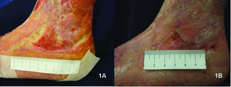
Case 1. A 75-year-old woman with a moderately exuding venous ulcer on the left lower leg was treated with the matrix dressing. The wound had been present for 11.5 months. She had a history of recurrent venous leg ulcers. Immediately prior to the trial she had been treated with multilayered compression bandaging without improvement.
Her past medical history included asthma, chronic obstructive pulmonary disease, and the venous stasis disease described. She had already required a number of skin grafts. Her regular medications were aspirin, furosemide, combination albuterol/ipratropium and fluticasone inhalers, nitrofurantoin, omeprazole, codeine, and paracetemol. She was a heavy long-term smoker with no known nutritional deficits. Mobility assessment showed a slight reduction in calf muscle pump.
The wound was 8.1cm2 at the start of therapy. It was treated with Keraderm (Blacksburg, Virginia), which is a robust matrix dressing derived from freeze-dried keratin protein. This matrix dressing is reabsorbed into the developing tissue without traumatic dressing changes, provides a resorbable “scaffold” for the rapid growth of new tissue, and helps maintain a moist wound environment.
The wound was 0.3cm2 after 16 weeks of treatment and completely healed after 30 weeks. The patient remained ulcer free after six months (Figure 1).
Figures 1A and 1B.
(A) Case 1: Ulcer at Day 0; (B) Case 1: Healed ulcer at Day 99
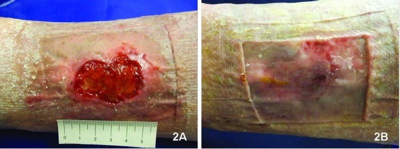
Case 2. A 62-year-old man with a highly exuding venous ulcer was treated with the foam dressing. The wound was present for six months at the start of the study. He had a history of chronic venous hypertension and, despite compression bandaging, had failed to heal.
His past medical history consisted of diet-controlled noninsulin-dependent diabetes, rheumatoid arthritis, bilateral total hip replacements, bilateral leg varicose vein surgery, and venous dermatitis. He was an ex-smoker whose only medications were nonsteroidal anti-inflammatory drugs.
The wound was 12cm2 at baseline. It was treated with Kerafoam (Onset Dermatologics), which is an absorbent polyurethane foam dressing with a laminated keratin film. The recalcitrant ulcer progressed from a nonhealing to a healing wound with the development of a healthy granulating base, and epithelializing margins completely healed after 10 weeks, but sustained trauma to that area two weeks later required further dressings. This wound then completely healed after 16 weeks and remained healed at six months (Figure 2).
Figure 2A and 2B.
(A) Case 2: Ulcer at Day 0; (B) Case 2: Healed ulcer at Day 78
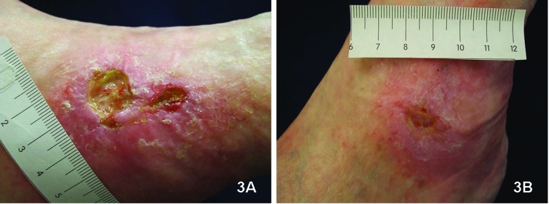
Case 3. A 74-year-old man with a mixed venous and arterial disease had an ulcer on the lateral malleolus of the left foot and was treated with the gel dressing. The wound was present for 20 years at the start of the study. He had a history of arterial disease resulting in a right leg below knee amputation for extensive ulceration. The current ulcer was being treated with 23mmHg compression bandaging, and this continued with the keratin dressing.
His past medical history consisted of abdominal aortic aneurysm and ischemic heart disease. He had tuberculosis in 1976 and rheumatic fever as a child. His regular medications were aspirin and simvastatin. He had ceased smoking in 1976.
The initial size of the wound was 2.2cm2. It was treated with the biodegradable “matrix” dressing. There was a rapid improvement of the wound that had not occurred with his previous dressings. Complete healing was achieved by Day 155. The area remained healed at six month follow up (Figure 3).
Figure 3A and 3B.
(A) Case 3: Ulcer at Day 0; (B) Case 3: Significant reduction in size of the ulcer at Day 85
Discussion
Leg ulcers affect 1.5 to 3 per 1,000 people and prevalence increases with age to 20 per 1,000 in people older than 80 years of age.23 Venous and mixed arterial and venous ulcers benefit from compression therapy to aid venous return24 with the level of compression being dependant upon the degree of arterial compromise.25 Over half of these ulcers heal within 12 weeks.26 A recent systematic review and meta-analysis has found that the type of dressing applied beneath compression bandaging does not affect ulcer healing.27
Chronic, recalcitrant wounds represent a particularly difficult subset of patients. Large wound size as well as long wound duration are associated with slower healing rates. Specifically, a wound duration of >6 months and a wound size of >5cm are associated with a much higher chance of failure to heal.28 As such, patients are recommended to advance to adjuvant therapies if only a minimal reduction in wound size occurs following standard of care therapy for four weeks.29
While this is a limited series, these three cases of recalcitrant leg wounds healed well after the introduction of keratin-based primary dressings, which provide an opportunity to consider the clinical application of recent knowledge advances regarding the role of keratin proteins in healing. All three patients had wounds that had become chronic despite appropriate compression and the use of a variety of primary dressings. The healing that occurred in these cases is noteworthy, particularly in Case 3 in which the wound had been present for 20 years. Although all three patients in the cases presented historically had large and refractory ulcers unresponsive to other treatment, the evidence is currently limited, and conclusively attributing the healing to the keratin-based dressings is not possible. As such, the exact mechanism of wound improvement in these cases is not certain, but it is possible that the improved healing potential of keratin suggested by in-vitro and animal studies may have been causal. Additionally, the dressings were found to be comfortable and easy to use by the patients and nurses.
There still remains a large group of patients who do not respond to compression alone, and therefore, investigation into new dressings is of the utmost importance. Keratin-based dressings may have the potential to improve healing outcomes and avoid the need for surgery on a range of recalcitrant, chronic leg ulcers, as demonstrated by the positive outcomes obtained in the three patients described herein. Controlled therapeutic trials are required to further investigate this issue.
Acknowledgment
The authors would like to acknowledge the registered nurses of the Specialist Wound Management Service, Nurse Maude, in Christchurch, New Zealand, for their involvement in the study and the New Zealand Institute of Community Health Care, at Nurse Maude, for their support and facilitation of the study and the preparation of this article.
Footnotes
DISCLOSURE:Ms. Hammond and Ms. Maderal report no relevant conflicts of interest. Dr. Than received a consultant fee from Keraplast Technologies. Dr. Smith is the Medical Director for Keraplast Technologies. Drs. Kelly and Marsh are employees of Keraplast Technologies. Dr. Kirsner is a consultant for Keraplast Technologies. This study was funded by Keraplast Technologies, LTD. An ethics approval was required and obtained by the Upper South Island Regional Ethics committee for the work done in this study.
References
- 1.Margolis DJ, Kantor J, Berlin JA. Healing of diabetic neuropathic foot ulcers receiving standard treatment. A meta-analysis. Diabetes Care. 1999;22(5):692–695. doi: 10.2337/diacare.22.5.692. [DOI] [PubMed] [Google Scholar]
- 2.Kirsner RS, Marston WA, Snyder RJ, et al. Spray-applied cell therapy with human allogeneic fibroblasts and keratinocytes for the treatment of chronic venous leg ulcers: a phase 2, multicentre, double-blind, randomised, placebo-controlled trial. Lancet. doi: 10.1016/S0140-6736(12)60644-8. 2012 Aug 2. [Epub ahead of print]. [DOI] [PubMed] [Google Scholar]
- 3.Veves A, Falanga V, Armstrong DG, et al. Graftskin, a human skin equivalent, is effective in the management of noninfected neuropathic diabetic foot ulcers: a prospective randomized multicenter clinical trial. Diabetes Care. 2001;24(2):290–295. doi: 10.2337/diacare.24.2.290. [DOI] [PubMed] [Google Scholar]
- 4.Roop D. Defects in the barrier. Science. 1995;267(5197): 474–475. doi: 10.1126/science.7529942. [DOI] [PubMed] [Google Scholar]
- 5.Morasso MI, Tomic-Canic M. Epidermal stem cells: the cradle of epidermal determination, differentiation and wound healing. Biol Cell. 2005;97(3):173–183. doi: 10.1042/BC20040098. [DOI] [PMC free article] [PubMed] [Google Scholar]
- 6.Ishida-Yamamoto A, Tanaka H, Nakane H, et al. Inherited disorders of epidermal keratinization. J Dermatol Sci. 1998;18(3):139–154. doi: 10.1016/s0923-1811(98)00041-3. [DOI] [PubMed] [Google Scholar]
- 7.Patel GK, Wilson CH, Harding KG, et al. Numerous keratinocyte subtypes involved in wound re-epithelialization. J Invest Dermatol. 2006;126(2):497–502. doi: 10.1038/sj.jid.5700101. [DOI] [PubMed] [Google Scholar]
- 8.Wojcik SM, Bundman DS, Roop DR. Delayed wound healing in keratin 6a knockout mice. Mol Cell Biol. 2000;20(14):5248–5255. doi: 10.1128/mcb.20.14.5248-5255.2000. [DOI] [PMC free article] [PubMed] [Google Scholar]
- 9.McGown KM, Tong X, Colucci-Guyon E, et al. Keratin 17 null mice exhibit age- and strain-dependent alopecia. Genes Dev. 2002;16(11):1412–1422. doi: 10.1101/gad.979502. [DOI] [PMC free article] [PubMed] [Google Scholar]
- 10.Kim S, Wong P, Coulombe PA. A keratin cytoskeletal protein regulates protein synthesis and epithelial cell growth. Nature. 2006;441(7091):362–365. doi: 10.1038/nature04659. [DOI] [PubMed] [Google Scholar]
- 11.Pechter PM, Gil J, Valdes J, et al. Keratin dressings speed epithelialization of deep partial-thickness wounds. Wound Repair Regen. 2012;20(2):236–242. doi: 10.1111/j.1524-475X.2012.00768.x. [DOI] [PubMed] [Google Scholar]
- 12.Davis S, Perez R, Rivas Y, et al. The effect of a keratin based dressing on the epithelialization of deep partial thickness wounds. Presented at: American Academy of Dermatology. 67th Annual Meeting; March 6-10, 2009; San Francisco, CA.
- 13.Perez R, Kirsner R, Gil J, et al. Evaluation of the effects of two keratin formulations on wound healing and keratin gene expression in a porcine model. Presented at: Symposium on Advanced Wound Care Conference; April 26–29, 2009; Dallas, TX.
- 14.Cassidy S, Than M. Improved healing of epidermolysis bullosa wounds using a novel keratin gel technology. In: Australian Wound Management Association Conference Proceedings; 2008; Darwin, NT, Australia.
- 15.Kelly R, Sigurjonsson F, Smith RA, et al. Keratin biopolymer dressings for wound care. Presented at: Symposium on Advanced Wound Care Conference Proceedings; April 30–May 6, 2006; San Antonio, TX.
- 16.Thompson W, Hanneke S, Compton M, et al. A keratin matrix interface in negative pressure wound therapy. Presented at: Clinical Symposium on Advances in Skin and Wound Care; 2008; Las Vegas, NV.
- 17.Dias GJ, Peplow PV, McLaughlin A, et al. Biocompatibility and osseointegration of reconstituted keratin in an ovine model. J Biomed Mater Res A. 2009;92(2):513–520. doi: 10.1002/jbm.a.32394. [DOI] [PubMed] [Google Scholar]
- 18.Aboushwareb T, Eberli D, Ward C, et al. A keratin biomaterial gel hemostat derived from human hair: evaluation in a rabbit model of lethal liver injury. J Biomed Mater Res B Appl Biomater. 2009;90(1):45–54. doi: 10.1002/jbm.b.31251. [DOI] [PubMed] [Google Scholar]
- 19.Apel PJ, Garrett JP, Sierpinski P, et al. Peripheral nerve regeneration using a keratin-based scaffold: long-term functional and histological outcomes in a mouse model. J Hand Surg Am. 2008;33(9):1541–1547. doi: 10.1016/j.jhsa.2008.05.034. [DOI] [PubMed] [Google Scholar]
- 20.Blanchard CR. Inventor; Timmons SF Inventor; Smith RA Inventor; KMS Ventures, Inc., assignee. Keratin-based hydrogel for biomedical applications and method of production. United States patent 5932552. 1999 Aug 3.
- 21.Kelly RJ. Inventor; Roddick-Lanzilotta AD Inventor; Ali Inventor; KMS Ventures, Inc., assignee. Wound care products containing keratin. United States patent 7732574. 2010 Jun 8.
- 22.Tachibana A, Nishikawa Y, Nishino M, et al. Modified keratin sponge: binding of bone morphogenetic protein-2 and osteoblast differentiation. J Biosci Bioeng. 2006;102(5): 425–429. doi: 10.1263/jbb.102.425. [DOI] [PubMed] [Google Scholar]
- 23.Callam MJ, Ruckley CV, Harper DR, Dale JJ. Chronic ulceration of the leg: extent of the problem and provision of care. Br Med J. 1985;290(6485):1855–1856. doi: 10.1136/bmj.290.6485.1855. [DOI] [PMC free article] [PubMed] [Google Scholar]
- 24.O’Meara S, Cullum NA, Nelson EA. Compression for venous leg ulcers. Cochrane Database Syst Rev. 2009;1:CD000265. doi: 10.1002/14651858.CD000265.pub2. [DOI] [PubMed] [Google Scholar]
- 25.Marston W, Vowden K. Compression therapy a guide to safe practice. In: European Wound Management Association Position Document: Understanding Compression Therapy. London, MEP Ltd.; 2003:11-17. [Google Scholar]
- 26.http://www.rcn.org.uk/__data/assets/pdf_file/0003/107940/003020.pdf Royal College of Nursing. The Nursing Management of Patients with Venous Leg Ulcers. In: Clinical Practice Guidelines. London. 2006. Publication code: 003 020.
- 27.Palfreyman S, Nelson EA, Michaels JA. Dressings for venous leg ulcers: systematic review and meta-analysis. BMJ. 2007;335(7613):244. doi: 10.1136/bmj.39248.634977.AE. [DOI] [PMC free article] [PubMed] [Google Scholar]
- 28.Margolis DJ, Berlin JA, Strom BL. Which venous leg ulcers will heal with limb compression bandages? Am J Med. 2000;109(1):15–19. doi: 10.1016/s0002-9343(00)00379-x. [DOI] [PubMed] [Google Scholar]
- 29.Sheehan P, Jones P, Caselli A, et al. Percent change in wound area of diabetic foot ulcers over a 4-week period is a robust predictor of complete healing in a 12-week prospective trial. Diabetes Care. 2003;26(6):1879–1882. doi: 10.2337/diacare.26.6.1879. [DOI] [PubMed] [Google Scholar]


