Abstract
Purpose
This study was performed to assess the compatibility of cone beam computed tomography (CBCT) synthesized cephalograms with conventional cephalograms, and to find a method for obtaining normative values for three-dimensional (3D) assessments.
Materials and Methods
The sample group consisted of 10 adults with normal occlusion and well-balanced faces. They were imaged using conventional and CBCT cephalograms. The CBCT cephalograms were synthesized from the CBCT data using OnDemand 3D software. Twenty-one angular and 12 linear measurements from each imaging modality were compared and analyzed using paired-t test.
Results
The linear measurements between the two imaging modalities were not statistically different (p>0.05) except for the U1 to facial plane distance. The angular measurements between the two imaging modalities were not statistically different (p>0.05) with the exception of the gonial angle, ANB difference, and facial convexity.
Conclusion
Two-dimensional cephalometric norms could be readily used for 3D quantitative assessment, if corrected for lateral cephalogram distortion.
Keywords: Cone-Beam Computed Tomography, Cephalometry
Introduction
Radiographic cephalometry is an essential clinical and research tool for diagnosing skeletal imbalance and for assessing skeletal growth and development. Lateral and frontal cephalograms have been well established and have resulted in several large databases of clinically normal and treated patient populations. Conventional cephalometric examination has, however, intrinsic limitations since it is a two-dimensional (2D) shadow of a three-dimensional (3D) structure, produced by a nonparallel beam that results in a distorted and enlarged image.1
The 3D imaging technique has provided a new possibility for orthodontic diagnosis and treatment evaluation.2,3 The application of cone beam computed tomography (CBCT) to the craniofacial region provides an alternative to traditional computed tomography (CT) systems with the advantages of less radiation and lower billing costs.
Because standard population norms have not been available for 3D CBCT volumes, patients from whom CBCT data are acquired may be subjected to further radiation exposure to acquire traditional lateral cephalograms and panoramic radiographs.4
The purpose of this study was to determine whether CBCT synthesized cephalograms could provide the same measurement of patients as conventional cephalograms and to find a method for obtaining normative values for 3D assessments.
Materials and Methods
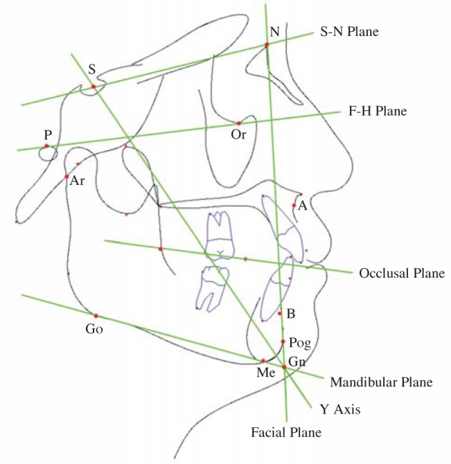
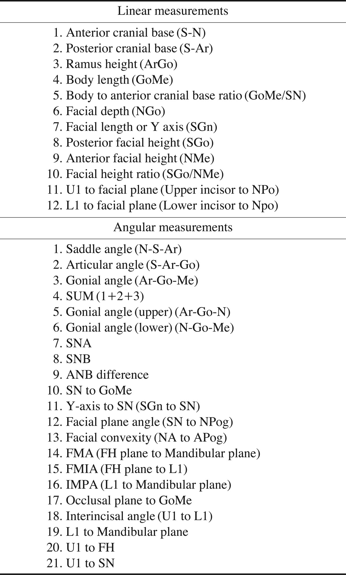
The sample group consisted of 10 adults (6 males and 4 females, 32.3±6.63 years old) with normal occlusions and well-balanced faces. The patients were imaged using conventional and CBCT synthesized cephalometric radiographs. Conventional cephalometric radiographs were taken using an X-ray equipment (Cranex 3+®, Sordex Orion Corp., Helsinki, Finland) set at 73 kVp, 10 mA, and 1.0 second and a cassette with an image plate (Fujifilm Holdings Corp., Tokyo, Japan). The image plates were scanned using a computed radiography system (FCR XG5000, Fujifilm Holdings Corp., Tokyo, Japan) and the conventional cephalometric images were then acquired. For CBCT cephalometry, CBCT data were obtained using CBCT equipment (Rayscan Symphony, Ray Co., Seoul, Korea). The scanning parameters were 90 kVp, 10 mA, 20 seconds of exposure time, a 0.38 mm slice thickness, and an 18 cm×15 cm field of view (FOV). The synthesized CBCT cephalometric radiographs were acquired using the CBCT data using 3D imaging software (OnDemand 3D, Cybermed Co., Seoul, Korea). Twenty-one angular and 12 linear measurements (Table 1) were performed based on the skeletal and dental analysis using imaging software (V-ceph, CyberMed Co., Seoul, Korea) (Fig. 1). The measurements from the conventional cephalometric radiographs were corrected, taking a 10% magnification rate into account. The measurements between the imaging modalities were compared and analyzed using a paired-t test using IBM SPSS Statistics 20.0 (IBM Corp., Armonk, NY, USA). The results were considered statistically significant at p<0.05.
Table 1.
Measurements used in the study
Fig. 1.

Anatomic landmarks and planes used in the cephalometric analysis.
Results
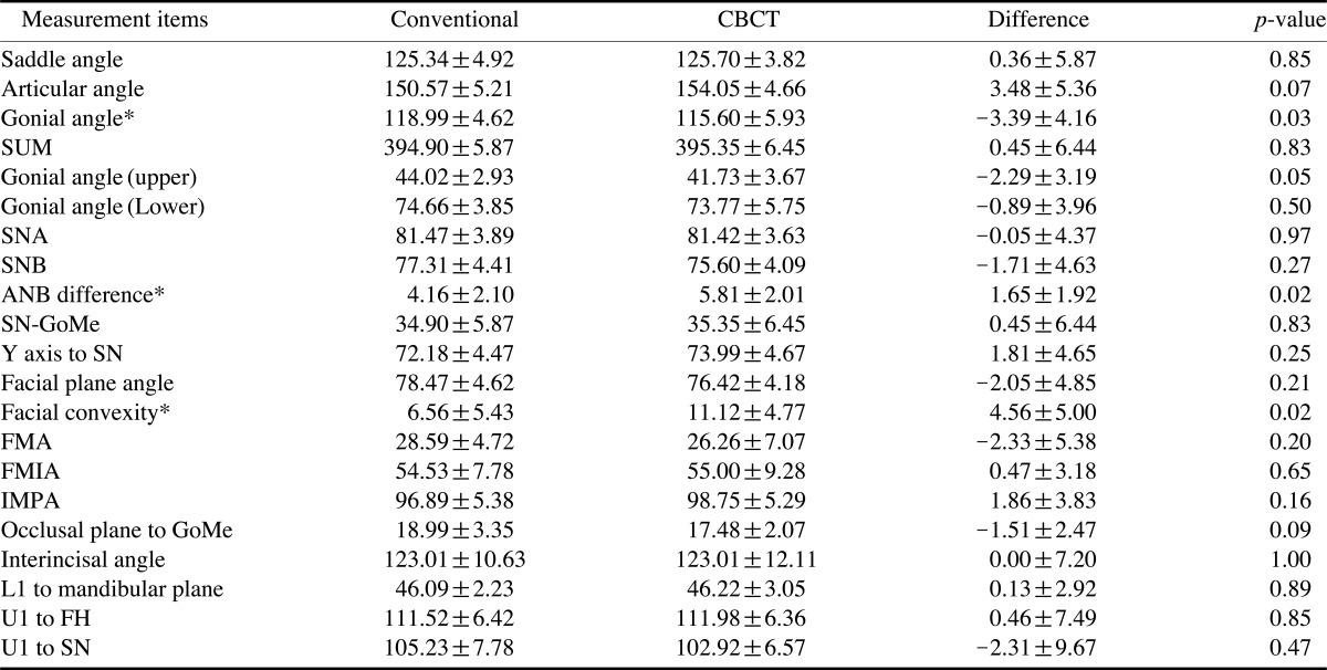
Table 2 shows that the differences between the linear measurements (mm) of the imaging modalities were not statistically different (p>0.05) except for the U1 to facial plane distance. Table 3 shows that differences between angular measurements from the two imaging modalities were not statistically different (p>0.05) with the exception of the gonial angle, ANB difference, and facial convexity.
Table 2.
Linear measurements from the two imaging modalities (mm)
*the statistically significant difference between the two imaging modalities at p<0.05.
Table 3.
Angular measurements from the two imaging modalities (degrees)
*the statistically significant difference between the two imaging modalities at p<0.05.
Discussion
Several large databases of lateral and frontal cephalograms of clinically normal and treated patient populations have been compiled. Since the time of Broadbent,5 researchers have proposed procedures for combining lateral, frontal, and submentovertex radiographs to obtain a 3D assessment of a patient.6 Although direct measurements on 3D images have marked advantages over other methods and measurements on the synthesized 2D images using 3D scans were similar with those of conventional radiographs proposed recently in the literatures,7,8 cephalometric analysis using 3D images still have the characteristics and limitations of a traditional cephalometric examination. Taking 3D measurements directly from 3D images such as CBCT or even 3D photographs allows an examiner to accurately quantify the right and left sides of the patient's jaw and face, separately. A diagnosis can then be reached by comparing the deviation of those measurements from the "normal values." Unfortunately, the exact nature of such "normal values" for 3D measurements remains undefined.9
Due to the high economic cost, high dose of exposure, and ethical issues, obtaining 3D normative values from a large number of people with normal occlusion is unlikely to occur in the near future.9 Thus three-dimensional measurements can be compared only to their contralateral side to evaluate asymmetries10 or to measurements taken at different times to monitor treatment effects.11 Normative values are essential to reach an appropriate diagnosis and to evaluate the net effects of the treatment. While a great deal of work is requested to demonstrate the added value of CBCT in standard orthodontic cases, it is not known whether data obtained from synthesized CBCT views can be compared with the current population norms and the existing databases obtained from conventional cephalograms.9
This study was performed to determine whether cephalometry could be synthesized from CBCT volumes and whether traditional cephalometry could be performed on these synthesized images with similar results. The results of this study showed that the linear measurements between the two imaging modalities were not statistically different except for U1 to facial plane distance; this distance difference was 2.27 mm, which was statistically and clinically significant (Table 2). The angular measurements between the two imaging modalities were not statistically significant except for the gonial angle, ANB difference, and facial convexity angle. These angular differences were -3.39, 1.65, and 4.56 degrees, respectively (Table 3). The differences of the gonial angle and facial convexity angle were not only statistically significant but also clinically significant. These results, although not definite, might have originated from the small sample size and the proficiency of the observer.
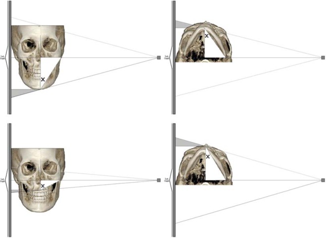
Gribel et al9 evaluated the accuracy of a simple algorithm that corrected 2D cephalometric measurements from landmarks both on and off the midsagittal plane into a 3D CBCT measurements with accuracy. By applying this algorithm to other existing cephalometric longitudinal growth studies, it was possible to derive normative values for 3D measurements without exposing new untreated subjects to radiation (Fig. 2).
Fig. 2.
Lateral cephalogram image construction diagram. 3D measurement=[(cephalometric measurement)-(cephalometric magnification)]/[cosine (X)].
In conclusion, there were significant differences in one linear and three angular measurements between the conventional lateral and CBCT synthesized cephalometric radiographs. These differences among the measurements might originate from the projection difference of the two imaging modalities. Two-dimensional cephalometric norms could be readily used for 3D quantitative assessment with corrections for lateral cephalogram distortion.
Footnotes
This study was supported by a faculty research grant of Yonsei University College of Dentistry for 2008-0060.
References
- 1.Baumrind S, Frantz RC. The reliability of head film measurements. 1. Landmark identification. Am J Orthod. 1971;60:111–127. doi: 10.1016/0002-9416(71)90028-5. [DOI] [PubMed] [Google Scholar]
- 2.Zamora N, Llamas JM, Cibrián R, Gandia JL, Paredes V. Cephalometric measurements from 3D reconstructed images compared with conventional 2D images. Angle Orthod. 2011;81:856–864. doi: 10.2319/121210-717.1. [DOI] [PMC free article] [PubMed] [Google Scholar]
- 3.Oz U, Orhan K, Abe N. Comparison of linear and angular measurements using two-dimensional conventional methods and three-dimensional cone beam CT images reconstructed from a volumetric rendering program in vivo. Dentomaxillofac Radiol. 2011;40:492–500. doi: 10.1259/dmfr/15644321. [DOI] [PMC free article] [PubMed] [Google Scholar]
- 4.Kumar V, Ludlow J, Soares Cevidanes LH, Mol A. In vivo comparison of conventional and cone beam CT synthesized cephalograms. Angle Orthod. 2008;78:873–879. doi: 10.2319/082907-399.1. [DOI] [PMC free article] [PubMed] [Google Scholar]
- 5.Broadbent BH. A new x-ray technique and its application to orthodontia. Angle Orthod. 1931;1:45–66. [Google Scholar]
- 6.Rousset MM, Simonek F, Dubus JP. A method for correction of radiographic errors in serial three-dimensional cephalometry. Dentomaxillofac Radiol. 2003;32:50–59. doi: 10.1259/dmfr/51868734. [DOI] [PubMed] [Google Scholar]
- 7.Farman AG, Scarfe WC. Development of imaging selection criteria and procedures should precede cephalometric assessment with cone-beam computed tomography. Am J Orthod Dentofacial Orthop. 2006;130:257–265. doi: 10.1016/j.ajodo.2005.10.021. [DOI] [PubMed] [Google Scholar]
- 8.Cattaneo PM, Bloch CB, Calmar D, Hjortshøj M, Melsen B. Comparison between conventional and cone-beam computed tomography-generated cephalograms. Am J Orthod Dentofacial Orthop. 2008;134:798–802. doi: 10.1016/j.ajodo.2008.07.008. [DOI] [PubMed] [Google Scholar]
- 9.Gribel BF, Gribel MN, Manzi FR, Brooks SL, McNamara JA., Jr From 2D to 3D: an algorithm to derive normal values for 3-dimensional computerized assessment. Angle Orthod. 2011;81:3–10. doi: 10.2319/032910-173.1. [DOI] [PMC free article] [PubMed] [Google Scholar]
- 10.Kwon TG, Park HS, Ryoo HM, Lee SH. A comparison of craniofacial morphology in patients with and without facial asymmetry - a three-dimensional analysis with computed tomography. Int J Oral Maxillofac Surg. 2006;35:43–48. doi: 10.1016/j.ijom.2005.04.006. [DOI] [PubMed] [Google Scholar]
- 11.Cevidanes LH, Bailey LJ, Tucker SF, Styner MA, Mol A, Phillips CL, et al. Three-dimensional cone-beam computed tomography for assessment of mandibular changes after orthognathic surgery. Am J Orthod Dentofacial Orthop. 2007;131:44–50. doi: 10.1016/j.ajodo.2005.03.029. [DOI] [PMC free article] [PubMed] [Google Scholar]


