


Laparoscopic sacropexy was found to be a safe and efficacious surgical treatment for genital prolapse with a low rate of perioperative complications and favorable anatomic results.
Keywords: Laparoscopy, Sacropexy, Prolapse, Complications
Abstract
Background and Objective:
The aim of this study was to evaluate the surgical outcomes and complications of laparoscopic sacropexy with regard to 3 varying mesh attachment points: the vaginal stump, the cervical stump, and the posterior side of the cervix in the case of uterus preservation.
Method:
A retrospective study was conducted among 310 women treated for descensus with laparoscopic sacropexy between January 2000 and December 2007. Information was obtained from medical files and follow-up examinations.
Results:
Sacropexies with mesh attachment to the cervical stump, to the vaginal stump, and with uterus preservation were performed in 213, 67, and 30 cases, respectively. In 40 cases, no concomitant interventions were necessary. One perioperative conversion and 2 terminations occurred. Short-term complications included fever in 15 cases and urinary incontinence in 7 cases. Average follow-up was 7.9 mo with 211 patients completing follow-up. Prolapse recurrence rate was 10.4%; the reoperation rate was 4%. No significant differences between groups were detected for cystocele recurrence. Rectocele recurrence was significantly higher (P < .05) for sacropexy with vaginal mesh attachment. A reduction of incontinence was observed, which was significant (P < .05) for those patients treated with simultaneous or previous hysterectomy.
Conclusion:
Laparoscopic sacropexy shows good short-term results with low reprolapse and complication rates.
INTRODUCTION
Genital prolapse is associated with a significant drop of women's quality of life and can lead to withdrawal from social activity1 due to its urinary, anorectal, and coital symptoms.2 The aim of prolapse surgery is to restore the normal anatomy of the vagina and descended organs to improve or even relieve symptoms and to restore regular physiological function. The satisfactory correction of genital prolapse is a considerable surgical challenge, and many abdominal and vaginal techniques are available.
While abdominal procedures are more likely to achieve an optimal outcome, vaginal procedures show improved recovery times compared with open abdominal surgery.3 Sacropexy is an abdominal-prolapse repair that restores pelvic anatomy by attaching graft material between the vagina and sacrum. It was first described by Arthure in 1957.4 Laparoscopic sacropexy has been reported by a number of authors and has shown comparable medium-term efficacy to traditional open approaches with reduced pain, decreased intraoperative blood loss and length of hospital stay, and shorter recovery time.5 Complications reported after sacropexy include ileus, intraoperative vessel injury, ureter injuries, recurrent descensus and mesh tearing.6 The use of mesh as a graft material results in higher success rates but also causes a higher number of complications, such as mesh erosions or chronic infections.7 Also, the selection of the correct mesh attachment point (apex, anterior, or posterior vaginal wall or sacrum) is crucial to minimize the risk of cystocele, rectocele, or enterocele recurrence.8 We report outcome differences and complications for laparoscopic sacropexy with regard to 3 different mesh attachment points: cervical stump, vaginal stump, and posterior side of the cervix in case of uterus preservation.
MATERIALS AND METHODS
Data Collection
The medical files of 310 patients who underwent laparoscopic sacropexy for treatment of descensus between January 2000 and December 2007 at the “Klinik für Minimal Invasive Chirurgie” (Berlin) were evaluated in this retrospective study. In total, 310 primary and 13 recurrent sacropexies were performed. The age of the patients, body mass index (BMI), menopausal status, parity, the classification of the patient according to the American Society of Anesthesiologists (ASA) score (I-IV), the classification of genital prolapse according to the DGGG guidelines issued by the German Association for Gynecology and Obstetrics,9 descensus of the anterior and posterior compartment, and leading symptoms were recorded.
Also, previous gynecological operations and the extent of concomitant surgical interventions during sacropexy, the weight of the removed uterus, surgery duration, the number and type of complications, the duration of hospitalization and the results of follow-up, including recurrences and attendant symptoms and residual surgeries were documented.
The indication for sacropexy and for simultaneous surgery was set by the respective surgeon preoperatively. Simultaneous operations were not considered as exclusion criteria.
Presurgical Preparations
No laxative preparation was given. On induction of anesthesia, the urinary bladder was catheterized; after vaginal disinfection, dressing forceps with a fixed swab or a blunt vaginal probe were inserted into the vagina.
Ten patients (3.2%) were permanently catheterized due to additional vaginal treatments. A total of 46 patients (14.2%) received prophylactically 1.5g ampicillin/sulbactam both intraoperatively and postoperatively.
Surgical Technique
After bladder catheterization and vaginal disinfection, dressing forceps with a fixed swab are inserted into the vagina. With the patient in a horizontal position, with stretched legs, a CO2 pneumoperitoneum is established utilizing a Veress needle. A 5-mm transumbilical trocar is used for the laparoscopy with 5mm/30° optics. The patient is then placed in a steep Trendelenburg position. The laparoscopic sacropexy then requires only 2 more 5-mm puncture sites in the lower abdomen. The trocar is inserted laterally to the epigastric vessels above the pubic hairline on the left side, and considerably higher, i.e., lateral to the promontorium, or even slightly higher on the right side (Figure 1).
Figure 1.

Trocar placement with patient in horizontal position with stretched legs (three 5-mm trocars).
To improve the exposure of the promontorium, positioning of the patient slightly to the left or removing the sigmoid colon from the operating area through a restraining suture on the mesosigmoid colon might be helpful. An existing right adnexa might also be temporarily removed from the operating area by using a PDS sling. After exposure and palpation of the promontorium using grasping forceps, bleeding is controlled by bipolar coagulation, and the peritoneum is opened up using scissors. To prevent injuries, the ureter and the lateral iliac vessels should be exposed on the right side.
The peritoneum is opened up along the right pelvic wall. Special caution must be observed regarding the ureter and the vessels of the mesosigmoid colon up to the cervix or the vaginal stump, respectively. The right ligamentum sacrouterinum serves as an orientation mark; that is, the peritoneum is opened up at the upper edge of the ligamentum sacrouterinum up to the cervix, after the partial dissection of the peritoneum from the posterior cervical wall (Figure 2).
Figure 2.

Illustration of the anatomical structures.
The presacral fat tissue is loosened and removed from the os sacrum by using atraumatic grasping forceps on the left, and bipolar grasping forceps on the right side after opening up the peritoneum above the promontorium. This happens rather bluntly and must be done very carefully so as not to rupture any vessels. Using this technique, the flat surface of the ligamentum longitudinale gradually becomes visible (Figure 3).
Figure 3.
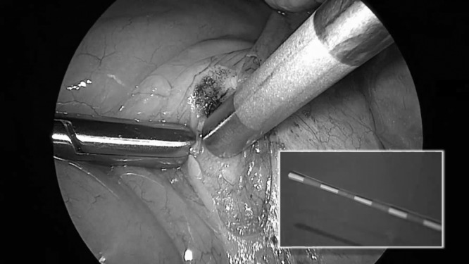
Blunt preparation with a blunt probe.
Here, it is of utmost importance to avoid a median sacral artery lesion. An 8-cm to 10-cm long, and 1.5-cm to 2-cm wide strip of Prolene mesh (SURGIPRO Mesh, Tyco Healthcare Switzerland) is then inserted into the abdomen. The atraumatic grasping forceps are then moved outward directed above the 5-mm trocar, after the valve of the trocar has been removed.
The folded mesh is grasped outside and is pulled into the abdominal cavity via the 5-mm trocar. The mesh is then attached on a broad-base to the cervical stump through a continuous suture, which requires several stitches into and out of the cervix and the mesh. The mesh is fixated by tying several knots, or through simple interrupted stitches. We generally use a POLYSORB 1. A second thread and needle (ETHIBOND) is inserted, and the ligamentum longitudinale is exposed once more. This usually requires holding aside the peritoneum or intestine with the grasping forceps.
On the right, the needle is clamped in the needle holder, and a Z-suture is stitched onto the ligamentum longitudinale (Figure 4). A sufficiently wide distance between stitches on the ligamentum longitudinale must be ensured to prevent the mesh from tearing after affixation.
Figure 4.
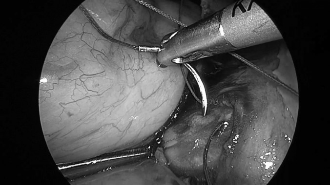
Mesh fixation with a Z-suture on the ligamentum longitudinale.
Using the swab or blunt vaginal probe placed in the vagina beforehand, the cervix is moved from a vaginal position to a position above the levator plate. The mesh is pulled cranially along the Z-suture on the ligamentum longitudinale using the atraumatic grasping forceps. The mesh is then stitched several times in line with the promontorium and tied to the ligamentum longitudinale (Figure 5).
Figure 5.

Mesh fixation on the cervical stump and ligamentum longitudinale.
Excess parts of the mesh and thread are cut off and disposed of. The cervical stump and the mesh are then peritonealized through a continuous suture (Figure 6). This technique of sacropexy may be carried out on the posterior side of the cervix in the case of uterus preservation, on the vaginal stump in the case of previous hysterectomy, or, as described, on the cervical stump during or after a supracervical hysterectomy. The affixation on the vaginal stump takes place after previous preparation at the posterior vaginal wall. However, while pushing up the vaginal stump, with the swab inside the vagina, it must be ensured that the needle does not grasp the swab and stitch it to the surface. In the case of uterus preservation, the mesh is fixated on the posterior cervical wall.10
Figure 6.

Mesh peritonealization.
Statistical Analysis
Continuous data collected from clinical records were listed as mean values with their standard deviations and 95% CI. The preoperative and postoperative assessments were compared using the paired-sample Wilcoxon's rank test. Spearmann's correlation coefficient was used to analyze the “duration of the operation.” All analyses were performed using Excel (Microsoft, Redmond, USA) and the Statistical Package for the Social Sciences (Windows version 15.0; SPSS Inc, Chicago, IL, USA). The significance level was set at a P < .05.
RESULTS
Demographic Data
From January 2000 to December 2007, 310 laparoscopic sacropexies were performed at the Klinik für Minimal Invasive Chirurgie (Berlin). Mean preoperative data from all patients can be found in Table 1.
Table 1.
Preoperative Data (n=310)
| Age (yrs, mean ± SD, range) | 56.7 ± 10.2 (33–81) |
| Parity | 1.85 |
| Body mass index (kg/m2, mean + SD) | 25.2 ± 3.51 |
| Menopause (n, %) | 51.4 |
| Previous pelvic floor repair surgery (n, %) | 43 (13.9) |
| Previous hysterectomy (n, %) | 79 (25.5) |
| Vaginal hysterectomy | 50 (16.1) |
| LASH | 18 (5.8) |
| Abdominal hysterectomy | 11 (3.5) |
| ASA (n, %) | |
| ASA I | 73 (23.5) |
| ASA II | 135 (43.5) |
| ASA III | 100 (32.3) |
| ASA IV | 2 (0.6) |
| Additional descensus (n, %) | |
| Anterior compartment | 112 (35.1) |
| Posterior compartment | 48 (15.5) |
| Urinary symptoms (n, %) | 164 (52.9) |
| Stress incontinence | 75 (45.7) |
| Urge incontinence | 6 (3.6) |
| Mixed incontinence | 10 (6.1) |
| No clear diagnosis | 73 (44.5) |
| Other symptoms (n, %) | |
| Pressure on bladder | 39 (12.6) |
| Pressure pain in vaginal area | 133 (42.9) |
| Dyspareunia | 23 (7.4) |
| Lower back pain | 100 (32.3) |
| Pressure downwards directed | 161 (51.9) |
Of the patients, 64.6% women were multipara, 92.6% of the children were delivered spontaneously, 2.8% were given birth by forceps delivery or vacuum extraction, respectively, and 1.7% caesarian deliveries were performed.
DGGG stages for all patients with regard to the mesh attachment point are shown in Table 2.
Table 2.
DGGG Staging with Regard to the Mesh Attachment Point
| Total (n, %) | Mesh Attachment to Cervical Stump (n, %) | Mesh Attachment to Vaginal Stump (n, %) | Sacropexy with Uterus Preservation (n, %) | |
|---|---|---|---|---|
| Stage I | 73 (23.5) | 55 (25.8) | 13 (19.4) | 4 (13.3) |
| Stage II | 158 (51) | 118 (55.4) | 23 (34.3) | 16 (53.3) |
| Stage III | 76 (24.5) | 38 (17.8) | 30 (44.7) | 10 (33.3) |
| Stage IV | 3 (1) | 2 (0.9) | 1 (1.6) | 0 |
Previous Operations
Of the study patients, 43 patients (13.9%) underwent previous pelvic floor repair surgery because of genital prolapse. In all cases, vaginal surgeries were performed; 25.5% (79 patients) had a previous hysterectomy (50 vaginal hysterectomies, 18 laparoscopic supracervical hysterectomies [LASH], and 11 abdominal hysterectomies). Of 50 vaginal hysterectomies, 20 surgeries were performed in combination with colporrhaphy, of which 13 were anterior, 6 were posterior colporrhaphies, and 1 was a simultaneous anterior and posterior colporrhaphy. Four vaginal sacrospinal fixations, 5 surgeries for urine incontinence, 1 Burch-colposuspension, and 4 tension-free vaginal tape (TVT) operations were previously performed.
Operative Procedures
All operations were performed by 3 surgeons, with a similar number of operations and recurrence rate for each surgeon. Of the study patients, 213 (68.7%), 67 (21.6%) and 30 (9.7%) underwent sacropexy with a mesh attachment to the cervical stump, to the vaginal stump, and to the posterior cervical side in case of uterus preservation, respectively.
Concomitant surgeries were necessary in 270 cases (87.1%); often numerous surgeries were performed (Table 3).
Table 3.
Concomitant Surgery (n=270 [87.1%])
| Surgery | n (%) |
|---|---|
| LASHa | 195 (62.9) |
| Vaginal hysterectomy | 5 (1.6) |
| LAVHa | 1 (0.3) |
| Adenectomy | 96 (31) |
| Salpingectomy | 19 (6.1) |
| Intestine and mesh adhesiolysis | 124 (40) |
| Anterior colporrhaphy surgery | 5 (1.6) |
| Posterior colporrhaphy surgery | 6 (1.9) |
| TVTa | 12 (3.8) |
| Ovarian cyst removal | 30 (9.6) |
LASH=laparoscopic supracervical hysterectomy, LAVH= laparoscopic-assisted vaginal hysterectomy, TVT=tension free vaginal tape.
The mean duration of operations can be found in Table 4.
Table 4.
Mean Duration of Operation
| min (95 % CI; min/max) | |
|---|---|
| All procedures | 118 ± 27.9 (115.3, 121.6; 25/263) |
| Mesh attachment to cervical stump | 121.4 ± 27.1 (117.7, 125.1; 60/263) |
| Mesh attachment to vaginal stump | 112.7 ± 30.4 (105.4, 120.1; 25/180) |
| Sacropexy with uterus preservation | 110.1 ± 25.4 (100.6, 119.6; 69/160) |
| Concomitant LASHa | 123.6 ± 26.8 min (119.7, 127.4; 70/263) |
| No concomitant operation (40 patients) | 99 ± 24.4 min (91.8, 106.7; 65/168) |
| Previous LASHa (9 patients) | 94.2 ± 9.4 min (84.4, 104.4; 80/111), |
| Previous total hysterectomy (19 patients) | 104.1 ± 26.8 min (89.5, 114.4; 65/168) |
| Sacropexy with uterus preservation (12 patients) | 98.3 ± 27.0 min (82.4, 115.3; 69/151) |
LASH = Laparoscopic supracervical hysterectomy.
The average weight of the extirpated uterus was 103.7 ± 10.86g (min/max, 12g/755g). A significant correlation between the duration of surgery and the weight of the removed tissue (P < .05) was found.
Complications
A conversion to laparotomy was required in 1 case (conversion rate 0.3%), due to extensive bowel and omental adhesions. Sacropexy was completed openly. Two terminations of the operation occurred (termination rate 0.6%), due to extensive lower abdominal adhesions in one case, and hemorrhage in the promontorium area during proximal fixation in the second case. New mesh implantation was not performed.
Duration of Hospitalization
The mean length of hospitalization was 4.1 ± 0.8 d (95% CI 4.01; 4.21, with a minimum and maximum duration of 3 d and 10 d, respectively. The length of hospitalization remained stable over the examined time period; 98% of the women were released from the hospital after 5 d. Longer hospitalization was required in one case due to a conversion to laparotomy, as described above (9 d hospitalization), and in another case, because of a revision with cervical extirpation due to uterine cancer (10 d hospitalization). One patient developed a hematoma in the area of trocar insertion, which had to be drained under local anesthesia. Three hundred patients (96.8%) were released free of pain.
Early Complications
Fifteen women (4.8%) suffered from postoperative fever, and 7 patients (2.3%) showed symptoms of urine incontinence one day after surgery (3 cases of de novo incontinence, 4 cases of persistent incontinence). Urine incontinence continued until the release from the hospital in 2 cases, in one of which the patient reported remaining symptoms during follow-up.
One patient (0.3%) was hospitalized again 5 d after surgery due to signs of intestinal obstruction, causing upper abdominal complaints. The patient was treated by infusion therapy. During the subsequent laparoscopy, small intestinal adhesions were identified as the cause of the symptoms and were removed. The patient was released the following day. One case of spondylitis in the junction area of lumbar vertebrae 5 and sacral vertebrae 1 occurred. The patient did not show prolapse symptoms; therefore, the mesh was not removed.
Follow-Up
The postoperative data of 211 women (68.1%) were collected mainly by the surgeon. The data are shown in Table 5.
Table 5.
Postoperative Data with Regard to Mesh Attachment Point (n = 211)
| Total | Mesh Attachment to Cervical Stump | Mesh Attachment to Vaginal Stump | Sacropexy with Uterus Preservation | |
|---|---|---|---|---|
| Patients, follow-up, n (%) | 211 (68.1) | 151 (77.4) | 40 (59.7) | 19 (63.3) |
| Post-OP examination (months) | 7.9 | 7.3 | 9 | 9.3 |
| Range (months) | 1–62.8 | 1–62.8 | 1–54.7 | 0.7–39.5 |
| Descensus rezidiv/ persistence, n (%) | 22 (10.4) | 14 (9.2) | 6 (14.6) | 2 (10.5) |
| Cystocele, n (%) | 32 (15.2) | 23 (15.2) | 6 (14.6) | 3 (15.7) |
| Rectocele, n (%) | 12 (6.6) | 9 (5.9) | 3 (7.3) | 0 |
| Preoperative Urinary Incontinence, n (%) | 115 (54.5) | 80 (52.9) | 26 (65) | 9 (47.4) |
| Postoperative Urinary Incontinence, n (%) | 47 (22.3) | 28 (18.5) | 11 (26.8) | 8 (42.1) |
| Reoperations, n (%) | 13 (4.2) | 7 (3.6) | 4 (6) | 2 (6.7) |
The mesh attachment on the middle compartment resulted in a significant reduction (P < .05) of descensus in the anterior and posterior compartment (see Table 6 for results). No significant differences between groups were detected for the recurrence of cystoceles. Rectoceles recurred significantly more often (P < .05) with patients who received sacropexy with vaginal mesh attachment.
Table 6.
Pre- and Postoperative Cystocele and Rectocele
| n = 211 | Preoperative (n, %) | Follow-up (Persistent) (n, %) | Follow-up (Newly Diagnosed) (n, %) |
|---|---|---|---|
| Cystocele | 84 (39.8 %) | 32 (15.2 %) | 15 (7.1 %) |
| Rectocele | 41 (19.4 %) | 12 (5.7 %) | 0 |
Four patients (1.9%) suffered from postoperative constipation. A reduction in urine incontinence from 115 patients (54.5%) to 47 patients (22.3%) was observed. One patient (2.1%) developed postoperative incontinence; for 2 patients (4.3%) incontinence symptoms worsened considerably after surgery. Patients who had a simultaneous or previous hysterectomy showed a significant reduction (P < .05) of incontinence symptoms. Only 1 of the incontinent patients undergoing sacropexy with uterus preservation reported no urine incontinence symptoms after surgery. All 12 patients (3.8%) receiving simultaneous operative correction of the anterior and posterior compartment were free of postsurgical symptoms of urinary incontinence symptoms. Follow-up examinations revealed a resolution of incontinence symptoms for 57.3% of the patients suffering from preoperative urine incontinence but not treated with concomitant incontinence surgery (110 preoperative, 47 postoperative).
Residual Operations
Of 323 sacropexies, 13 resacropexies (4.0%) were performed. One reresacropexy was necessary after the mesh tore on the distal pole 2 mo after residual operation. The mean intermediate time between operations was 26.6 ± 18.3 mo (min/max, 1 mo/60 mo). Indications for reoperations were descensus level IV in 6 cases (41.7%) and descensus level III in 7 cases (58.3%). Eight surgeries were performed without concomitant intervention, and 5 resacropexies were accompanied by simultaneous surgeries.
In all cases, the distal fixation pole was torn. Reaffixation of the original mesh was possible in 6 cases. Seven patients were supplied with a new mesh of the same kind, proximally fixated on the original mesh and in the respective distal pole (vaginal or cervical stump). After complete prolapse, affixation was performed using the old mesh.
The mean duration of operation was 89.2 ± 28.3 min (95% CI 87.3; 92.4, min/max 55 min/150 min). Patients were released after 3 ± 0.62 d (95% CI 2.8; 3.3, min/max 2 d/4 d). No complications occurred.
DISCUSSION
In accordance with previous studies,11,12 we found laparoscopic sacropexy to be a safe and effective surgical treatment for genital prolapse. Persistent or recurrent prolapse was found in 22 women on examination at a postoperative mean of 7.9 mo, resulting in a total persistence/recurrence rate of 10.4%. For sacropexy with mesh attachment to the vaginal stump the persistence/recurrence rate slightly, but not significantly, increased to 14.6%. This might be due to the decreased support of the extensive Z-suture on the basal pole by attachment to the vaginal stump, in comparison to mesh attachment to the cervix. Our data present a comparable recurrence rate to that reported in other studies.11,13
Recurrent cystoceles and rectoceles were found in 32 (15.2%) and 12 (5.7%) of patients, respectively, with significantly more recurrent rectoceles after mesh attachment to the vaginal stump. This represents a significant reduction in descensus in the anterior and posterior compartment through elevation of the middle compartment in all groups. Previous studies showed rectocele recurrence rates of between 0% and 55%.14–16 Fifteen cystoceles were newly diagnosed after surgery in our study. However, because the preoperative extent of cystocele and rectocele were not documented, these might represent recurrent cystoceles, as preoperative descensus of the middle compartment covered existing cystoceles in these 15 cases.
Thirteen reoperations were necessary, resulting in a reoperation rate of 4.1. This represents a comparable reoperation rate to that reported by other studies.11,17 In all cases, recurrent prolapse was due to the mesh tearing at the basal fixation pole.
Simultaneously with the 310 laparoscopic sacropexies, 270 concomitant procedures were performed. Even so, the duration of the operation and the length of hospital stay was comparable to, or even lower than, reports about laparoscopic procedures in the literature.11,13,18
We report a conversion rate of 0.3%, in comparison to the 2.2% rate reported by Rozet.13 Two operations were terminated to avoid injuries due to an unclear operating field. Only one case of relevant bleeding caused by the suture occurred. It did not require blood transfusion. Severe complications, defined as injury to nerves and organs, did not occur.
No suture or mesh erosion occurred in our study, in comparison to other reports documenting erosion totals as high as 11%.18,19 A reason for the reduced numbers of cases of mesh erosion might be the restrained fixation on the basal section of the mesh. This reduces the occurrence of mesh erosion, but mesh tearing on this location is more probable. Retroperitonealization of the mesh material seems to have proved successful.
Immediate postoperative complications included 1 case of intestinal obstruction and spondylitis, respectively. Even though Rozet13 also report 1 case of spondylitis, the link to the graft insertion is doubtful. Immediate postoperative complications also included 15 cases (4.8%) of fever, maybe due to infections, and 7 cases of incontinence 1 d after surgery, being persistent in 1 case during follow-up. One patient was hospitalized again 5 d after surgery due to signs of intestinal obstruction. This very low number of perioperative and immediate postoperative complications suggests that the laparoscopic approach is safe.
Four patients suffered from constipation after surgery, which was also reported by Mellgren20 after rectocele repair. Incontinence recurred in 59.1% of the patients. Although patients undergoing simultaneous or previous hysterectomy reported a significant reduction (P < .05) in incontinence symptoms, only 1 patient undergoing sacropexy with uterus preservation showed no incontinence symptoms after surgery.
One patient (2.1%) developed postoperative incontinence, and for 2 patients (4.3%) incontinence symptoms deteriorated considerably after surgery. Thus, de novo incontinence is very rare in our study, compared to the 14% reported by Chaikin.21 Our findings of a significant reduction in incontinence for patients who had undergone a simultaneous or previous hysterectomy are surprising; although discussed controversially, different studies show an increased risk for stress incontinence after hysterectomy.22 Through the elevation of the middle compartment, the better anatomical functional positioning of the urethra possibly improves urine incontinence symptoms. The less pronounced effect on patients who had undergone sacropexy with uterus preservation might be due to a less pronounced elevation of the anterior compartment, because mesh attachment was performed at the posterior cervix side.
In our clinic, the first-line treatment for most patients suffering from prolapse of the middle compartment consists of descensus treatment without concomitant incontinence surgery, because the postoperative improvement of incontinence is possible for 57% of patients. Only if the recurrence of incontinence symptoms is observed are further treatments considered. This way, exposure to unnecessary morbidity is prevented.
Our results show that laparoscopic sacropexy provides similar success rates in comparison to abdominal sacropexy, which is considered the gold standard in the surgical treatment of vaginal vault prolapse.3,8,23 Vaginal sacropexy is the method of choice for specialized experts. However, our findings prove that laparoscopic sacropexy, performed by surgeons with excellent endoscopic skills, offers effective vault support and similar safety results.11
The major weakness of this study was its retrospective nature. Not all data could be collected for all patients. Additionally, there is a low response rate, with postoperative data representative of only 68.1% of the patients treated. However, this response rate compares to data from other reports.8,11 This presents a source of potential bias, although it is hard to know which way the results might be influenced. Another drawback is the relatively short follow-up period of 7.9 mo and the varying interval for postsurgical examination (1 mo to 62.8 mo). To draw definitive conclusions about the rate of recurrent prolapse, a longer follow-up is needed. Furthermore, mesh complications can occur several years after surgery. For example, extrusion of mesh into the vagina has been reported up to 3 y following surgery.11
CONCLUSION
Our results confirm previous findings that laparoscopic sacropexy is a safe and efficacious surgical treatment for genital prolapse with a very low rate of perioperative complications and favorable anatomic results. It provides excellent vault support and good functional outcome, concerning incontinence. Compared to similar studies, the demonstrated standardized surgical technique has been shown to have a short operation time and hospitalization. An extended period of follow-up is needed to confirm these findings over the longer term.
Contributor Information
Bernd Bojahr, Klinik für Minimal Invasive Chirurgie, Kurstraße 11, 14129 Berlin-Zehlendorf, Germany..
Garri Tchartchian, Klinik für Minimal Invasive Chirurgie, Kurstraße 11, 14129 Berlin-Zehlendorf, Germany..
Matthias Waldschmidt, Klinik für Minimal Invasive Chirurgie, Kurstraße 11, 14129 Berlin-Zehlendorf, Germany..
Thoralf Schollmeyer, Universitätsfrauenklinik Kiel, Michaelisstrasse 16, 24105 Kiel, Germany..
Rudy L. De Wilde, Pius-Hospital, Klinik für Frauenheilkunde, Georgstraße 12, 26121 Oldenburg, Germany..
References:
- 1. Dowell CJ, Bryant CM, Moore KH, Simons AM. Calculating the direct cost of urinary incontinence: a new test instrument. BJU Int. 1999; 83 (6): 596–606 [DOI] [PubMed] [Google Scholar]
- 2. Hendrix SL, Clark A, Nygaard I, Aragaki A, Barnabei V, McTiernan A. Pelvic organ prolapse in the Women's Health Initiative: gravity and gravidity. Am J Obstet Gynecol. 2002(June);186(6):1160–1166 [DOI] [PubMed] [Google Scholar]
- 3. Benson J, Lucente M, McClellan E. Vaginal versus abdominal reconstructive surgery for the treatment of pelvic support defects: a prospective randomized study with long-term outcome evaluation. Am J Obstet Gynecol. 175(6): 1418–1421, 1996. December; discussion 1421–1422 [DOI] [PubMed] [Google Scholar]
- 4. Arthure HG, Savage D. Uterine prolapse and prolapse of the vaginal vault treated by sacral hysteropexy. J Obstet Gynaecol Br Emp. 1957; 64 (3): 355–360 [DOI] [PubMed] [Google Scholar]
- 5. Ganatra AM, Rozet F, Sanchez-Salas R, et al. The current status of lapaorscopic sacrocolpopexy: a review. Eur Urol. 2009. May; 55(5): 1089–1103 Epub 2009 Feb 4. Review [DOI] [PubMed] [Google Scholar]
- 6. Ross JW, Preston M. Laparoscopic sacropexy for severe vaginal vault prolapse: 5-year outcome. J Minim Invasive Gynecol. 12(3): 221–226, 2005. May-Jun [DOI] [PubMed] [Google Scholar]
- 7. Fatton B, Amblard J, Debodinance P, Cosson M, Jacquetin B. Transvaginal repair of genital prolapse: preliminary results of a new tension-free vaginal mesh (Prolift technique) – a case series multicentric study. Int Urogynecol J Pelvic Floor Dysfunct. 2007. July; 18(7): 743–752 Epub 2006 Nov 28 [DOI] [PubMed] [Google Scholar]
- 8. Huebner M, Krzonkalla M, Tunn R. Abdominal sacrocolpopexy – standardized surgical technique, perioperative management and outcome in women with posthysterectomy vaginal vault prolapse. Gynäkol Geburtshilfliche Rundsch. 2009; 49 (4): 308–314 Epub 2010 May 19 [DOI] [PubMed] [Google Scholar]
- 9.DGGG. 2010. [Accessed May 24, 2011]. http://www.dggg.de/fileadmin/public_docs/Leitlinien/1-3-2-descensus-2010.pdf [online]
- 10. Bojahr B. Laparoskopische Sakropexie. Erfahrungen mit der modifizierten Technik nach Raatz. Gynecol. Prax. 2003; 33: 95–106 (article in German). [Google Scholar]
- 11. Higgs PJ, Chua HL, Smith AR. Long term review of laparoscopic sacropexy. BJOG. 112(8): 1134–1138, 2005. August [DOI] [PubMed] [Google Scholar]
- 12. North C, Ali-Ross N, Smith A, Reid FM. A prospective study of laparoscopic sacropexy for the management of pelvic organ prolapse. BJOG. 2009. August; 116(9): 1251–1257 Epub 2009 May 28 [DOI] [PubMed] [Google Scholar]
- 13. Rozet F, Mandron E, Arroyo C, et al. Laparoscopic sacral colpopexy approach for genito-urinary prolapse: experience with 363 cases. Eur Uro. 47(2): 230–236, 2005. February [DOI] [PubMed] [Google Scholar]
- 14. Costantini E, Mearini L, Bini V, Zucchi A, Mearini E, Porena M. Uterus preservation in surgical correction of urogenital prolapse. Eur Urol. 48(4): 642–649, 2005. October [DOI] [PubMed] [Google Scholar]
- 15. Schettini M, Fortunato P, Gallucci M. Abdominal sacral colpopexy with Prolene mesh. Int Urogynecol J Pelvic Floor Dysfunct. 1999; 10 (5): 295–299 [DOI] [PubMed] [Google Scholar]
- 16. Baessler K, Schuessler B. Abdominal sacrocolpopexy and anatomy and function of the posterior compartment. Obstet Gynecol. 97(5 Pt 1): 678–684, 2001. May [DOI] [PubMed] [Google Scholar]
- 17. Nygaard IE, McCreery R, Brubaker L, et al. Abdominal sacropexy: a comprehensive review. Obstet Gynecol. 104(4): 805–823, 2004. October Review [DOI] [PubMed] [Google Scholar]
- 18. Misrai V, Rouprêt M, Cour F, Chartier-Kastler E, Richard F. De novo urinary stress incontinence after laparoscopic sacral colpopexy. BJU Int. 2008. March; 101(5): 594–597 Epub 2007 Nov 8 [DOI] [PubMed] [Google Scholar]
- 19. Iglesia CB, Fenner DE, Brubaker L. The use of mesh in gynecologic surgery. Int Urogynecol J Pelvic Floor Dysfunct. 8(2): 105–115, 1997. Review [DOI] [PubMed] [Google Scholar]
- 20. Mellgren A, Anzén B, Nilsson BY, et al. Results of rectocele repair. A prospective study. Dis Colon Rectum. 1995. January; (1)38: 7–13 [DOI] [PubMed] [Google Scholar]
- 21. Chaikin DC, Groutz A, Blaivas JG. Predicting the need for anti-incontinence surgery in continent women undergoing repair of severe urogenital prolapse. J Urol. 163(2): 531–534, 2000. February [PubMed] [Google Scholar]
- 22. Altman D, Granath F, Cnattingius S, Falconer C. Hysterectomy and risk of stress-urinary-incontinence surgery: nationwide cohort study. Lancet. 370(9597): 1494–1499, 2007. October 27 [DOI] [PubMed] [Google Scholar]
- 23. Maher CF, Qatawneh AM, Dwyer PL, Carey MP, Cornish A, Schluter PJ. Abdominal sacral colpopexy or vaginal sacrospinous colpopexy for vaginal vault prolapse: A prospective randomized study. Am J Obstet Gynecol 2004. January; 190(1): 20–26 [DOI] [PubMed] [Google Scholar]