Abstract
Long-term stability is a desired characteristic of vaccines, especially anthrax vaccines, which must be stockpiled for large-scale use in an emergency situation; however, spontaneous deamidation of purified vaccine antigens has the potential to adversely affect vaccine immunogenicity over time. In order to explore whether spontaneous deamidation of recombinant protective antigen (rPA)—the major component of new-generation anthrax vaccines—affects vaccine immunogenicity, we created a “genetically deamidated” form of rPA using site-directed mutagenesis to replace six deamidation-prone asparagine residues, at positions 408, 466, 537, 601, 713, and 719, with either aspartate, glutamine, or alanine residues. We found that the structure of the six-Asp mutant rPA was not significantly altered relative to that of the wild-type protein as assessed by circular dichroism (CD) spectroscopy and biological activity. In contrast, immunogenicity of aluminum-adjuvanted six-Asp mutant rPA, as measured by induction of toxin-neutralizing antibodies, was significantly lower than that of the corresponding wild-type rPA vaccine formulation. The six-Gln and six-Ala mutants also exhibited lower immunogenicity than the wild type. While the wild-type rPA vaccine formulation exhibited a high level of immunogenicity initially, its immunogenicity declined significantly upon storage at 25°C for 4 weeks. In contrast, the immunogenicity of the six-Asp mutant rPA vaccine formulation was low initially but did not change significantly upon storage. Taken together, results from this study suggest that spontaneous deamidation of asparagine residues predicted to occur during storage of rPA vaccines would adversely affect vaccine immunogenicity and therefore the storage life of vaccines.
INTRODUCTION
Anthrax toxin is a major virulence factor of Bacillus anthracis, the etiologic agent of anthrax disease. The toxin is composed of a receptor-binding component known as protective antigen (PA) and two enzymatically active components, lethal factor (LF), a zinc-dependent metalloprotease, and edema factor (EF), a calmodulin-sensitive adenylate cyclase. PA transports LF and EF into the host cell cytosol, where the enzymatically active moieties exert their toxicities (1). Animal studies have shown that protective immunity to anthrax disease correlates with induction of neutralizing anti-PA antibodies (2–4). Therefore, new-generation anthrax vaccines are being developed that are composed of purified recombinant PA (rPA) adsorbed to aluminum adjuvant. Unfortunately, efforts to develop rPA vaccines have been hampered by a lack of stability of this vaccine antigen (5).
Instability of recombinant protein vaccines can be due, at least in part, to spontaneous modifications such as deamidation, oxidation, isomerization, disulfide bond formation, and/or hydrolysis. Of these modifications, deamidation of asparagine residues is the most common and is a major source of amino acid sequence heterogeneity that occurs under neutral or slightly alkaline pH conditions (6). Deamidation of Asn occurs by nucleophilic attack of the α-amino group of the C-terminal-adjacent amino acid on the Asn side chain, which leads to formation of an unstable succinimide intermediate that then hydrolyzes to either Asp or isoAsp, in a ratio of approximately 1:3 (7). Deamidation of Asn to Asp introduces a negative charge into the protein, while the change to isoAsp results in the addition of both a negative charge and an extra methylene residue in the peptide backbone. Gln residues can also undergo deamidation, although they are less susceptible than Asn residues (8, 9). Deamidation of Asn and Gln residues occurring in vivo (10–12) has been hypothesized to play an important role as a molecular clock that controls the rates of protein turnover (13, 14). However, the deamidation that occurs in vitro during the isolation of proteins and subsequent storage can adversely affect the biological properties of the protein. Effects on epitope structure, antigen processing, and antigen presentation represent negative outcomes that are particularly relevant to vaccine development (15, 16). The rate of deamidation of any given Asn or Gln residue depends on a number of parameters, including primary structure (neighboring amino acids), pH, temperature, and ionic strength.
Previously, others have demonstrated that certain Asn residues within rPA deamidate on a time scale relevant to vaccine dating periods (17, 18). In a comprehensive study, Powell et al. (18) detected measurable deamidation of 7 of the 68 Asn residues of rPA that had been subjected to conditions conducive to deamidation. The extent of deamidation of these seven residues occurred in the following order: N537> N713 > N466 > N719 > N601 > N408 > N602. In another report, evidence was also presented for deamidation of Asn162 during storage of rPA (17).
While deamidation of Asn residues in rPA has been demonstrated, little is known about the effect that deamidation might have on the immunogenicity of the protein. Previously, Ribot et al. (19) examined the immunogenicity and protective efficacy of isoforms of rPA that differed in deamidation levels (18, 19). The two isoforms compared in that study exhibited comparable immunogenicity and protective immunity; however, the difference in the extent of deamidation between the two isoforms used in that study was minimal, as judged by the incremental difference in charge between the two isoforms (18, 19). Thus, this difference in deamidation most likely did not adequately mimic the extent of deamidation that might be expected upon long-term vaccine storage.
In order to further explore whether deamidation could affect the immunogenicity of rPA, we genetically engineered rPA so as to model a deamidated form of the protein that could be expected to result upon prolonged vaccine storage. To do so, we mutated the pagA gene of B. anthracis such that six deamidation-prone Asn residues of rPA were substituted with Asp residues. The six Asn residues that we chose to mutate, Asn537, Asn713, Asn466, Asn719, Asn601, and Asn408, were those rPA residues that exhibited the highest levels of deamidation as determined by Powell et al. (18). We examined the structure and immunogenicity of this “genetically deamidated” form of rPA and compared its properties to those of wild-type rPA.
MATERIALS AND METHODS
Materials.
B. anthracis recombinant PA83 (NR-140), recombinant LF (NR-142), anti-rPA rabbit reference polyclonal serum (NR-3839), and murine macrophage-like J774A.1 cells (NR-28) were from the NIH Biodefense and Emerging Infections Research Resources Repository, NIAID, NIH (Bethesda, MD). The aluminum hydroxide adjuvant Alhydrogel was obtained from Brenntag Biosector (Denmark). Cell culture reagents were obtained from Invitrogen (Carlsbad, CA). Phenyl-Sepharose 6 fast-flow (high-sub) resin and gel filtration chromatography column Superdex 200 10/300GL were from GE Healthcare (Sweden). Strong anion-exchange spin columns were from Pierce (Rockford, IL).
Cloning and expression of wild-type and mutant rPA genes.
Expression of the genes encoding wild-type rPA and mutant derivatives was accomplished using a host strain and expression plasmid recently developed in our laboratory. The host strain BA822 is derived from the avirulent B. anthracis Sterne 7702 strain by multiple rounds of markerless allelic exchange using the method of Janes and Stibitz (20). In this way, 12 genes predicted to encode secreted proteases have been deleted. In addition, the pagA gene has been deleted to avoid contamination of mutant proteins with trace amounts of wild-type protein. The expression vector pSS4570 is based on the Gram-positive plasmid pIP404 replicon, as derived from pJIR751 (21), and uses spectinomycin resistance as a selectable marker. Expression of cloned genes is driven by a mutant derivative of the E. coli gapA promoter that contains a consensus −35 region, a consensus −10 region, and an extended −10 region (22). Details of the derivation of BA822 and the construction of pSS4570 will be presented in a separate manuscript. The entire rPA gene, including signal sequence, was PCR amplified using the primers CGCGCGGCCGCAGAAAGGAGGAACGTATATGAAAAAACGAAAAGTG and CGCGGATCCTTATCCTATCTCATAGCCTTTTTTAG and subsequently cloned as a BamHI-NotI fragment into pSS4570 to create pBAM136. The six-Asp, six-Gln, and six-Ala versions of the gene were synthesized by GenScript, Inc. (Piscataway, NJ), and similarly cloned into pSS4570 to create pBAM168, pBAM164, and pQC1804, respectively. For production of rPA, plasmids were transferred to BA822 by conjugation.
Production and purification of wild-type and six-Asp mutant rPA.
Wild-type and mutant rPA proteins were purified from the culture supernatants of the avirulent strains constructed as described above using a slight modification of a previously described purification procedure (23). Briefly, culture supernatant containing secreted rPA protein was filtered and treated with EDTA (5 mM) and phenylmethylsulfonyl fluoride (1 mM), followed by the addition of ammonium sulfate (final concentration = 2 M) and phenyl-Sepharose 6 fast flow resin (10 ml/liter of culture supernatant). After 1 to 2 h of gentle mixing at 4°C, the resin was collected on a 0.22-μm filter and washed with a volume of cold wash buffer (10 mM HEPES [pH 7.5], 1 mM EDTA containing 1.5 M ammonium sulfate) equal to that of the culture supernatant. rPA was eluted from the resin with elution buffer (10 mM HEPES [pH 7.5], 1 mM EDTA containing 0.3 M ammonium sulfate). rPA was then further purified by ammonium sulfate precipitation (47 g/100 ml of eluted protein) and subjected to anion-exchange chromatography using strong anion-exchange spin columns (Pierce, Rockford, IL). Finally, the rPA preparation was purified by size exclusion chromatography on a Superdex 200 10/300GL column (GE Healthcare, Sweden). Purified rPA proteins obtained using this procedure were approximately 90 to 95% pure, as determined by SDS-PAGE.
CD of wild-type and six-Asp mutant rPA.
Far-UV circular dichroism (CD) spectra of solutions containing wild-type or six-Asp mutant rPA were recorded at 185 to 260 nm using a 1-mm path length quartz cuvette in a Jasco J-815 spectrophotometer at 20°C. CD spectra were measured for rPA (0.3 mg/ml) in 20 mM sodium phosphate buffer, pH 7.8. Raw data measured in millidegrees were converted into ellipticity (degrees cm2/dmol). The ellipticity (degrees cm2/dmol) was plotted as a function of wavelength (nm) using Microsoft Office Excel.
rPA storage.
Purified rPA protein samples were stored at 0.2 mg/ml in 100 mM sodium phosphate buffer (pH 7.8) for up to 4 weeks at 25°C. Aliquots were removed each week and stored at −30°C until analyzed. After storage, aliquots were subjected to native polyacrylamide gel electrophoresis (PAGE) in order to visualize the negatively charged isoforms that had spontaneously generated over time and to SDS-PAGE to verify protein integrity.
Native and SDS-PAGE.
Aliquots of wild-type and six-Asp mutant rPA proteins were analyzed either on native PAGE gels (4 to 16% Bis-Tris gels; Invitrogen, Carlsbad, CA) in 1× native PAGE buffer or on SDS-PAGE gels (NuPAGE 4 to 12% Bis-Tris gels; Invitrogen, Carlsbad, CA) in 1× morpholinepropanesulfonic acid (MOPS) running buffer. Gels were stained with Coomassie blue for visualization.
Cell culture, TNA assay, and macrophage lysis assay.
Sera obtained from mouse immunization studies were analyzed by the toxin-neutralizing antibody (TNA) assay using J774A.1 cells essentially as described previously (24). The murine macrophage-like cell line J774A.1 was grown in Dulbecco's modified Eagle medium (DMEM; containing a high glucose concentration and sodium pyruvate) supplemented with 5% heat-inactivated fetal bovine serum, 2 mM glutamine, penicillin (25 U/ml), streptomycin sulfate (25 μg/ml), and 10 mM HEPES. Briefly, cells were plated in 96-well flat-bottomed plates (40,000 cells/well) and incubated for 17 to 19 h at 37°C in a 5% CO2 incubator. Neutralization of lethal toxin cytotoxicity was measured by assessing cell viability with 2-fold serial dilutions of the test sample and rabbit polyclonal serum (NR-3839) as the reference serum sample. Serum samples were prepared in a separate 96-well microtiter plate using an appropriate starting dilution followed by 2-fold dilutions for a total of seven dilutions per sample. The samples were then incubated with a constant concentration of lethal toxin (PA at 50 ng/ml plus LF at 40 ng/ml) for 30 min prior to the addition to the cells. This concentration of toxin kills approximately 95% of the cells in the absence of any neutralizing serum sample. The cell-serum-toxin mixtures were transferred to the 96-well cell plate, and cells were incubated for 4 h at 37°C. Following incubation, 3-(4,5-dimethylthiazol-2-yl)-2, 5-diphenyltetrazolium bromide (MTT) was added to the plates. After 2 h of incubation, cells were lysed by the addition of solubilization buffer (90% isopropanol, 0.5% sodium dodecyl sulfate [wt/vol], and 38 mM HCl). The plates were read for optical density using a microplate reader at A570. A four-parameter logistic regression model was used to fit the data points generated when the optical density was plotted versus the reciprocal of the serum dilution. The inflection point, which indicates 50% neutralization, was reported as the mean effective dilution (ED50).
Macrophage lysis assays were performed to assess the cytotoxic activity of wild-type and mutant rPA using J774A.1 cells as described previously (25). Briefly, the cytotoxicity of rPA samples was measured by using various concentrations of rPA samples (2-fold dilutions) mixed with a fixed concentration of LF (40 ng/ml). The rPA-LF mixture was added to J774A.1 cells. After 4 h, cell viability was measured by the addition of MTT as described above for the TNA assay. Percent viability of J774A.1 was calculated by using the untreated cell control (cells without rPA-LF) and plotted against rPA protein concentration (ng/ml) using a four-parameter logistic regression model. The inflection point, which indicates 50% survival, was reported as the mean effective concentration (EC50).
Preparation of rPA-Alhydrogel formulations.
All rPA-Alhydrogel formulations used for mouse immunization studies were prepared using rPA samples either at a 30-μg/ml concentration with 0.9 mg/ml of aluminum adjuvant or at 50 μg/ml with 1.5 mg/ml of aluminum adjuvant in normal saline solution (0.9% NaCl [wt/vol]), as indicated. The mixture was gently vortexed and allowed to stand for 1 h at room temperature (RT) for adsorption. The degree of adsorption of rPA to Alhydrogel was determined after centrifugation to pellet the adjuvant and collection of the supernatant which was examined both for protein content using the Pierce BCA protein assay (Pierce, Rockford, IL) and for rPA bioactivity using the macrophage lysis assay. Adsorbed preparations were subsequently stored at 25°C for up to 4 weeks.
Immunization of mice.
All mouse immunization studies (immunizations and serum collections) were carried out either at an in-house animal facility (CBER/FDA) or by Cocalico Biologicals, Inc. (Reamstown, PA) in compliance with the guidelines of their respective Institutional Animal Care and Use Committee. Mice (six-week-old female CD-1 mice) were immunized once intraperitoneally with vaccine (equivalent to either 9 μg or 10 μg of rPA/mouse, as indicated). Groups of 20 mice were immunized either with freshly prepared rPA-Alhydrogel formulations or with formulations stored at 25°C for up to 4 weeks. A control group of 20 mice was simultaneously immunized with Alhydrogel in normal saline solution. Mice were bled 28 days postimmunization. Serum samples collected from the various groups of mice were analyzed individually using the TNA assay.
Data analysis.
All data were plotted using GraphPad Prism software (version 5; GraphPad Software, La Jolla, CA). For the TNA assay, a four-parameter logistic (4-PL) regression model was used to analyze optical density versus the reciprocal of the serum dilution. ED50s were determined as described by Ngundi et al. (26).
RESULTS
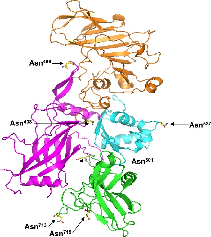
We used site-directed mutagenesis to alter specific Asn residues of PA in order to produce a form of rPA that modeled a relevant deamidated form of the protein. We designed our deamidated rPA such that the six Asn residues of rPA that had previously been reported to be the most prone to deamidation, Asn408, Asn466, Asn537, Asn601, Asn713, and Asn719 (18), were mutated to Asp residues. The locations of these six Asn residues within the protein are indicated in Fig. 1. We then compared the structure and immunogenicity of the wild-type and six-Asp mutant forms of rPA.
Fig 1.
Location of six deamidation-prone Asn residues within the crystal structure of PA (18). Shown are the six Asn residues that were mutated in this study. PA domains are indicated. Domain I, orange; domain II, pink; domain III, blue; domain IV, green.
Secondary structure of wild-type and six-Asp mutant rPA.
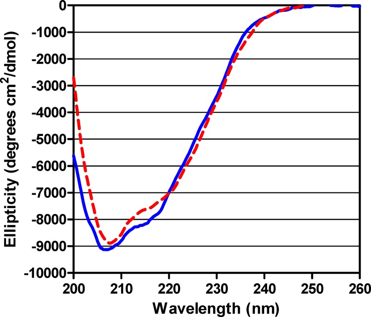
CD analysis of wild-type rPA and the six-Asp mutant rPA was performed in order to compare the secondary structures of the two rPA proteins. As shown in Fig. 2, the far-UV CD spectrum of the six-Asp mutant rPA was similar to that of wild-type rPA. Ellipticity data obtained for wild-type rPA and the six-Asp mutant rPA were further analyzed using the DICROWEB Web server, http://dichroweb.cryst.bbk.ac.uk, to calculate secondary structure content (27). This analysis yielded a composition for the wild-type rPA of 13% ± 3% α-helix, 30% ± 2% β-sheet, 22% ± 2% β-turn, and 35% ± 4% random coil (mean and standard deviation [SD]), which is very similar to that of the composition calculated for the six-Asp mutant rPA (15% ± 1% α-helix, 30% ± 3% β-sheet, 24% ± 2% β-turn, and 32% ± 4% random coil). For none of these secondary structure forms was there a difference in composition between the wild-type rPA and the six-Asp mutant rPA (Student's t test, P > 0.1). These results indicate that substituting six deamidation-prone Asn residues with Asp residues does not significantly affect the secondary structure of the protein.
Fig 2.
Comparison of ellipticity plots of wild-type and six-Asp mutant rPA. Far-UV CD spectra of wild-type (solid blue line) and six-Asp mutant rPA (dashed red line) were recorded over the wavelength range of 200 to 260 nm. Ellipticity (degrees cm2/dmol) is plotted as a function of wavelength (nm).
Biological activity of wild-type and six-Asp mutant rPA.
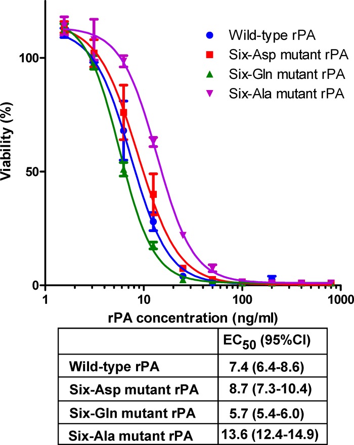
The biological activity of the six-Asp mutant rPA was measured using the macrophage lysis assay and compared to that of the wild-type protein. This assay measures the ability of PA, when combined with LF, to elicit cytotoxic activity on J774A.1 cells. As shown in Fig. 3, the EC50 of the six-Asp mutant rPA was similar to that of wild-type rPA, indicating that substitution of the six deamidation-prone Asn residues with Asp in the mutant rPA resulted in little or no impairment of the biological activity of the protein.
Fig 3.
Cytotoxic activity of wild-type rPA and rPA mutants. Cytotoxic activity was determined by combining the indicated concentrations of rPA with a fixed concentration of LF (40 ng/ml). Viability of the cells, normalized to that of untreated cells, is shown as a function of rPA concentration. Samples were analyzed on two independently prepared plates, with the means shown by the symbols and the ranges indicated by error bars. A four-parameter logistic model was fit to the data, and the table reports the EC50 and 95% confidence interval for each of the preparations tested. Very similar results were obtained when the experiment was repeated.
Analysis of wild-type and six-Asp mutant rPA isoforms by native PAGE.
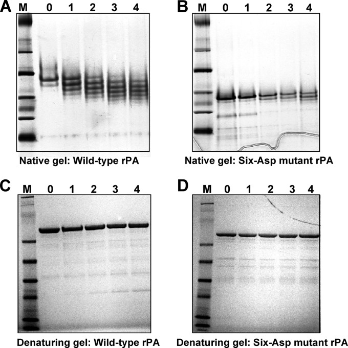
Samples of wild-type rPA that were either freshly prepared or which had been stored at 25°C in order to accelerate deamidation were analyzed by native PAGE. As shown in Fig. 4A, wild-type rPA initially migrated as one major isoform and a minor, faster-migrating isoform. Upon storage, a gradual increase in the number of negatively charged isoforms was observed, consistent with an accumulation of negative charge expected from deamidation of multiple Asn residues. After 4 weeks of storage at 25°C, seven isoforms were evident.
Fig 4.
Native PAGE and SDS-PAGE profiles of wild-type and six-Asp mutant rPA after storage for various periods of time. Equal amounts of rPA samples that had been stored for up to 4 weeks at 25°C as described in Materials and Methods were analyzed on native PAGE and SDS-PAGE gels. (A) Wild-type rPA, native PAGE; (B) six-Asp mutant rPA, native PAGE; (C) wild-type rPA, SDS-PAGE; (D) six-Asp mutant rPA, SDS-PAGE. Lanes are marked with time of storage (in weeks). Lane M, molecular weight markers.
Before storage at 25°C, the six-Asp mutant rPA appeared as one predominant isoform that migrated faster than wild-type rPA on native gels (Fig. 4B). This more rapid migration is expected due to the introduction of six negative charges that occurred upon mutation of six Asn residues to Asp residues. After storage of the six-Asp mutant rPA at 25°C for 4 weeks, additional isoforms were evident, but there were clearly fewer than the number observed with wild-type rPA.
SDS-PAGE analysis of the wild-type and six-Asp mutant rPA samples did not reveal any change in protein migration profile over time (Fig. 4C and D). These results demonstrate that neither the wild-type nor the six-Asp mutant rPA exhibited significant proteolytic degradation upon storage for up to 4 weeks at 25°C.
Immunogenicity of wild-type and six-Asp mutant rPA.
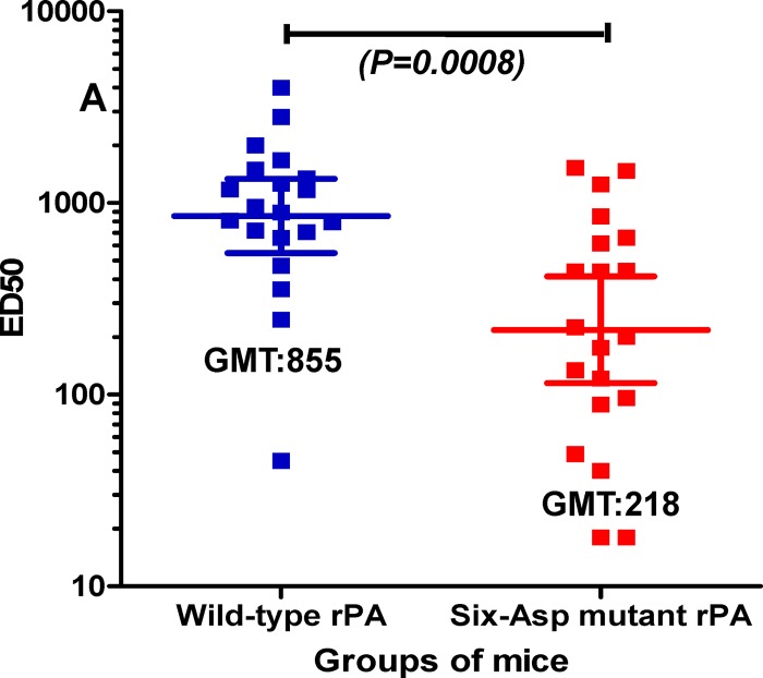
In order to assess the immunogenicity of wild-type and six-Asp mutant rPA in vaccine formulations, groups of mice were immunized with rPA proteins adsorbed to the aluminum hydroxide adjuvant, Alhydrogel. As shown in Fig. 5, toxin-neutralizing titers of mice immunized with the wild-type rPA-Alhydrogel formulation were significantly higher (P = 0.0008; unpaired t test) than those of mice immunized with the six-Asp mutant rPA-Alhydrogel formulation. These data indicate that substitution of six deamidation-prone Asn residues with Asp significantly decreases the immunogenicity of rPA.
Fig 5.
TNA titers of mice immunized with freshly prepared wild-type or six-Asp mutant rPA formulated with Alhydrogel. Two groups of 20 mice each were immunized with rPA-Alhydrogel formulations (10 μg of rPA/mouse). Twenty-eight days postimmunization, mice were bled and serum samples were analyzed using the TNA assay. Graph shows the TNA titer (expressed as ED50) for each mouse and geometric mean titer (GMT) for each group. Horizontal lines represent the geometric means with 95% confidence intervals for the group. P value is indicated (unpaired t test).
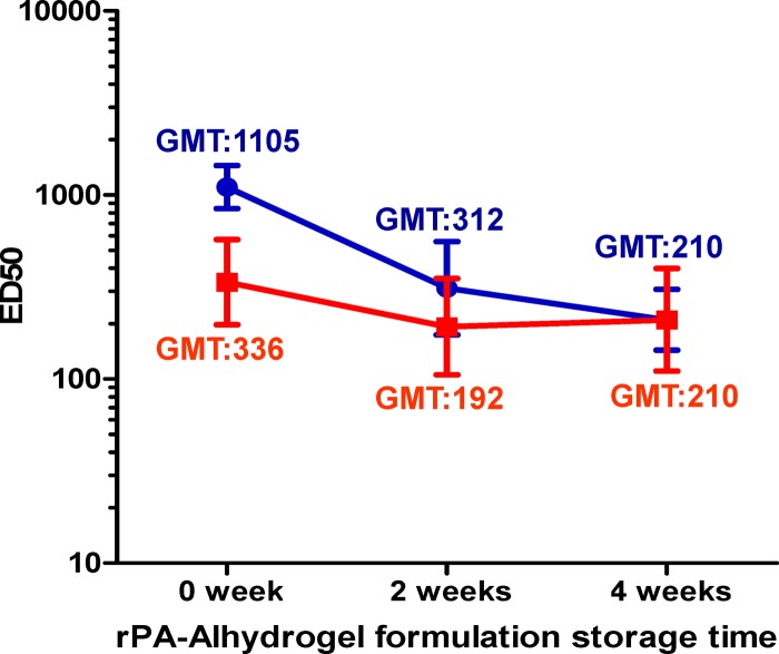
The immunogenicities of rPA-Alhydrogel formulations that had been stored at 25°C for up to 4 weeks were also examined. As shown in Fig. 6, the wild-type rPA-Alhydrogel formulation exhibited a significant decrease in immunogenicity over time (P < 0.05; ANOVA with Tukey posttest for trend) as has been reported previously (28). In contrast, the immunogenicity of the six-Asp mutant rPA-Alhydrogel formulation did not change significantly upon storage. Of note, while the immunogenicity of the wild-type rPA-Alhydrogel formulation was initially higher than that of the six-Asp mutant rPA-Alhydrogel formulation (P = 0.0002; unpaired t test), the immunogenicities of the two formulations were similar after storage for 4 weeks.
Fig 6.
TNA titers of mice immunized with wild-type (blue circles) and six-Asp mutant (red squares) rPA-Alhydrogel formulations either freshly prepared or stored at 25°C for up to 4 weeks. Groups of 20 mice each were immunized with rPA-Alhydrogel formulations (equivalent to 9 μg of rPA/mouse). Twenty-eight days postimmunization, mice were bled and serum samples were analyzed using the TNA assay. Graph shows geometric mean of TNA titers (GMTs) for each group with 95% confidence interval. This figure is representative of two independent immunization studies.
Biological activity and immunogenicity of other mutant forms of rPA.
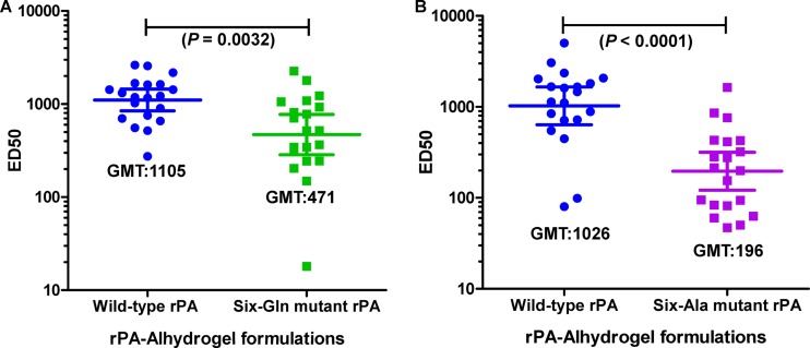
We also examined the biological activity and immunogenicity of rPA in which the six deamidation-prone Asn residues were changed to either Gln or Ala residues. We chose to explore these particular substitutions since Gln differs from Asn only by one additional methylene (CH2) group in its side chain and Ala lacks the amide group present in the side chain of Asn that undergoes deamidation. Thus, mutation of Asn to either Gln or Ala would represent minimal structural perturbations of Asn and, notably, would not introduce the negative charge that occurs upon deamidation that might have contributed to the loss in immunogenicity observed with the six-Asp mutant rPA. Our hope was to obtain a more stable mutant of rPA that retained its immunogenicity. As seen in Fig. 3, purified preparations of both the six-Gln mutant rPA and the six-Ala mutant rPA retained much of the biological activity of the wild-type rPA protein, indicating that the structures of these mutant proteins were not significantly perturbed. However, when we examined the immunogenicity of these proteins (Fig. 7), we found that both the six-Gln mutant rPA-Alhydrogel formulation (panel A) and the six-Ala mutant rPA-Alhydrogel formulation (panel B) exhibited significantly lower immunogenicity than that of the wild-type rPA-Alhydrogel formulation, indicating that these minimal changes in amino acid structure were sufficient to affect immunogenicity of the protein.
Fig 7.
TNA titers of mice immunized with freshly prepared wild-type, six-Gln mutant, or six-Ala mutant rPA formulated with Alhydrogel. Groups of 20 mice each were immunized intraperitoneally with rPA-Alhydrogel formulations (equivalent to 10 μg of rPA/mouse). Twenty-eight days postimmunization, mice were bled and serum samples were analyzed using the TNA assay. Each graph shows individual TNA titers (expressed as ED50) as well as geometric mean titer (GMT) for each group. (A) Mice immunized with freshly prepared wild-type or six-Gln mutant rPA-Alhydrogel formulations; (B) mice immunized with freshly prepared wild-type or six-Ala mutant rPA-Alhydrogel formulations. Horizontal lines represent the geometric mean with 95% confidence interval for the group. This figure is a representative of two independent immunization studies. P values are indicated (unpaired t test).
DISCUSSION
In this study, we used site-directed mutagenesis to create a mutant rPA that mimics a deamidated form of rPA predicted to be generated upon long-term storage of rPA vaccines. We believe the mutant that we created, in which six deamidation-prone Asn residues were changed to Asp residues, represents a good mimic that can provide valuable information about the effects that deamidation may have on the immunogenicity of rPA.
The six Asn residues of rPA targeted here are those reported by Powell et al. (18) to be the most prone to this spontaneous modification. When we examined the isoforms of wild-type rPA that appeared upon storage for 4 weeks at 25°C, we found that multiple new isoforms of wild-type rPA were generated during storage, each with an increasing cumulative negative charge that approached that of the six-Asp mutant by 4 weeks (Fig. 4). In contrast, only a few new isoforms of rPA were generated during storage of the six-Asp mutant rPA. From these results, we can conclude that the six-Asp mutant rPA captures much, but not all, of the natural deamidation that occurs during storage of the wild-type rPA protein. The appearance of additional isoforms of the six-Asp mutant rPA during storage is consistent with the finding of others that, in addition to the Asn residues that we altered to generate the six-Asp mutant rPA, at least two additional Asn residues, Asn602 and Asn162, have been shown to exhibit measurable deamidation upon storage of rPA (17, 18). Of note, immunogenicity of the six-Asp mutant rPA-Alhydrogel formulation did not change significantly upon storage (Fig. 6). Thus, deamidation of these specific Asn residues may not alter immunogenicity. Alternatively, the deamidation seen upon storage of the six-Asp mutant rPA (Fig. 4) may represent deamidation of a small percentage of each of the remaining 62 Asn residues of the mutant rPA, which together would add up to observable deamidation. If such were the case, it would not be surprising that immunogenicity is not significantly affected, since the majority of rPA molecules would retain Asn at any given position.
Given these results, we believe that the six-Asp mutant rPA is a good mimic of a relevant deamidated form of rPA. Clearly, however, the six-Asp mutant rPA is not a perfect mimic of deamidated rPA since it does not capture all Asn residues that are prone to deamidate in rPA, and it was created by substituting Asn with Asp rather than the mixture of Asp and isoAsp seen with natural deamidation. However, the changes that are not represented by the six-Asp mutant, namely, the deamidation of additional Asn residues or introduction of an isoAsp residue instead of an Asp residue with the resultant incorporation of an additional methylene residue into the polypeptide backbone, would be expected, if anything, to affect protein structure more severely. Thus, natural deamidation may have more pronounced effects on the protein and its immunogenicity than the “deamidation” that we introduced genetically.
We assessed whether mutation to Asp of deamidation-prone Asn residues at positions 408, 466, 537, 601, 713, and 719 perturbed the structure of the protein. Surprisingly, even though six amino acids located throughout the molecule were altered, the structure of the protein was minimally affected. Neither the secondary structure of the protein (Fig. 2) nor its biological activity (Fig. 3) was markedly affected by substitution of Asn residues with Asp residues at these six positions. Biological activity was also not affected by introduction of Gln residues at these positions and only slightly affected (approximately 2-fold) by introduction of Ala residues (Fig. 3). Thus, the structure of the protein exhibits remarkable resiliency to mutation of these six deamidation-prone residues. Perhaps the same qualities that make Asn residues at these positions prone to deamidation are the qualities that make biological activity relatively insensitive to changes at those positions. Examination of the crystal structure of rPA (Fig. 1) shows that these deamidation-prone residues tend to be located in extended loop regions that may have the flexibility that favors deamidation of Asn residues (29). The relative flexibility of extended loop structures, compared to the rigidity of α-helices or β-sheets, might allow changes in charge (introduction of Asp residues), changes in side chain length (introduction of Gln residues), and changes in polarity (introduction of Ala residues) to occur without affecting critical structural motifs of the protein essential for its complex biological functions (binding LF/EF, binding cellular receptors, forming membrane pores, and delivering LF/EF to the cytoplasm). We should note that others have previously reported a loss of biological activity of rPA upon storage (17, 18). We also observed a gradual decline in biological activity of both the wild-type and mutant rPA forms upon storage in solution at 25°C for 4 weeks, with approximately a 5-fold increase in EC50 observed for the wild-type and a 3-fold increase for the mutant. Spontaneous deamidation of Asn residues to isoAsp residues in addition to Asp residues, which occurs with natural deamidation, may contribute to such a loss of biological activity. Also, this loss of activity might be due, at least in part, to deamidation of Asn162, which was not altered in our mutants, but which has been shown to deamidate during storage of rPA and to be important for its biological activity (17).
In contrast to the lack of effect of mutation of six deamidation-prone Asn residues on protein structure and function, mutation of these residues significantly affected the immunogenicity of the protein. The six-Asp mutant rPA exhibited significantly lower immunogenicity than that of the wild-type rPA as measured by its ability to induce toxin-neutralizing antibodies (Fig. 5). The striking difference between the immunogenicity of the wild-type and that of the six-Asp mutant rPA provides strong evidence that deamidation of susceptible Asn residues would adversely impact immunogenicity of rPA. The six-Gln and six-Ala rPA mutants were also less immunogenic than wild-type rPA (Fig. 7). These results suggest that immunogenicity of the protein, unlike its biological activity, is quite sensitive to changes at these positions. The lack of a correlation between immunogenicity and biological activity, while perhaps surprising, is not necessarily unexpected, since residues important for biological activity may not be the same as those important for immunogenicity.
In Fig. 6, we show that a wild-type rPA-Alhydrogel vaccine formulation exhibited a high level of immunogenicity initially, but upon storage at 25°C, immunogenicity decreased over time. In contrast, the six-Asp mutant rPA-Alhydrogel formulation exhibited low immunogenicity initially but did not lose significant immunogenicity during storage. After 4 weeks of storage of the vaccine formulations, the immunogenicity of the wild-type rPA-Alhydrogel formulation approached that of the mutant rPA-Alhydrogel formulation. These results are consistent with the possibility that deamidation of wild-type aluminum-adsorbed rPA occurs during storage of the vaccine that adversely affects its immunogenicity. We must caution, however, that we could not measure the extent of deamidation of rPA that occurred during storage of the vaccine formulations, because rPA becomes irreversibly bound to the adjuvant. This irreversible binding interferes with methodologies that could be used to assess deamidation. Factors other than deamidation, such as structural destabilization of rPA protein upon adsorption to aluminum adjuvant (28), may have contributed to the loss of immunogenicity that we observed with the wild-type rPA formulation. Of note, however, is the observation that after 4 weeks of storage, the wild-type rPA and six-Asp mutant vaccine formulations exhibited similar immunogenicities, consistent with the idea that deamidation of susceptible Asn residues played a role in the loss of immunogenicity seen over time with the wild-type rPA formulation.
To our knowledge, this study is the first to demonstrate that deamidation of Asn residues prone to this modification would be expected to adversely affect the immunogenicity of the rPA. Several possible mechanisms may be the basis for this loss of immunogenicity. One possibility is that deamidation-prone Asn residues of rPA are part of immunodominant epitopes that are critical for induction of toxin-neutralizing antibodies, although our preliminary results indicate that this may not be the case since we found that sera from mice immunized with wild-type rPA neutralized both wild-type rPA and the six-Asp mutant rPA equally well, and likewise, sera from mice immunized with the six-Asp mutant rPA neutralized wild-type and six-Asp mutant rPA to similar extents (A. Verma and D. Burns, unpublished results). Another possibility is that deamidated forms of the protein might be processed and presented to the immune system in a way that differs from that of the nondeamidated protein. Whatever the mechanism, this study suggests that deamidation of Asn residues of rPA may have contributed to the lack of long-term stability of rPA vaccines that has been encountered during their development and further suggests that efforts should be made to explore conditions that would slow spontaneous deamidation of rPA in order to increase vaccine shelf life.
ACKNOWLEDGMENTS
This work was supported by the Intramural Research Programs of the National Institute of Allergy and Infectious Diseases, National Institutes of Health, and the Center for Biologics Evaluation and Research, Food and Drug Administration, as well as an interagency agreement between the National Institute of Allergy and Infectious Diseases, National Institutes of Health, and the Food and Drug Administration.
The following reagents were obtained from the NIH Biodefense and Emerging Infections Research Resources Repository, NIAID, NIH: anthrax PA, recombinant from Bacillus anthracis, NR-140; anthrax LF, recombinant from Bacillus anthracis, NR-142; J774A.1 monocyte/macrophage (mouse) working cell bank, NR-28; rabbit anti-PA reference serum pool, NR-3839.
We thank Leslie Wagner for help with mouse immunization studies, Dominique Jones for expert technical assistance with the CD studies, and David Narum for helpful discussions.
Footnotes
Published ahead of print 31 October 2012
REFERENCES
- 1. Collier RJ, Young JA. 2003. Anthrax toxin. Annu. Rev. Cell Dev. Biol. 19:45–70 [DOI] [PubMed] [Google Scholar]
- 2. Fellows PF, Linscott MK, Ivins BE, Pitt ML, Rossi CA, Gibbs PH, Friedlander AM. 2001. Efficacy of a human anthrax vaccine in guinea pigs, rabbits, and rhesus macaques against challenge by Bacillus anthracis isolates of diverse geographical origin. Vaccine 19:3241–3247 [DOI] [PubMed] [Google Scholar]
- 3. Little SF, Ivins BE, Fellows PF, Friedlander AM. 1997. Passive protection by polyclonal antibodies against Bacillus anthracis infection in guinea pigs. Infect. Immun. 65:5171–5175 [DOI] [PMC free article] [PubMed] [Google Scholar]
- 4. Pitt ML, Little SF, Ivins BE, Fellows P, Barth J, Hewetson J, Gibbs P, Dertzbaugh M, Friedlander AM. 2001. In vitro correlate of immunity in a rabbit model of inhalational anthrax. Vaccine 19:4768–4773 [DOI] [PubMed] [Google Scholar]
- 5. Baillie LW. 2009. Is new always better than old? The development of human vaccines for anthrax. Hum. Vaccin. 5:806–816 [DOI] [PubMed] [Google Scholar]
- 6. Aswad D. (ed). 1995. Deamidation and isoaspartate formation in peptides and proteins. CRC Press, Boca Raton, FL [Google Scholar]
- 7. Reissner KJ, Aswad DW. 2003. Deamidation and isoaspartate formation in proteins: unwanted alterations or surreptitious signals? Cell. Mol. Life Sci. 60:1281–1295 [DOI] [PMC free article] [PubMed] [Google Scholar]
- 8. Robinson NE, Robinson ZW, Robinson BR, Robinson AL, Robinson JA, Robinson ML, Robinson AB. 2004. Structure-dependent nonenzymatic deamidation of glutaminyl and asparaginyl pentapeptides. J. Pept. Res. 63:426–436 [DOI] [PubMed] [Google Scholar]
- 9. Hains PG, Truscott RJ. 2010. Age-dependent deamidation of lifelong proteins in the human lens. Invest. Ophthalmol. Vis. Sci. 51:3107–3114 [DOI] [PMC free article] [PubMed] [Google Scholar]
- 10. Lapko VN, Purkiss AG, Smith DL, Smith JB. 2002. Deamidation in human gamma S-crystallin from cataractous lenses is influenced by surface exposure. Biochemistry 41:8638–8648 [DOI] [PubMed] [Google Scholar]
- 11. Watanabe A, Takio K, Ihara Y. 1999. Deamidation and isoaspartate formation in smeared tau in paired helical filaments. Unusual properties of the microtubule-binding domain of tau. J. Biol. Chem. 274:7368–7378 [DOI] [PubMed] [Google Scholar]
- 12. Robinson NE, Robinson AB. 2004. Amide molecular clocks in Drosophila proteins: potential regulators of aging and other processes. Mech. Ageing Dev. 125:259–267 [DOI] [PubMed] [Google Scholar]
- 13. Robinson AB, Robinson LR. 1991. Distribution of glutamine and asparagine residues and their near neighbors in peptides and proteins. Proc. Natl. Acad. Sci. U. S. A. 88:8880–8884 [DOI] [PMC free article] [PubMed] [Google Scholar]
- 14. Robinson NE, Robinson AB. 2001. Molecular clocks. Proc. Natl. Acad. Sci. U. S. A. 98:944–949 [DOI] [PMC free article] [PubMed] [Google Scholar]
- 15. Moss CX, Matthews SP, Lamont DJ, Watts C. 2005. Asparagine deamidation perturbs antigen presentation on class II major histocompatibility complex molecules. J. Biol. Chem. 280:18498–18503 [DOI] [PubMed] [Google Scholar]
- 16. McAdam SN, Fleckenstein B, Rasmussen IB, Schmid DG, Sandlie I, Bogen B, Viner NJ, Sollid LM. 2001. T cell recognition of the dominant I-A(k)-restricted hen egg lysozyme epitope: critical role for asparagine deamidation. J. Exp. Med. 193:1239–1246 [DOI] [PMC free article] [PubMed] [Google Scholar]
- 17. Zomber G, Reuveny S, Garti N, Shafferman A, Elhanany E. 2005. Effects of spontaneous deamidation on the cytotoxic activity of the Bacillus anthracis protective antigen. J. Biol. Chem. 280:39897–39906 [DOI] [PubMed] [Google Scholar]
- 18. Powell BS, Enama JT, Ribot WJ, Webster W, Little S, Hoover T, Adamovicz JJ, Andrews GP. 2007. Multiple asparagine deamidation of Bacillus anthracis protective antigen causes charge isoforms whose complexity correlates with reduced biological activity. Proteins 68:458–479 [DOI] [PubMed] [Google Scholar]
- 19. Ribot WJ, Powell BS, Ivins BE, Little SF, Johnson WM, Hoover TA, Norris SL, Adamovicz JJ, Friedlander AM, Andrews GP. 2006. Comparative vaccine efficacy of different isoforms of recombinant protective antigen against Bacillus anthracis spore challenge in rabbits. Vaccine 24:3469–3476 [DOI] [PubMed] [Google Scholar]
- 20. Janes BK, Stibitz S. 2006. Routine markerless gene replacement in Bacillus anthracis. Infect. Immun. 74:1949–1953 [DOI] [PMC free article] [PubMed] [Google Scholar]
- 21. Bannam TL, Rood JI. 1993. Clostridium perfringens-Escherichia coli shuttle vectors that carry single antibiotic resistance determinants. Plasmid 3:233–235 [DOI] [PubMed] [Google Scholar]
- 22. Thouvenot B, Charpentier B, Branlant C. 2004. The strong efficiency of the Escherichia coli gapA P1 promoter depends on a complex combination of functional determinants. Biochem. J. 383:371–382 [DOI] [PMC free article] [PubMed] [Google Scholar]
- 23. Rosovitz MJ, Schuck P, Varughese M, Chopra AP, Mehra V, Singh Y, McGinnis LM, Leppla SH. 2003. Alanine-scanning mutations in domain 4 of anthrax toxin protective antigen reveal residues important for binding to the cellular receptor and to a neutralizing monoclonal antibody. J. Biol. Chem. 278:30936–30944 [DOI] [PubMed] [Google Scholar]
- 24. Quinn CP, Dull PM, Semenova V, Li H, Crotty S, Taylor TH, Steward-Clark E, Stamey KL, Schmidt DS, Stinson KW, Freeman AE, Elie CM, Martin SK, Greene C, Aubert RD, Glidewell J, Perkins BA, Ahmed R, Stephens DS. 2004. Immune responses to Bacillus anthracis protective antigen in patients with bioterrorism-related cutaneous or inhalation anthrax. J. Infect. Dis. 190:1228–1236 [DOI] [PubMed] [Google Scholar]
- 25. Verma A, Wagner L, Stibitz S, Nguyen N, Guerengomba F, Burns DL. 2008. Role of the N-terminal amino acid of Bacillus anthracis lethal factor in lethal toxin cytotoxicity and its effect on the lethal toxin neutralization assay. Clin. Vaccine Immunol. 15:1737–1741 [DOI] [PMC free article] [PubMed] [Google Scholar]
- 26. Ngundi MM, Meade BD, Lin TL, Tang WJ, Burns DL. 2010. Comparison of three anthrax toxin neutralization assays. Clin. Vaccine Immunol. 17:895–903 [DOI] [PMC free article] [PubMed] [Google Scholar]
- 27. Whitmore L, Wallace BA. 2004. DICHROWEB, an online server for protein secondary structure analyses from circular dichroism spectroscopic data. Nucleic Acids Res. 32:W668–W673 [DOI] [PMC free article] [PubMed] [Google Scholar]
- 28. Wagner L, Verma A, Meade BD, Reiter K, Narum DL, Brady RA, Little SF, Burns DL. 2012. Structural and immunological analysis of anthrax recombinant protective antigen adsorbed to aluminum hydroxide adjuvant. Clin. Vaccine Immunol. 19:1465–1473 [DOI] [PMC free article] [PubMed] [Google Scholar]
- 29. Kosky AA, Dharmavaram V, Ratnaswamy G, Manning MC. 2009. Multivariate analysis of the sequence dependence of asparagine deamidation rates in peptides. Pharm. Res. 26:2417–2428 [DOI] [PubMed] [Google Scholar]


