Abstract
Coinfection of a cell by two different strains of a segmented virus can give rise to a “reassortant” with phenotypic characteristics that might differ from those of the parental strains. Bluetongue virus (BTV) is a double-stranded RNA (dsRNA) segmented virus and the cause of bluetongue, a major infectious disease of livestock. BTV exists as at least 26 different serotypes (BTV-1 to BTV-26). Prompted by the isolation of a field reassortant between BTV-1 and BTV-8, we systematically characterized the process of BTV reassortment. Using a reverse genetics approach, our study clearly indicates that any BTV-1 or BTV-8 genome segment can be rescued in the heterologous “backbone.” To assess phenotypic variation as a result of reassortment, we examined viral growth kinetics and plaque sizes in in vitro experiments and virulence in an experimental mouse model of bluetongue disease. The monoreassortants generated had phenotypes that were very similar to those of the parental wild-type strains both in vitro and in vivo. Using a forward genetics approach in cells coinfected with BTV-1 and BTV-8, we have shown that reassortants between BTV-1 and BTV-8 are generated very readily. After only four passages in cell culture, we could not detect wild-type BTV-1 or BTV-8 in any of 140 isolated viral plaques. In addition, most of the isolated reassortants contained heterologous VP2 and VP5 structural proteins, while only 17% had homologous VP2 and VP5 proteins. Our study has shown that reassortment in BTV is very flexible, and there is no fundamental barrier to the reassortment of any genome segment. Given the propensity of BTV to reassort, it is increasingly important to have an alternative classification system for orbiviruses.
INTRODUCTION
A characteristic of segmented viruses is their ability to “reassort” genomic segments with related members of the same species. Reassortment has been observed in many virus families, including members of Bunyaviridae (1, 2), Orthomyxoviridae (3), and Reoviridae (4–6). Numerous pandemic outbreaks of influenza throughout history have been associated with reassortant events among influenza viruses, including the pandemics of H2N2 (in 1957), H3N2 (in 1968), and H1N1 (in 2009) lineages (7–9).
The process of reassortment has the potential to cause fundamental shifts in the phenotypic characteristics of a virus. When both pathogenic and nonpathogenic strains of a virus exist, reassortment can lead to an increase in the pathogenicity of a previously avirulent strain (and vice versa) (10, 11). Such a scenario is of great importance with regard to the development of live-attenuated vaccines for segmented viruses. The possibility exists for vaccines and field viruses to reassort, leading to viruses with unwanted phenotypes. For this reason, efforts to develop reassortment-incompetent viruses, such as influenza and Rift Valley fever viruses, have been made (12, 13). Similarly, reassortment can lead to a virus with limited transmissibility becoming more easily transmissible, as is widely demonstrated by the influenza virus (14, 15) and orthoreovirus (16). Reassortment can also result in viruses being able to jump the species barrier. Group A rotaviruses are known to reassort (4), and there is extensive evidence for reassortment between strains of different animal species, including viruses of feline, porcine, canine, and bovine origins (17–19). Reassortment can also be of key epidemiological significance. For example, the reassortment of an exotic virus with an endemic strain may favor the subsequent persistence of genes from the exotic strain (20, 21).
Bluetongue virus (BTV), the type species of the genus Orbivirus within the family Reoviridae, is the causative agent of bluetongue, a major disease of ruminants. BTV is an arbovirus that is spread between mammalian hosts by competent species of biting midges (Culicoides spp.) (22, 23). BTV possesses a segmented genome comprising 10 segments of double-stranded RNA (dsRNA) that encode 11 proteins. The BTV particle comprises 7 structural proteins (VP1 to VP7), with an additional 4 nonstructural proteins (NS1 to NS4) observed during the infection and replication stages (24–26). The virus core comprises the dsRNA genomic segments associated with transcriptase complexes made up of VP1 (RNA-dependent RNA polymerase), VP4 (capping enzyme), and VP6 (the viral helicase), encased within successive layers of VP3 and VP7 (24, 27, 28). In turn, the core is surrounded by an outer capsid layer comprising the variable proteins VP2 and VP5. NS1 is heavily expressed during infection and forms abundant cytoplasmic tubules, which may be associated with cytopathogenicity (29–31). NS2 is an RNA-interacting phosphoprotein and is the major constituent of viral inclusion bodies (VIBs), where viral transcription and morphogenesis take place (32–35). NS3 (and a shorter form, NS3A, missing 13 N-terminal amino acid residues) is a glycoprotein that is heavily involved in virus egress and cell exit (36–40). NS3 contains a canonical late domain and has been shown to interact with the ESCRT (endosomal-sorting complexes required for transport) pathway to mediate budding (41). NS4 is the most recently discovered BTV protein, has a nucleolar localization, and confers a replication advantage to cells pretreated with interferon (25, 26).
In recent years, Europe has experienced multiple incursions of different serotypes, topotypes, and strains of BTV. Initially, as a result of the northward expansion of the Afro-Asiatic vector Culicoides imicola, several bluetongue disease outbreaks occurred throughout the Mediterranean basin. BTV incursions have entered Europe predominantly via three distinct routes. Strains of BTV-1 (eastern topotype), BTV-4 (western topotype), BTV-9, and BTV-16 have all entered the eastern Mediterranean region. In addition, other strains of BTV-1, BTV-2, BTV-4, and BTV-9 (all western topotypes) have entered Europe from northern Africa, most likely as a result of wind-blown infected midges. However, BTV-8 was discovered in the Netherlands in 2006. In contrast to the previous strains, the origin of BTV-8 could not be defined precisely. BTV-8 spread rapidly throughout northern Europe and subsequently to other regions of Europe, including the Mediterranean basin; this represents the largest outbreak of BTV that has been recorded in the Palearctic ecozone and the first-ever recorded outbreak in northern Europe (42). Throughout most of this period, BTV strains derived from modified live-attenuated vaccines (MLVs) have also been used and isolated in the field (43, 44). The cocirculation of a diverse array of BTV strains within the same population of animals and midges provides multiple opportunities for reassortment, resulting in the possibility of novel viruses with unknown or altered serological and/or pathogenic characteristics.
The segment 2 (Seg-2) gene (which encodes VP2) is the most variable of the BTV segments and varies in a manner that correlates with virus serotype (45–47). Thus, Seg-2 was the original focus of sequence analyses for molecular epidemiology studies of BTV throughout the Mediterranean basin and northern Europe. However, full-genome sequencing studies have increasingly revealed evidence of reassortment between multiple strains of BTV in the field (5, 47). Reassortment has been reported for BTV in both arthropod and mammalian hosts and, experimentally, in vitro (48–53). In 2002, a BTV-16 vaccine strain was found to be circulating in Italy and was subsequently shown to contain Seg-5 of a BTV-2 vaccine strain (5). Similarly, a strain of BTV-6 isolated in 2008 in the Netherlands was shown to contain segments from two other viruses, including the BTV-8 strain, which was cocirculating in the area (47).
In 2007, a strain of BTV-1 (54) entered southern Spain from northern Africa and spread up through Spain and into France, reaching as far as Brittany in the north; this demonstrated that it can be transmitted by north European Culicoides populations. The ensuing cocirculation of BTV-1 and BTV-8 in Spain and France has provided opportunities for these two strains to reassort in the field.
Reassortment has been used as a tool for studying the biological aspects of a variety of orbiviruses (55–62). However, the advent of reverse genetics, and thus the ability to generate reassortants de novo, allows a novel approach with which to look at the reassortment process of BTV using selected viruses. In this study, we used BTV-1 and BTV-8 in an experimental framework within which we could study the potential for reassortment between two different BTV serotypes. We used both forward and reverse genetics to determine the contribution of each individual viral genomic segment to phenotypic characteristics, such as viral growth in vitro, virulence, and serotype determination.
MATERIALS AND METHODS
Cells and viruses.
BSR cells (kindly provided by Karl Conzelmann) were derived from BHK-21 cells and were maintained in Dulbecco's modified Eagle's medium (DMEM) (Gibco) supplemented with 5% fetal bovine serum (FBS) and 25 μg/ml penicillin-streptomycin (P/S) (Gibco). Vero cells (ATCC CCL-81) were maintained in DMEM supplemented with 5% FBS and 25 µg/ml P/S. CPT-Tert cells, an immortalized line of sheep choroid plexus cells, were maintained in Iscove's modified Dulbecco's medium (IMDM) (Gibco) supplemented with 10% FBS and 25 µg/ml P/S (63). All mammalian cells were incubated at 37°C in a humidified incubator with 5% CO2. The virus isolates were obtained from the Orbivirus reference collection (ORC) at The Pirbright Institute (see http://www.reoviridae.org/dsRNA_virus_proteins/ReoID/BTV-Nos.htm). These include the South African BTV-1 reference strain (RSArrrr/01) and the European BTV-8 strain (NET2006/04). BTV-1 and BTV-8 are both pathogenic in a mouse model of BTV infection (26, 64). The field isolate BTV-1 (FRA2007/18) was isolated from the spleen of a nonvaccinated sheep from Sare in the Basque province of Labourd, in southwestern France in 2007; BTV-1 (FRA2008/24) was isolated from the blood of an unvaccinated cow in Bardigues, Midi-Pyrénées, France.
Virus stocks were produced by infecting BSR cells at a low multiplicity of infection (MOI) and incubating them at 37°C until a cytopathic effect (CPE) was advanced. The supernatant was collected by centrifugation at 500 × g for 5 min and further clarified by 0.2-μm filtration.
Virus sequencing.
The French isolates of BTV were sequenced using the full-length amplification of cDNA (FLAC) method as described previously (45). Raw sequences were assembled using DNAStar (Lasergene) and analyzed further using the CLC Genomics Workbench (CLC bio, Aarhus, Denmark). The nucleotide identities for each segment of FRA2008/24 were determined relative to BTV-1 (FRA2007/18) (this study) and the European 2006 BTV-8 strain (GenBank accession numbers FJ183374 to FJ183383 [65]). Additional full-length genomic sequences were obtained from GenBank.
Coinfection assays.
CPT-Tert cells were grown in a 12-well plate and coinfected (in duplicate wells) with BTV-1 and BTV-8 at an MOI of 1 for 2 h. The inocula were then discarded, the cells were washed, and 1 ml fresh growth medium was added. At 24 h postinfection (PI), a CPE was evident, whereupon the supernatant and cellular debris were collected (referred to here as the reassortant mix). A 5-μl aliquot of the reassortant mix was passaged four times in CPT-Tert cells, each time using 5 μl of the previous passage and harvesting the supernatant and cellular debris at 48 h PI. This experiment was performed in triplicate. At the end of the 4th passage, the supernatant was collected, filtered through a 0.2-μm filter in order to remove aggregated particles associated with cellular debris, and used to infect BSR cells in 6-well plates, followed by an agar overlay. Individual well-isolated plaques were picked through the agar overlay and resuspended in 350 μl of unmodified DMEM and stored at 4°C. Viral RNA was then extracted from 140 μl of the plaque resuspension using the QIAamp viral RNA minikit (Qiagen) according to the manufacturer's instructions.
Characterization of BTV reassortants.
Each segment of the isolated reassortants was characterized using reverse transcription-PCR (RT-PCR) assays with the ability to differentiate between BTV-1 and BTV-8. For segments encoding VP1, VP3, VP4, VP6/NS4, VP7, and NS1 through NS3/3A, forward and reverse primers common to both BTV-1 and BTV-8 were used in conjunction with probes specific to BTV-1 or BTV-8 in duplex real-time RT-PCR assays (Table 1). For the segment encoding VP5, separate BTV-1 and BTV-8 primer-probe sets were used. BTV-1–specific probes were labeled with 5′ FAM (6-carboxyfluorescein) and 3′ BHQ-1 (black hole quencher 1), and BTV-8-specific probes were labeled with 5′ HEX (hexachloro-fluorescein) and 3′ BHQ-1. Quantitative RT-PCR was carried out using the Agilent Brilliant III Ultra-Fast one-step RT-PCR master mix. The reaction mixtures contained 5 μl 2× master mix, 5 pmol of each primer, 0.25 pmol of each probe, 0.5 μl RT-block, 1 μl sample RNA, and water to a final volume of 10 μl. The reaction mixtures were cycled in an Agilent Mx3005P PCR system using the following program: 10 min at 50°C, 3 min at 95°C and 40 cycles of 20 s at 95°C, and 22 s at 60°C. The BTV-1 or BTV-8 origins of each segment were determined based upon the amplification signal in either the FAM (BTV-1) or HEX (BTV-8) channels. Additional confirmation of the origin of the VP5 segment for selected plaques was obtained by using conventional gel-based RT-PCR assays with strain-specific primers and the Invitrogen SuperScript III one-step RT-PCR system with Platinum high-fidelity Taq according to the manufacturer's instructions. For Seg-2, which encodes VP2, a SYBR green assay was used with a BTV-1 and BTV-8 universal reverse primer and forward primers specific to either BTV-1 or BTV-8 (Table 1). The reaction mixtures contained 5 μl 2× Agilent Brilliant III Ultra-Fast one-step RT-PCR SYBR green master mix and 5 pmol of each primer, 0.5 μl RT-block, 1 μl sample RNA, and water in a final volume of 10 μl. The reaction mixtures were cycled in an Agilent Mx3005P using the following program: 10 min at 50°C, 3 min at 95°C and 40 cycles of 20 s at 95°C, and 22 s at 60°C, followed by a dissociation curve. BTV-1–BTV-8 differentiation was based upon the melting temperature of the resulting amplicons. In order to determine the association between genomic segments in the reassortants we analyzed, we performed chi-squared analyses with Bonferroni's correction using R (Comprehensive R Archive Network; http://www.R-project.org).
Table 1.
Primers and probes used for the detection of BTV-1 and BTV-8 genome segments
| Segment | Oligonucleotide position (strain(s))a | Sequence (5′–3′) | 5′ fluor |
|---|---|---|---|
| 1 (VP1) | 2261–2279 (1/8) | GCCAGGGATTCTCACTTTC | |
| 2335–2308 (1/8) | GTAGTGTGTAGTTTTGTGTAAAACAGTG | ||
| 2305–2284 (1) | TCATCTCCCACGTATTGCTCCG | FAM | |
| 2307–2284 (8) | TGTCGTCCCCTACATATTGTTCCG | HEX | |
| 2 (VP2) | 2865–2846 (1/8) | CAAAAACGTGTCCAGAAAAC | |
| 2670–2689 (1) | ATTAGAGCGGGGCGTATAAG | ||
| 2758–2778 (8) | AGGTATCGGTTCAAACAGAGG | ||
| 3 (VP3) | 1652–1673 (1/8) | AGACGATATGTTCAACGCATTA | |
| 1736–1715 (1/8) | CCAGCTCACATCTAGTACGACT | ||
| 1683–1700 (1) | CTAATCGCCGGGGCGCAT | FAM | |
| 1678–1698 (8) | CCGACCTAATTGCTGGAGCGC | HEX | |
| 4 (VP4) | 472–492 (1/8) | AGATACCACCGCTATACATGG | |
| 555–532 (1/8) | GCATCGATACTAACTTTTCATCAG | ||
| 498–522 (1) | CGTCAAATCTAGTTCCAATCTCCGC | FAM | |
| 498–522 (8) | CGTCAAATTTAGCTCCGATCTCTGC | HEX | |
| 5 (NS1) | 1465–1485 (1/8) | GGATTGGAATAACCTTGGAAG | |
| 1556–1537 (1/8) | ACATCGTAGCATAAGCCCTC | ||
| 1525–1506 (1) | CTCGCTGAGCTCCGGTCCTT | FAM | |
| 1525–1506 (8) | TTCACTGAGTTCCGGCCCCT | HEX | |
| 6 (VP5) | 519–517 (1) | AAGATCTACAGATGCGCCG | |
| 657–632 (1) | CCATCTCTTTCTACCTCAATCGCATT | ||
| 566–590 (1) | GGAGAAAGAACACATGCGGAGACGG | FAM | |
| 517–537 (8) | GAGGAGGAAAAACAAATGAGG | ||
| 681–659 (8) | CTGAATCGCCTCTTCCTGCATTC | ||
| 562–581 (8) | GAGGTGAAGGAGCGGACGGG | HEX | |
| 300–322 (1)b | AGCGAGGGATACAAGCTAAGTTG | ||
| 893–867 (1)b | GGTACTCAAGAATAGTTGAAACAACAC | ||
| 301–324 (8)b | GAACGTGGAATGCAGACAAAGATC | ||
| 894–869 (8)b | ATGATCCAACACAGTCGATACTATCG | ||
| 7 (VP7) | 951–970 (1/8) | CTTTTGTCTACGCTTGCTGA | |
| 1071–1053 (1/8) | TGGACTACACATAGGCGGC | ||
| 1009–991 (1) | CCGTGGATCGCGAACTCAG | FAM | |
| 1011–991 (8) | CGCCATGAATCGCAAACTCAG | HEX | |
| 8 (NS2) | 704–728 (1/8) | GATGAGAAAGATGAAGAGGATAGAG | |
| 829–803 (1/8) | CTTAGTTATATGAGTTCTTGGGTGTTC | ||
| 776–750 (1) | CATCATCGTCACTCAGAGTCTTCACCT | FAM | |
| 774–751 (8) | TCATCATCGCTCAGGTTCTTCACC | HEX | |
| 9 (VP6/NS4) | 637–656 (1/8) | CGTAAACAAGGGAAGAAGCC | |
| 731–712 (1/8) | ATAGTGATCCCGATACTCGC | ||
| 690–668 (1) | TTCCTCGGTACCCTCTCTCTGCG | FAM | |
| 689–667 (8) | TTTTCGGTGTCCTCTTTCTGCGC | HEX | |
| 10 (NS3) | 265–282 (1/8) | GGAAGCGTTTCGTGATGA | |
| 361–339 (1) | CTTTAAGCCTCCTAGGTCACTTT | ||
| 364–340 (8) | CTTCTTCAATCCACTTAGATCACTT | ||
| 304–330 (1) | ACGACATGTAAATGAACAAATTCTGCC | FAM | |
| 305–329 (8) | CGCCATGTGAACGAGCAGATTTTAC | HEX |
Plasmids.
The plasmids used for the rescue of BTV-1 and BTV-8 have been described previously (26). The BTV-1 rescue plasmids were based upon the BTV-1 reference strain, and the BTV-8 rescue plasmids were based upon the BTV-8 Netherlands strain. Each plasmid contained a single cDNA copy of a BTV genomic segment flanked by a 5′ T7 promoter and a 3′ restriction site.
Reverse genetics.
BTV reassortants were rescued essentially as described previously (26, 66). Briefly, 2 × 10⁵ BSR cells were plated in 12-well plates in growth medium without antibiotics 24 h prior to transfection. The cells were transfected twice with RNA using Lipofectamine 2000 (Invitrogen). For the first transfection, 1 × 10¹¹ copies of in vitro-transcribed BTV-like capped RNA encoding VP1, VP3, VP4, VP6/NS4, NS1, and NS2 were mixed with Opti-MEM (Gibco) containing RNasin Plus (0.5 U/μl; Promega) in a final volume of 10 μl. Two microliters of Lipofectamine 2000 was mixed with 10 μl of Opti-MEM (with RNasin Plus) and incubated for 5 min prior to combination with RNA. The transfection complexes were allowed to form for 20 min before the transfection mixture was added dropwise to the cells. Approximately 18 h after the first transfection, the medium was replaced, and the cells were transfected a second time with RNA for all 10 genomic segments using Lipofectamine 2000. Four hours after the second transfection, the medium was replaced with an agar overlay, and the cells were incubated at 37°C. The reassortants were then picked from virus plaques between 24 and 72 h after the final transfection.
Reassortant nomenclature.
A standard nomenclature is used throughout this article: the uppercase section of the name refers to the backbone of the virus (i.e., the majority of the viral genomic segments), followed by a subscript comprising the serotype origin of the segment and the protein substituted. For example, BTV-18VP1 refers to a reassortant that has all the genomic segments derived from BTV-1 with the exception of the genomic segment encoding VP1, which was derived from BTV-8.
Viral growth curves.
For the analysis of virus growth in vitro, 2 × 10⁵ CPT-Tert cells/well were plated in 12-well plates in 1 ml of growth medium the day prior to infection. The cells were then incubated with 1 ml medium containing the relevant virus at the specified MOI for 2 h at 37°C. The inocula were then discarded, and the cells were washed once with DMEM and then incubated with 1 ml of the appropriate fresh growth medium. One-hundred-microliter samples of the supernatant were removed at 0, 8, 24, 48, and 72 h postinfection and replaced with 100 μl fresh growth medium. The collected supernatants were clarified by centrifugation for 5 min at 500 × g and then stored at 4°C. The samples were then titrated using limiting-dilution assays (67) in BSR cells, and the virus titers are expressed as log10 50% tissue culture infective doses (TCID50)/ml.
Plaque assays.
CPT-Tert cells were seeded at a density of 4 × 10⁵ cells/well in 6-well plates 24 h prior to infection with BTV. The cells were infected for 2 h at 37°C. Afterward, medium containing the virus was discarded, and the cells were washed with DMEM and incubated with 3 ml of a semisolid overlay (1.2% Avicel in DMEM supplemented with 2% FBS and 25 µg/ml P/S) at 37°C. After 72 h, the overlay was discarded, and the cells were washed with phosphate-buffered saline (PBS) and stained for >1 h with crystal violet in formaldehyde. Approximately 15 plaques per reassortant were measured using a ruler (with a 500-μm scale) with magnification. The overall variance among plaque diameters was assessed using one-way analysis of variance (ANOVA), and individual comparisons were performed using Student's t test.
Neutralization assays.
Three-fold serial dilutions of BTV-1- or BTV-8-specific antiserum were prepared in quadruplicate in 96-well plates using 50 μl of each test serum diluted in DMEM containing 3% FBS. One hundred TCID50 of BTV-1, BTV-8, or selected reassortants was then added to the corresponding wells and incubated at 37°C for 1 h. Finally, 2.5 × 10⁴ Vero cells were added to each well, and the plate was incubated at 37°C for 5 days in a humidified incubator with 5% CO2, whereupon the viral CPE was visually assessed. The 50% protective dose (PD50) for each serum sample, defined as the serum dilution that inhibits BTV infection in 50% of Vero cell cultures, was then determined using the Reed and Muench method (67). BTV-1- and BTV-8-neutralizing antisera were derived from animals vaccinated against BTV-1 or BTV-8 and subsequently challenged with the homologous strain as published previously (68).
In vivo studies.
Animal experiments were carried out at the Istituto G. Caporale (Teramo, Italy) in accordance with locally and nationally approved protocols regulating the experimental use of animals. The established type I interferon (IFN) receptor knockout mouse model (IFN alpha Ro/o [IFNAR−/−] 129/Sv) was used to determine the in vivo pathogenicity of selected reassortant viruses as indicated in Results. Two groups of age-matched mice (5 per group) were inoculated intraperitoneally with 300 PFU of the appropriate virus in a volume of 200 μl as described previously (69). Another group inoculated with 200 μl DMEM served as a mock-infected control.
Nucleotide sequence accession numbers.
The sequences of BTV-1 FRA2007/18 and BTV-1 FRA2008/24 have been deposited in GenBank (accession numbers JX861487 to JX861506), as have the sequences for the BTV-1 and BTV-8 rescue plasmids (accession numbers JX680447 to JX690466).
RESULTS
A field reassortant between BTV-1 and BTV-8.
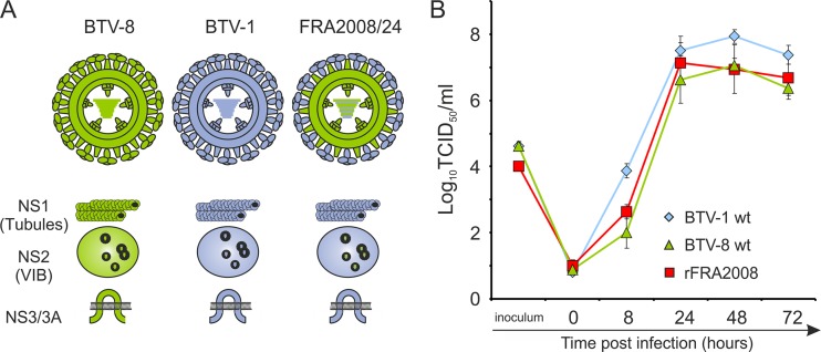
In 2007, while France was already experiencing ongoing outbreaks of BTV-8, a north African strain of BTV-1 entered Spain and spread into France, eventually reaching as far north as Brittany (54). Thus, the cocirculation of these two serotypes raised the possibility of the emergence of reassortants between BTV-1 and BTV-8. Indeed, through full-genome sequencing and pairwise comparisons of field isolates, we identified a reassortant circulating in 2008 (FRA2008/24) that contained multiple segments from BTV-1 in a backbone of BTV-8 (Fig. 1A; Table 2). We compared the sequences of the reassortant virus FRA2008/24 to those of the BTV-1 and BTV-8 field strains circulating in Europe (FRA2007/18 and BTV-8 NET2006/04 [65]) and to the sequences of the BTV-1 and BTV-8 genomic segments used for reverse genetics in our study (Table 2). Interestingly, the two outer capsid proteins, VP2 and VP5, were derived from BTV-1 and BTV-8, respectively. At the amino acid level, Seg-2 of FRA2008/24 is 99.9% identical to Seg-2 of BTV-1 (FRA2007/18) and 95.2% identical to Seg-2 of the BTV-1 South African reference strain (RSArrrr/01). It is notable that all of the segments of FRA2008/24 encoding nonstructural proteins alone (those encoding NS1, NS2, and NS3) were of BTV-1 origin. The nucleotide identities of individual segments to the relevant segment of the parental virus were ≥99.33% (Table 2).
Fig 1.
FRA2008/24 is a reassortant virus between BTV-1 and BTV-8. (A) The wild-type BTV-1 field strain (FRA2007/18) and a reassortant virus (FRA2008/24) were sequenced in order to determine the parental origin of each segment. The cartoons of the BTV particles (VP1 to VP7) and the nonstructural proteins (NS1 to NS3) illustrate each protein as being of either BTV-8 (green) or BTV-1 (blue) origin. With the exception of VP2, FRA2008/24 is composed primarily of BTV-8 proteins, whereas NS1, NS2, and NS3 are all of BTV-1 origin. (B) The combination of BTV-1 and BTV-8 segments of the FRA2008/24 genotype was rescued using reverse genetics and plasmids based upon the European BTV-8 strain and the BTV-1 reference strain. The growth kinetics of the rescued reassortant virus (indicated as rFRA2008 in the panel) was similar to the parental wild-type BTV-1 and BTV-8 in sheep choroid plexus cells.
Table 2.
Nucleotide identities between the BTV-1 and BTV-8 isolates used in this study
| Segment | % Identity with FRA2008/24a |
|||
|---|---|---|---|---|
| BTV-1 (FRA2007/18b) | BTV-1 (referencec) | BTV-8 (NET2006/04d) | BTV-8 (NET2006/04c) | |
| Seg-1 (VP1) | 94.42 | 95.44 | 99.82 | 99.85 |
| Seg-2 (VP2) | 99.9 | 95.24 | 55.49 | 55.49 |
| Seg-3 (VP3) | 95.27 | 95.63 | 99.78 | 99.75 |
| Seg-4 (VP4) | 98.03 | 96.47 | 99.95 | 99.95 |
| Seg-5 (NS1) | 100 | 93.80 | 92.45 | 94.32 |
| Seg-6 (VP5) | 73.61 | 73.00 | 99.88 | 99.76 |
| Seg-7 (VP7) | 96.71 | 96.97 | 99.83 | 99.74 |
| Seg-8 (NS2) | 99.82 | 96.09 | 95.64 | 95.56 |
| Seg-9 (VP6/NS4) | 97.04 | 94.85 | 99.33 | 99.43 |
| Seg-10 (NS3/3A) | 100 | 97.08 | 84.06 | 84.06 |
The European BTV-1 and BTV-8 strains, while clearly distinct, are not the most distantly related BTV strains at the nucleotide level. We performed pairwise comparisons of full-length BTV genomic segments in order to assess the diversity among all of the full-length sequences available in GenBank (Table 3). Table 3 also shows pairwise comparisons between BTV-1 and BTV-8 and selected other BTV strains, such as BTV-2, BTV-4, and BTV-9. The maximum level of divergence (35.09 to 100%) was observed for Seg-2. Seg-6 (which encodes VP5) was also highly variable, with identity levels ranging between 54.47 and 100% (Table 3). The remaining segments all showed levels of identity ranging between 64.02% (Seg-9) and 100%, with a serotype 26 strain from Kuwait being, in general, the most distantly related strain of BTV in most genomic segments (Table 4).
Table 3.
Nucleotide identities between the global BTV sequences present in GenBank or between European BTV strains
| Segment | GenBanka |
BTV-1 (FRA2007/18)b |
BTV-8 (NET2006/04)c |
|||||
|---|---|---|---|---|---|---|---|---|
| Max. | Min. | BTV-2 | BTV-4 | BTV-9 | BTV-2 | BTV-4 | BTV-9 | |
| Seg-1 (VP1) | 100 | 74.52 | 98.63 | 98.76 | 80.15 | 94.57 | 94.62 | 79.82 |
| Seg-2 (VP2) | 100 | 35.09 | 59.46 | 48.36 | 48.36 | 56.68 | 47.99 | 48.32 |
| Seg-3 (VP3) | 100 | 74.68 | 95.09 | 95.09 | 81.85 | 95.20 | 95.20 | 81.10 |
| Seg-4 (VP4) | 100 | 72.70 | 96.11 | 95.91 | 80.26 | 95.86 | 95.76 | 80.21 |
| Seg-5 (NS1) | 100 | 71.17 | 97.69 | 96.67 | 82.75 | 92.39 | 92.51 | 81.69 |
| Seg-6 (VP5) | 100 | 54.47 | 78.41 | 68.74 | 69.41 | 72.57 | 69.47 | 69.96 |
| Seg-7 (VP7) | 100 | 66.44 | 80.88 | 94.38 | 80.10 | 80.28 | 94.64 | 80.71 |
| Seg-8 (NS2) | 100 | 69.07 | 92.98 | 95.82 | 80.18 | 93.33 | 96.27 | 80.09 |
| Seg-9 (VP6/NS4) | 100 | 64.02 | 96.95 | 97.62 | 77.30 | 97.15 | 96.67 | 76.85 |
| Seg-10 (NS3/3A) | 100 | 76.28 | 99.03 | 98.54 | 84.79 | 83.82 | 83.70 | 83.82 |
Table 4.
Minimum and maximum nucleotide identity values between BTV genomic segments deposited in GenBank
| Segment | GenBank accession number (serotype)a | Section | % nucleotide identity at indicated section | ||||
|---|---|---|---|---|---|---|---|
| 1 | GQ506526a (BTV-6) | 1 | 1 | 2 | 3 | 4 | 5 |
| GQ506506 (BTV-6) | 2 | 100 | |||||
| FJ183374b (BTV-8) | 3 | 95.1 | 95.1 | ||||
| JX861488 (BTV-1) | 4 | 94.7 | 94.7 | 94.5 | |||
| GU390658 (BTV-12) | 5 | 79.7 | 79.7 | 79.5 | 80.2 | ||
| JN255156 (BTV-26) | 6 | 75.4 | 75.4 | 75.5 | 75.7 | 74.5 | |
| 2 | JN255923 (BTV-2) | 1 | 1 | 2 | 3 | 4 | 5 |
| JN255933 (BTV-2) | 2 | 100 | |||||
| JX861489 (BTV-1) | 3 | 59.9 | 59.9 | ||||
| AJ585134 (BTV-13) | 4 | 51.2 | 51.2 | 51.8 | |||
| FJ183375 (BTV-8) | 5 | 56.4 | 56.4 | 55.2 | 51.0 | ||
| AJ585136 (BTV-15) | 6 | 37.9 | 37.9 | 38.7 | 36.0 | 38.6 | |
| 3 | JX861490 (BTV-1) | 1 | 1 | 2 | 3 | 4 | 5 |
| FJ183376 (BTV-8) | 2 | 95.4 | |||||
| JN255924 (BTV-2) | 3 | 95.1 | 95.3 | ||||
| JN255934 (BTV-2) | 4 | 95.1 | 95.3 | 100 | |||
| AY322428 (BTV-1) | 5 | 81.2 | 80.3 | 80.5 | 80.5 | ||
| HM590643 (BTV-26) | 6 | 75.6 | 76.1 | 75.9 | 75.9 | 74.7 | |
| 4 | JN255925 (BTV-2) | 1 | 1 | 2 | 3 | 4 | 5 |
| JN255935 (BTV-2) | 2 | 100 | |||||
| L08637 (BTV-2) | 3 | 98.2 | 98.2 | ||||
| FJ183377 (BTV-8) | 4 | 95.2 | 95.2 | 94.0 | |||
| JX861491 (BTV-1) | 5 | 95.6 | 95.6 | 94.4 | 98.1 | ||
| JN255157 (BTV-26) | 6 | 73.1 | 73.1 | 72.7 | 73.7 | 73.6 | |
| 5 | GQ506476 (BTV-6) | 1 | 1 | 2 | 3 | 4 | 5 |
| GQ506510 (BTV-6) | 2 | 100 | |||||
| GQ506482 (BTV-6) | 3 | 99.2 | 99.2 | ||||
| FJ183378 (BTV-8) | 4 | 92.7 | 92.7 | 92.0 | |||
| JX861492 (BTV-1) | 5 | 92.1 | 92.1 | 91.3 | 92.5 | ||
| JN255158 (BTV-26) | 6 | 72.6 | 72.6 | 71.8 | 72.3 | 72.8 | |
| 6 | JX861493a (BTV-1) | 1 | 1 | 2 | 3 | 4 | 5 |
| AJ631215 (BTV-9) | 2 | 78.6 | |||||
| AJ586673 (BTV-2) | 3 | 78.2 | 77.1 | ||||
| AJ586674 (BTV-2) | 4 | 78.2 | 77.1 | 100 | |||
| FJ183379 (BTV-8) | 5 | 73.5 | 75.0 | 72.5 | 72.5 | ||
| AJ586716 (BTV-15) | 6 | 58.7 | 57.1 | 56.8 | 56.8 | 59.2 | |
| 7 | FJ183380 (BTV-8) | 1 | 1 | 2 | 3 | 4 | 5 |
| JX861494 (BTV-1) | 2 | 96.9 | |||||
| AY776331 (BTV-1) | 3 | 97.0 | 96.6 | ||||
| FJ969725 (BTV-1) | 4 | 97.0 | 96.6 | 100 | |||
| AY855284 (BTV-17) | 5 | 87.0 | 86.7 | 87.4 | 87.4 | ||
| L11724 (BTV-15) | 6 | 70.9 | 70.7 | 70.8 | 70.8 | 68.1 | |
| 8 | JN255939 (BTV-2) | 1 | 1 | 2 | 3 | 4 | 5 |
| JN255929 (BTV-2) | 2 | 100 | |||||
| JX861495 (BTV-1) | 3 | 97.2 | 97.1 | ||||
| FJ183381 (BTV-8) | 4 | 97.0 | 97.0 | 97.0 | |||
| L08676 (BTV-17) | 5 | 89.3 | 89.3 | 88.6 | 88.7 | ||
| JN255160 (BTV-26) | 6 | 69.4 | 69.4 | 68.9 | 69.6 | 64.9 | |
| 9 | JN255870 (BTV-2) | 1 | 1 | 2 | 3 | 4 | 5 |
| JN255880 (BTV-2) | 2 | 100 | |||||
| FJ183382 (BTV-8) | 3 | 97.2 | 97.1 | ||||
| JX861496 (BTV-1) | 4 | 97.0 | 97.0 | 97.0 | |||
| D10905 (BTV-1) | 5 | 89.3 | 89.3 | 88.6 | 88.7 | ||
| JN255161 (BTV-26) | 6 | 69.4 | 69.4 | 68.9 | 69.6 | 64.9 | |
| 10 | AF044383 (BTV-11) | 1 | 1 | 2 | 3 | 4 | 5 |
| AF044382 (BTV-10) | 2 | 100 | |||||
| FJ183383 (BTV-8) | 3 | 92.8 | 92.8 | ||||
| JX861487 (BTV-1) | 4 | 83.3 | 83.3 | 84.1 | |||
| FJ713333 (BTV-22) | 5 | 77.3 | 77.3 | 78.1 | 92.1 | ||
| JN255162 (BTV-26) | 6 | 79.7 | 79.7 | 80.2 | 80.4 | 73.7 | |
GenBank accession number (serotype).
BTV-1 and BTV-8 European strains are in bold type.
We next sought to characterize possible phenotypic changes which may arise from a novel rearrangement of segments in FRA2008/24. Through reverse genetics, we rescued rFRA2008, a reassortant virus based upon the BTV-1 reference strain and BTV-8 Netherlands strain, which has the same genomic segment composition as the field reassortant FRA2008/24. When assessed for virus growth in CPT-Tert cells, BTV-1 was found to reach a higher titer at 8 h postinfection than both BTV-8 and rFRA2008 (Fig. 1B). However, by 24 h and at subsequent time points, all three viruses displayed comparable titers, suggesting that the overall growth kinetics of the three viruses are largely similar.
Coinfection of sheep cells with BTV-1 and BTV-8.
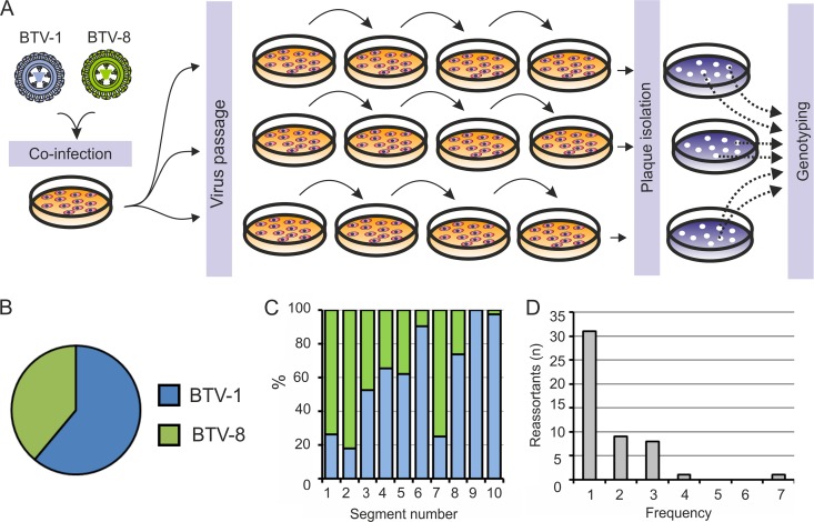
Reassortment studies using a variety of viruses have previously suggested that reassortment is not necessarily a random event, and different compatibilities exist between segments of distinct parental viruses (70–72). In light of this, and the discovery in the field of FRA2008/24, we performed coinfection experiments using BTV-1 and BTV-8 in order to determine, by forward genetics, the type of reassortants generated by these two viruses in vitro. Using real-time RT-PCR, we characterized 140 plaques that were derived from coinfection experiments and had been passaged four times in sheep cells (Fig. 2A). These coinfections were carried out in two independent experiments, and in each experiment, virus mixtures were subsequently passaged in triplicate (Fig. 2A). While every plaque contained evidence of the BTV genome, for 22 plaques, one or more segments could not be amplified and were therefore discarded from our analysis. In addition, 34 plaques contained one or more genome segments of both BTV-1 and BTV-8, suggesting that these plaques were formed by more than one virus; therefore, these were also eliminated from our analysis. In contrast, 84 plaques could be completely characterized and were considered for further analysis. Interestingly, 100% of the characterized plaques were reassortants. The characterized plaques could be divided into 50 unique combinations of segments (Table 5). The majority (n = 31) of the unique combinations occurred just once. However, certain segment combinations were found at frequencies greater than 1, including the genotype 1 combination, which occurred 7 times within our data set (Table 5; Fig. 2D). Interestingly, some segment combinations were found in two separate coinfection experiments; for example, genotype 1 was found in 3 of the 6 virus mixtures that were passaged independently.
Fig 2.
Forward genetic analysis of BTV-1–BTV-8 coinfection. (A) Schematic diagram of the procedure followed. CPT-Tert cells were coinfected at a 1:1 ratio of BTV-1 and BTV-8 (MOIs of 1). After 24 h of incubation, the supernatant was harvested and serially passaged four times in three separate series of CPT-Tert cells. The reassortant viruses were isolated by plaque assays, and the resulting plaques (n = 140) were genotyped by RT-PCR. This experiment was performed in duplicate. (B) Pie chart representing the proportions of BTV-1 and BTV-8 genomic segments identified in the reassortants characterized in these experiments. Sixty-one percent of the segments identified in 84 reassortants originated from BTV-1 and the remaining 39% from BTV-8. (C) Chart representing the frequencies of the genotypes identified in this study. Among the isolated reassortants, most genotypes (31/50) were present only once. In contrast, the numbers of genotypes occurring at a frequency of 2, 3, or 4 represented just 9/50, 8/50, and 1/50 of the genotypes, respectively. A particular genotype was represented in 7 isolated reassortants (see Table 3). (D) Chart representing the origin of each segment within the total number of reassortants identified in this study. Seg-1, Seg-2, and Seg-7 in more than 75% of the isolated reassortants originated from BTV-8. In the majority of reassortants, the remaining genomic segments originated from BTV-1.
Table 5.
Genotypes of reassortants isolated after coinfection of CPT-Tert cells
| Genotype | Sequencea | Frequency |
|---|---|---|
| 1 | 8811118111 | 7 |
| 2 | 8811818111 | 4 |
| 3 | 1818818111 | 3 |
| 4 | 1881118111 | 3 |
| 5 | 8811111811 | 3 |
| 6 | 8818818111 | 3 |
| 7 | 8881111111 | 3 |
| 8 | 8881118111 | 3 |
| 9 | 8881818111 | 3 |
| 10 | 8888118111 | 3 |
| 11 | 1818118111 | 2 |
| 12 | 1881118811 | 2 |
| 13 | 8111818111 | 2 |
| 14 | 8818111111 | 2 |
| 15 | 8818118111 | 2 |
| 16 | 8881111811 | 2 |
| 17 | 8881188111 | 2 |
| 18 | 8881818811 | 2 |
| 19 | 8888111111 | 2 |
| 20 | 1111888811 | 1 |
| 21 | 1181818111 | 1 |
| 22 | 1188818111 | 1 |
| 23 | 1811118118 | 1 |
| 24 | 1811118811 | 1 |
| 25 | 1811818111 | 1 |
| 26 | 1881811111 | 1 |
| 27 | 1881811811 | 1 |
| 28 | 1881818111 | 1 |
| 29 | 1888111111 | 1 |
| 30 | 1888118111 | 1 |
| 31 | 1888118811 | 1 |
| 32 | 8111118111 | 1 |
| 33 | 8111188111 | 1 |
| 34 | 8111811111 | 1 |
| 35 | 8111811811 | 1 |
| 36 | 8111888811 | 1 |
| 37 | 8118111111 | 1 |
| 38 | 8118118811 | 1 |
| 39 | 8181111811 | 1 |
| 40 | 8181188111 | 1 |
| 41 | 8181818111 | 1 |
| 42 | 8811118818 | 1 |
| 43 | 8811888111 | 1 |
| 44 | 8818118811 | 1 |
| 45 | 8818811111 | 1 |
| 46 | 8818818811 | 1 |
| 47 | 8881118811 | 1 |
| 48 | 8888118811 | 1 |
| 49 | 8888188111 | 1 |
| 50 | 8888811111 | 1 |
Numbers refer to the strain of BTV (1 indicates BTV-1; 8, BTV-8) from which each segment originated. The sequence of segments is provided in the order Seg-1 to Seg-10.
BTV-1 contributed 60% of the total segments of the reassortants that were analyzed (Fig. 2B). The majority of the reassortants had Seg-1, Seg-2, and Seg-7 that originated from BTV-8 (Fig. 2C). The remaining segments were derived in the majority of cases from BTV-1, although no clear bias was observed for Seg-3, Seg-4, and Seg-5 (Fig. 2C). Of particular note, almost all of the reassortants isolated had Seg-6, Seg-9, and Seg-10 that originated from BTV-1 (Fig. 2C). Pairwise comparisons of each segment combination by chi-squared analysis (with Bonferroni's correction) revealed no significant association between segments in the reassortant progeny mix, with the exception of an association between Seg-2 (VP2) and Seg-6 (VP5) (P = 0.04 prior to Bonferroni's correction, assuming a 5% significance level). A z test analysis revealed that, for Seg-6 of BTV-1, a statistically significant difference existed between the proportion of Seg-2s that are of BTV-1 origin (n = 11) and the proportion of Seg-2s that are of BTV-8 origin (n = 65) (BTV-8 − BTV-1 = 0.71, 95% confidence interval = 0.49 to 0.95, P < 0.001). On the other hand, for Seg-6 of BTV-8, there were equivalent proportions of Seg-2 of BTV-1 (n = 4) and BTV-8 (n = 4) origins.
Monoreassortants between BTV-1 and BTV-8 can be formed with any combination of genomic segments.
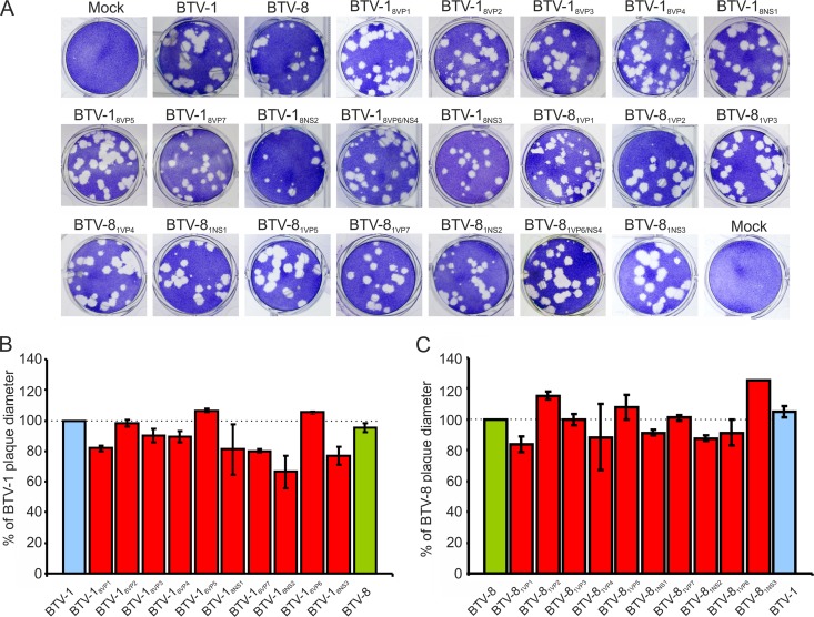
Next, we used reverse genetics in order to determine whether it is possible to reassort any individual segment of either BTV-1 or BTV-8 into an alternative backbone. Interestingly, we successfully rescued each combination of monoreassortants with a single segment of BTV-8 in the backbone of BTV-1, and vice versa. We then assessed the replication characteristics of each monoreassortant in vitro using both plaque assays and in vitro growth assays in CPT-Tert cells. The average plaque diameters were determined from two independent rounds of plaque assays by measuring 10 to 15 individual plaques in each experiment. In the majority of cases, the plaque sizes of the various monoreassortants were relatively uniform and comparable to those of the parental backbones (Fig. 3A to C). Some reassortants consistently yielded smaller or larger plaques relative to the parental backbones that, in some cases, were statistically significant (ANOVA, P < 0.05). The sizes of the plaques induced by the reassortant viruses were never greater or smaller than 30% of the sizes of the plaques induced by the parental viruses. The substitution of the Seg-8 of BTV-1 (which encodes NS2) with that of BTV-8 (BTV-18NS2) resulted in plaques that were consistently smaller than those of wild-type BTV-1 (P < 0.005) (Fig. 3B). The sizes of the plaques of the opposite reassortant, i.e., BTV-81NS2, were also reduced, although to a lesser extent (P = 0.11) (Fig. 3C). Other monoreassortants that consistently yielded plaques with smaller diameters than those of the parental viruses were those with reassorted segments encoding VP1, VP4, and NS1. For certain segments, the backbone into which the segment was being reassorted impacted the resulting plaque diameter. For example, while the substitution of Seg-7 of BTV-1 for that of BTV-8 within the BTV-1 backbone (BTV-18VP7) was consistently found to be deleterious (P = 0.002), both wild-type (wt) BTV-8 and BTV-81VP7 produced plaques of comparable diameters (P = 0.43) (Fig. 3C).
Fig 3.
Analysis of plaque diameter for BTV-1–BTV-8 monoreassortants. Monoreassortants containing 9 segments from either BTV-1 or BTV-8 and a single segment derived from the heterologous BTV strain were rescued as indicated in Materials and Methods. (A) Representative pictures of the plaques generated by each of the monoreassortant viruses. (B and C) Graphs illustrating the relative sizes of the plaques induced in CPT-Tert cells by the monoreassortants indicated. The average diameters (± standard deviation) of the plaques are expressed as percentages of the plaque size induced by the wild-type BTV-1 (panel B) or BTV-8 (panel C).
The most notable alterations in plaque sizes were achieved when Seg-10 of BTV-1 or BTV-8 was inserted into the alternative backbone (BTV-81NS3 and BTV-18NS3, respectively). BTV-18NS3 generated plaques with a 23% decrease in diameter relative to those of wild-type BTV-1 (Fig. 3A). Conversely, BTV-81NS3 produced plaques that were 25% greater in diameter than those of wild-type BTV-8 (Fig. 3B).
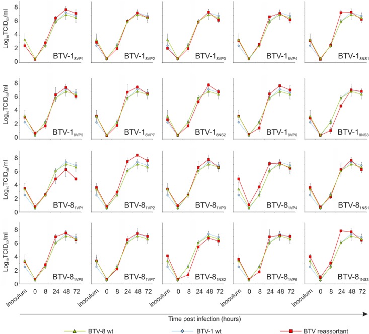
We then performed growth curve analyses in order to assess the growth kinetics of each reassortant. Overall, the majority of the reassortants demonstrated growth kinetics similar to that of the corresponding wild-type virus that contributed the backbone. At 8 h postinfection, some of the reassortants had a lower titer than those of wild-type BTV-1 and BTV-8. In particular, BTV-18VP6 and BTV-18NS3 produced lower titers than those of the wild-type viruses (Fig. 4). The Seg-8 (NS2) monoreassortants (BTV-18NS2 and BTV-81NS2) both had reduced plaque diameters (Fig. 3B and C) and lower titers (Fig. 4). Only two reassortant viruses (BTV-18NS3 and BTV-81VP1) gave substantially lower titers than those of the wild-type BTV-1 or BTV-8 at 24 h postinfection (Fig. 4). However, by 24 h postinfection, BTV-18NS3 had a titer comparable to those of the parental viruses (Fig. 4). Of particular note, BTV-18NS3 also showed a delayed growth curve in comparison to wild-type BTV-1. In contrast, the corresponding reassortant BTV-81NS3 also replicated efficiently at early time points. Very few reassortants showed a substantially higher titer than the wild-type strain of BTV-1 or BTV-8 at 24 h postinfection.
Fig 4.
In vitro growth kinetics of each monoreassortant identified in this study. Viral growth curves (MOIs of 0.05) were created as described in Materials and Methods. The majority of reassortants showed growth kinetics similar to those of the wild-type BTV-1 and BTV-8 parental viruses. Differences between the wild-type and monoreassortant viruses were observed primarily at 8 h postinfection. Most monoreassortants with a substituted Seg-8, Seg-9, or Seg-10 showed reduced titers at 8 h postinfection.
Pathogenicity of BTV-1 and BTV-8 monoreassortants in IFNAR−/− mice.
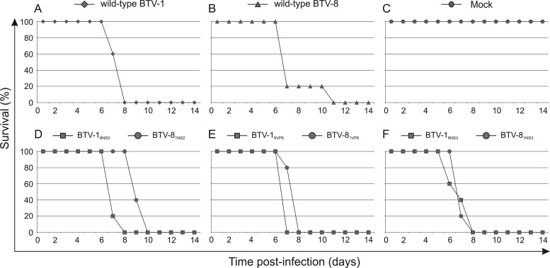
The data shown above indicate that some monoreassortants have delayed early growth and/or produce smaller (or larger) plaques in vitro than those of the parental viruses. In light of these data, we used a selection of reassortants with altered phenotypes (BTV-18NS2, BTV-18NS3, BTV-81NS3) or with characteristics similar to those of the parental viruses (BTV-18VP6, BTV-81NS2, BTV-81VP6) in order to determine whether some of these differences could be correlated with differences in pathogenicity in vivo in an experimental model of BTV infection. In line with our previously published data (26), both wild-type BTV-1 and BTV-8 killed 100% of the interferon receptor-deficient (IFNAR−/−) mice by 11 days postinoculation, whereas the mock-infected mice all survived (Fig. 5A to C). Interestingly, monoreassortants that showed a small plaque phenotype or slower growth kinetics, such as those containing heterologous Seg-8 (BTV-18NS2 and BTV-81NS2), Seg-9 (BTV-18VP6 and BTV-81VP6), or Seg-10 (BTV-18NS3 and BTV-81NS3), killed mice with efficiency similar to that of the parental viruses (Fig. 5D to F). Thus, the variations observed in early growth kinetics of the monoreassortants in vitro are not correlated with pathogenicity in vivo, at least in this experimental model of bluetongue disease.
Fig 5.
Experimental infection of IFNAR−/− mice with selected monoreassortants. Survival plots of adult 129/Sv IFNAR−/− mice (5 per group) inoculated intraperitoneally with 300 PFU of BTV-1–BTV-8 monoreassortants containing heterologous Seg-8, Seg-9, or Seg-10. Wild-type BTV-1 and BTV-8 strains were used as positive controls, while a mock-infected group included mice inoculated with supernatant of noninfected cells. All monoreassortants killed 100% of infected mice by 10 days postinfection.
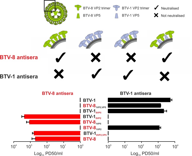
VP2 alone determines the BTV serotype.
Several studies have indicated that VP2 is inextricably linked to BTV serotype (62, 73, 74). However, it has also been suggested that VP5 can contribute to defining the serotype of each particular strain (75). To investigate the relative contributions of VP2 and VP5 to serotype determination, we performed serum neutralization assays using reassortants with the backbone of either BTV-1 or BTV-8 and the heterologous VP2, either alone (BTV-18VP2 and BTV-81VP2) or with the corresponding VP5 (BTV-18VP2,VP5 and BTV-81VP2,VP5). As expected, BTV-8 antiserum completely neutralized wild-type BTV-8 but showed no neutralizing capacity against BTV-1. On the other hand, BTV-1 antiserum neutralized wt BTV-1 but failed to block the infectivity of BTV-8 (Fig. 6). The BTV-1 reassortant with both VP2 and VP5 (BTV-18VP2,VP5) or with only the VP2 of BTV-8 (BTV-18VP2) was neutralized by the BTV-8 antiserum, while BTV-18VP5 was not neutralized by the BTV-8 antiserum (Fig. 6). Conversely, BTV-8 reassortants with either VP2 and VP5 (BTV-81VP2,VP5) or only the VP2 of BTV-1 (BTV-81VP2) were neutralized by BTV-1 antiserum, whereas no neutralization of these reassortants was observed when BTV-8 antiserum was used. These data clearly confirm that VP2 alone determines the BTV serotype.
Fig 6.
The outer capsid protein VP2 determines the serotype of bluetongue virus. Serum neutralization assays were carried out as described in Materials and Methods using BTV-8 (left) or BTV-1 (right) monospecific antiserum and wild-type BTV-1, BTV-8, or reassortant viruses. The reassortants included BTV-1 viruses containing both VP2 and VP5 derived from BTV-8 (BTV-18VP2,VP5) or containing only the heterologous VP2 (BTV-18VP2) or VP5 (BTV-18VP5). In addition, BTV-8 reassortants with only the VP2 protein (BTV-81VP2), the VP5 protein (BTV-81VP5), or both proteins (BTV-81VP2,VP5) derived from BTV-1 were also used. The data are expressed as the PD50 for each serum sample, defined as the serum dilution that inhibits BTV infection in 50% of the infected cells. The data shown clearly indicate that, in the case of BTV-1–BTV-8, VP2 (and not VP5) is the sole protein that determines the BTV serotype.
DISCUSSION
Coinfection of a cell with two different strains of a segmented virus can give rise to reassortants with phenotypic characteristics that might differ from those of their parental strains. The cocirculation of BTV-1 and the highly pathogenic BTV-8 in northern Europe has offered ample opportunities for reassortment between these two serotypes in the field. Indeed, we isolated a BTV reassortant (FRA2008/24) from an outbreak in France possessing Seg-2, Seg-5, Seg-8, and Seg-10 derived from BTV-1 and the remaining genomic segments from BTV-8. This finding prompted us to systematically investigate the process of reassortment in BTV using both forward and reverse genetics approaches. By coinfecting cells with two different virus strains, it is possible to propagate mixed virus populations in culture, including viable and “fit” reassortants that might compete with the parental wild-type viruses (forward genetics). While this may reflect the biological and random (or otherwise) nature of reassortment, it is extremely difficult to derive a specific constellation of segments, therefore restricting a detailed analysis of whether specific barriers exist within the reassortment process. However, reverse genetics allows the precise and directed generation of reassortant viruses that can be further assessed phenotypically.
In this study, we established that reassortment in BTV is an extremely flexible process. Viable monoreassortants can be rescued with any individual segment in the alternative parental backbone when using BTV-1 and BTV-8. We noticed some relatively minor differences in the growth patterns and plaque sizes in vitro, suggesting various degrees of compatibility between the segments of these two strains. It remains possible that it is more difficult to achieve reassortment between BTV strains other than BTV-1 and BTV-8.
We assessed the plaque diameters and multistep growth curve patterns of the rescued reassortants in a comparison of the phenotypic variations resulting from reassortment. The diameters of the plaques induced by various reassortants were found to vary for specific segments and to be impacted to differing extents according to the backbone of the reassortant. This suggests that differences in the interactions between different combinations of gene products may influence the relative efficiency of the replication and transmission mechanisms in which they are involved. The major differences between reassortants in the replication cycle in vitro were observed at early time points postinfection. However, a selection of reassortants with relatively slow early growth in vitro were as pathogenic as wild-type BTV in an experimental model of disease in vivo.
Our results build upon previous studies on reassortment within the Reoviridae family (57, 62, 75–77). Studies on rotaviruses have revealed various degrees of compatibility between different virus strains, with evidence of a nonrandom segregation of genome segments (57, 78, 79). Studies using BTV-10 and BTV-17 have also shown that not all segments reassort equally well, at least when using BTV-10 and BTV-17 (57). However, these studies have used only a forward genetics approach. Using reverse genetics, we have shown that there is great plasticity in the ability of these viruses to reassort, at least when using BTV-1 and BTV-8. It is possible that BTV strains with greater levels of divergence, compared to those of the BTV-1 and BTV-8 strains used in this study, might show a less flexible process of reassortment.
The forward genetics approaches reported previously for the study of reassortment have largely relied upon the migration of each segment during electrophoresis; this necessarily restricts the strains that can be used for study, as each segment must be distinguishable using this technique (57). Another approach to the characterization of segments is the use of radioactively labeled probes (56, 80–82). However, while simple hybridization allows the distinction of sufficiently divergent sequences, cross-reactions can occur, resulting in difficulty in interpreting the results based upon signal levels. Indeed, in our forward genetics screen, we observed evidence of multiple cases of plaques containing one or more segments arising from both parental viruses, something which may be unclear when a hybridization approach is used. In contrast, the use of RT-PCR has the advantage of increasing throughput, giving a clear binary output and furthermore allowing analysis of any virus strain. Complete genome sequencing offers the highest level of resolution, and it is increasingly high throughout. As such, for the analysis of field isolates, full genomic sequencing is certainly the method of choice for reassortant analysis.
Given the previous observation that the BTV outer core proteins contribute to serotype (75, 76), as well as the possibility of structural constraints, it may be hypothesized that VP2 and VP5 of a particular serotype preferentially reassort together. Our study suggests that this is not always the case. First of all, in the field reassortant isolated in this study, VP2 derives from BTV-1, while VP5 derives from BTV-8. In addition, our coinfection assays showed that 78% of the reassortants propagated in culture had VP2 derived from BTV-8, while VP5 derived from BTV-1. Only 17% of the reassortants had the homologous VP2–VP5 (12% from BTV-1 and 4.7% from BTV-8). Thus, at least for BTV-1 and BTV-8, there is no fundamental barrier for VP2 and VP5 to segregate with the homologous counterpart in coinfected cells.
Importantly, in this study, using VP2 monoreassortants generated by reverse genetics, we confirmed that the serotype determination of BTV-1 versus BTV-8 is linked solely to VP2. There are contrasting data in the literature as to whether only VP2 or both VP2 and VP5 determine the BTV serotype (62, 73–76). The data presented in our study show clearly that VP5 had no influence on BTV serotype, at least in the cases of BTV-1 and BTV-8.
From our coinfection assays, it was striking that none of the 140 plaques analyzed had the genotype of the parental BTV-1 or BTV-8 in its entirety. At most, the reassortants had 7 of the 10 segments from one of the two serotypes. Thus, there are no structural constraints in BTV that favor packaging of the 10 genomic segments of an individual strain.
Besides VP2 and VP5, the proteins forming the polymerase complex were also not shown to reassort preferentially within the homologous serotype. Only 16% of reassortants contained the VP1, VP4, and VP6 proteins of BTV-1, while none of them contained all the enzymatic proteins deriving from BTV-8.
During our plaque assay experiments, we also observed that a heterologous NS2 was detrimental to plaque formation. BTV-18NS2 was found to be affected to a greater extent, producing plaques that were smaller than those of the parental BTV-1 and the reassortant BTV-81NS2. Interestingly, Seg-8 of BTV-1 was represented at a higher frequency in the analyzed reassortants during our forward genetics screen than in the reverse genetics screen. More studies will be needed in order to clarify how the NS2 may affect virus-induced plaque size. It is likely that this is not due to structural constraints within the virus particle, because NS2 is a nonstructural protein.
As more full-genome sequences of BTV are becoming available, it is becoming clear that the occurrence of Orbivirus reassortment in the field is extensive, with evidence of reassortment in both mammalian hosts and Culicoides (5, 6, 20, 49, 50, 52, 53, 80, 83–85). All of our experiments were performed using mammalian cells in vitro. Obviously, in contrast to the controlled experimental procedures in vitro (e.g., equal MOIs in cells largely devoid of antiviral activity), the process of reassortment in the field (in either the insect or mammalian vector) is likely to be governed by both stochastic and deterministic events, such as host immunity, vector specificity, the existing distribution of parental virus strains, and vaccination campaigns with both live and multivalent vaccines. However, our study has shown that reassortment in BTV is a very plastic event, and there is no inherent or structural barrier for any genomic segment to reassort. In areas where several serotypes of BTV cocirculate, reassortment could be the rule rather than the exception (20, 49, 83).
BTV has historically been classified from the serological point of view. Serology was, and remains, a key parameter for differentiating BTV strains. Molecular methods for “typing” a virus have concentrated on reconstructing the serological relationships and consequently focused upon the genomic segments that express the outer capsid proteins (46, 86). Sequence analyses have shown that the majority of the BTV proteins group according to their geographic origins (topotype). If considered within the serotype, this is also true for both of the outer capsid proteins VP2 and VP5. Studies of virus topotypes have shown that the majority of BTV genome segments and/or proteins can be divided into two major topotypes, “eastern” and “western,” with evidence of further subgroups that also depend on the geographic origins of the isolates. However, recent studies of BTV-25 and BTV-26 have suggested that they may represent two additional major groups, and the VP7 and NS3 genes do not appear to fit this pattern, showing multiple eastern and western groups (87). Although these groupings provide a useful guide, the introduction of exotic virus strains, the reassortment of genome segments, and the selective pressures on the progeny viruses in the field have, in some cases, altered the distribution of specific gene variants and blurred the topotype boundaries. It appears likely that, although reassortment can occur between any BTV genome segment, subsequent environmental pressures and negative selection will have important influences on the survival of individual progeny strains and, therefore, on individual genes and gene combinations (88–90).
Using reverse genetics, we have confirmed in this study that VP2 is indeed the key determinant of neutralizing antibody specificity. Obviously, understanding to which serotype a BTV strain belongs is critical in designing the appropriate control strategies. However, simply defining a BTV strain on the basis of its serotype (and/or Seg-2 sequence) excludes important information on the remaining nine segments. As more detailed analyses of the properties and impacts of individual proteins (for example, associations with pathogenicity [69] or transmission) become available, using an alternative classification system will become increasingly important. Similar issues have been encountered with rotaviruses, leading to the proposal for creating standard classification systems (91, 92). An analogous approach for orbiviruses, taking into account virus serotypes and topotypes, as well as different groupings of the VP7 and NS3 genes, may provide a way forward to construct a more comprehensive nomenclature for individual BTV strains.
ACKNOWLEDGMENTS
This work was funded by the BBSRC via the EMIDA program and the Wellcome Trust.
We are grateful to Karl Conzelmann and David Griffiths for reagents and to Sema Nickbakhsh for statistical analysis.
Footnotes
Published ahead of print 24 October 2012
REFERENCES
- 1. Liu J, Liu DY, Chen W, Li JL, Luo F, Li Q, Ling JX, Liu YY, Xiong HR, Ding XH, Hou W, Zhang Y, Li SY, Wang J, Yang ZQ. 2012. Genetic analysis of hantaviruses and their rodent hosts in central-south China. Virus Res. 163:439–447 [DOI] [PubMed] [Google Scholar]
- 2. Sall AA, Zanotto PM, Sene OK, Zeller HG, Digoutte JP, Thiongane Y, Bouloy M. 1999. Genetic reassortment of Rift Valley fever virus in nature. J. Virol. 73:8196–8200 [DOI] [PMC free article] [PubMed] [Google Scholar]
- 3. Liu Q, Ma J, Liu H, Qi W, Anderson J, Henry SC, Hesse RA, Richt JA, Ma W. 2012. Emergence of novel reassortant H3N2 swine influenza viruses with the 2009 pandemic H1N1 genes in the United States. Arch. Virol. 157:555–562 [DOI] [PubMed] [Google Scholar]
- 4. Watanabe M, Nakagomi T, Koshimura Y, Nakagomi O. 2001. Direct evidence for genome segment reassortment between concurrently-circulating human rotavirus strains. Arch. Virol. 146:557–570 [DOI] [PubMed] [Google Scholar]
- 5. Batten CA, Maan S, Shaw AE, Maan NS, Mertens PP. 2008. A European field strain of bluetongue virus derived from two parental vaccine strains by genome segment reassortment. Virus Res. 137:56–63 [DOI] [PubMed] [Google Scholar]
- 6. Allison AB, Holmes EC, Potgieter AC, Wright IM, Sailleau C, Breard E, Ruder MG, Stallknecht DE. 2012. Segmental configuration and putative origin of the reassortant orbivirus, epizootic hemorrhagic disease virus serotype 6, strain Indiana. Virology 424:67–75 [DOI] [PubMed] [Google Scholar]
- 7. Scholtissek C, Rohde W, Von Hoyningen V, Rott R. 1978. On the origin of the human influenza virus subtypes H2N2 and H3N2. Virology 87:13–20664248 [Google Scholar]
- 8. Nelson MI, Viboud C, Simonsen L, Bennett RT, Griesemer SB, St. George K, Taylor J, Spiro DJ, Sengamalay NA, Ghedin E, Taubenberger JK, Holmes EC. 2008. Multiple reassortment events in the evolutionary history of H1N1 influenza A virus since 1918. PLoS Pathog. 4:e1000012 doi:10.1371/journal.ppat.1000012 [DOI] [PMC free article] [PubMed] [Google Scholar]
- 9. Garten RJ, Davis CT, Russell CA, Shu B, Lindstrom S, Balish A, Sessions WM, Xu X, Skepner E, Deyde V, Okomo-Adhiambo M, Gubareva L, Barnes J, Smith CB, Emery SL, Hillman MJ, Rivailler P, Smagala J, de Graaf M, Burke DF, Fouchier RA, Pappas C, Alpuche-Aranda CM, Lopez-Gatell H, Olivera H, Lopez I, Myers CA, Faix D, Blair PJ, Yu C, Keene KM, Dotson PD, Jr, Boxrud D, Sambol AR, Abid SH, ST. George K, Bannerman T, Moore AL, Stringer DJ, Blevins P, Demmler-Harrison GJ, Ginsberg M, Kriner P, Waterman S, Smole S, Guevara HF, Belongia EA, Clark PA, Beatrice ST, Donis R, Katz J, Finelli L, Bridges CB, Shaw M, Jernigan DB, Uyeki TM, Smith DJ, Klimov AI, Cox NJ. 2009. Antigenic and genetic characteristics of swine-origin 2009 A(H1N1) influenza viruses circulating in humans. Science 325:197–201 [DOI] [PMC free article] [PubMed] [Google Scholar]
- 10. Shelton H, Smith M, Hartgroves L, Stilwell P, Roberts K, Johnson B, Barclay W. 2012. An influenza reassortant with polymerase of pH1N1 and NS gene of H3N2 influenza A virus is attenuated in vivo. J. Gen. Virol. 93:998–1006 [DOI] [PMC free article] [PubMed] [Google Scholar]
- 11. Song MS, Pascua PN, Lee JH, Baek YH, Park KJ, Kwon HI, Park SJ, Kim CJ, Kim H, Webby RJ, Webster RG, Choi YK. 2011. Virulence and genetic compatibility of polymerase reassortant viruses derived from the pandemic (H1N1) 2009 influenza virus and circulating influenza A viruses. J. Virol. 85:6275–6286 [DOI] [PMC free article] [PubMed] [Google Scholar]
- 12. Hai R, Garcia-Sastre A, Swayne DE, Palese P. 2011. A reassortment-incompetent live attenuated influenza virus vaccine for protection against pandemic virus strains. J. Virol. 85:6832–6843 [DOI] [PMC free article] [PubMed] [Google Scholar]
- 13. Brennan B, Welch SR, McLees A, Elliott RM. 2011. Creation of a recombinant Rift Valley fever virus with a two-segmented genome. J. Virol. 85:10310–10318 [DOI] [PMC free article] [PubMed] [Google Scholar]
- 14. Bouvier NM, Rahmat S, Pica N. 2012. Enhanced mammalian transmissibility of seasonal influenza A/H1N1 viruses encoding an oseltamivir-resistant neuraminidase. J. Virol. 86:7268–7269 [DOI] [PMC free article] [PubMed] [Google Scholar]
- 15. Lakdawala SS, Lamirande EW, Suguitan AL, Jr, Wang W, Santos CP, Vogel L, Matsuoka Y, Lindsley WG, Jin H, Subbarao K. 2011. Eurasian-origin gene segments contribute to the transmissibility, aerosol release, and morphology of the 2009 pandemic H1N1 influenza virus. PLoS Pathog. 7:e1002443 doi:10.1371/journal.ppat.1002443 [DOI] [PMC free article] [PubMed] [Google Scholar]
- 16. Keroack M, Fields BN. 1986. Viral shedding and transmission between hosts determined by reovirus L2 gene. Science 232:1635–1638 [DOI] [PubMed] [Google Scholar]
- 17. Nakagomi O, Ohshima A, Aboudy Y, Shif I, Mochizuki M, Nakagomi T, Gotlieb-Stematsky T. 1990. Molecular identification by RNA-RNA hybridization of a human rotavirus that is closely related to rotaviruses of feline and canine origin. J. Clin. Microbiol. 28:1198–1203 [DOI] [PMC free article] [PubMed] [Google Scholar]
- 18. Mukherjee A, Dutta D, Ghosh S, Bagchi P, Chattopadhyay S, Nagashima S, Kobayashi N, Dutta P, Krishnan T, Naik T, Chawla-Sarkar M. 2009. Full genomic analysis of a human group A rotavirus G9P[6] strain from Eastern India provides evidence for porcine-to-human interspecies transmission. Arch. Virol. 154:733–746 [DOI] [PubMed] [Google Scholar]
- 19. Browning GF, Snodgrass DR, Nakagomi O, Kaga A, Sarasini A, Gerna G. 1992. Human and bovine serotype G8 rotaviruses may be derived by reassortment. Arch. Virol. 125:121–128 [DOI] [PubMed] [Google Scholar]
- 20. de Mattos CC, de Mattos CA, MacLachlan NJ, Giavedoni LD, Yilma T, Osburn BI. 1996. Phylogenetic comparison of the S3 gene of United States prototype strains of bluetongue virus with that of field isolates from California. J. Virol. 70:5735–5739 [DOI] [PMC free article] [PubMed] [Google Scholar]
- 21. Pritchard LI, Daniels PW, Melville LF, Kirkland PD, Johnson SJ, Lunt R, Eaton BT. 2004. Genetic diversity of bluetongue viruses in Australasia. Vet. Ital. 40:438–445 [PubMed] [Google Scholar]
- 22. Du Toit RM. 1944. The transmission of blue-tongue and horse sickness by Culicoides. Onderstepoort J. Vet. Res. 19:7–16 [Google Scholar]
- 23. Price DA, Hardy WT. 1954. Isolation of the bluetongue virus from Texas sheep—Culicoides shown to be a vector. J. Am. Vet. Med. Assoc. 124:255–258 [PubMed] [Google Scholar]
- 24. Roy P, Noad R. 2006. Bluetongue virus assembly and morphogenesis. Curr. Top. Microbiol. Immunol. 309:87–116 [DOI] [PubMed] [Google Scholar]
- 25. Belhouchet M, Mohd Jaafar F, Firth AE, Grimes JM, Mertens PP, Attoui H. 2011. Detection of a fourth orbivirus non-structural protein. PLoS One 6:e25697 doi:10.1371/journal.pone.0025697 [DOI] [PMC free article] [PubMed] [Google Scholar]
- 26. Ratinier M, Caporale M, Golder M, Franzoni G, Allan K, Nunes SF, Armezzani A, Bayoumy A, Rixon F, Shaw A, Palmarini M. 2011. Identification and characterization of a novel non-structural protein of bluetongue virus. PLoS Pathog. 7:e1002477 doi:10.1371/journal.ppat.1002477 [DOI] [PMC free article] [PubMed] [Google Scholar]
- 27. Grimes J, Basak AK, Roy P, Stuart D. 1995. The crystal structure of bluetongue virus VP7. Nature 373:167–170 [DOI] [PubMed] [Google Scholar]
- 28. Grimes JM, Burroughs JN, Gouet P, Diprose JM, Malby R, Zientara S, Mertens PP, Stuart DI. 1998. The atomic structure of the bluetongue virus core. Nature 395:470–478 [DOI] [PubMed] [Google Scholar]
- 29. Owens RJ, Limn C, Roy P. 2004. Role of an arbovirus nonstructural protein in cellular pathogenesis and virus release. J. Virol. 78:6649–6656 [DOI] [PMC free article] [PubMed] [Google Scholar]
- 30. Monastyrskaya K, Booth T, Nel L, Roy P. 1994. Mutation of either of two cysteine residues or deletion of the amino or carboxy terminus of nonstructural protein NS1 of bluetongue virus abrogates virus-specified tubule formation in insect cells. J. Virol. 68:2169–2178 [DOI] [PMC free article] [PubMed] [Google Scholar]
- 31. Monastyrskaya K, Gould EA, Roy P. 1995. Characterization and modification of the carboxy-terminal sequences of bluetongue virus type 10 NS1 protein in relation to tubule formation and location of an antigenic epitope in the vicinity of the carboxy terminus of the protein. J. Virol. 69:2831–2841 [DOI] [PMC free article] [PubMed] [Google Scholar]
- 32. Lymperopoulos K, Noad R, Tosi S, Nethisinghe S, Brierley I, Roy P. 2006. Specific binding of Bluetongue virus NS2 to different viral plus-strand RNAs. Virology 353:17–26 [DOI] [PMC free article] [PubMed] [Google Scholar]
- 33. Horscroft NJ, Roy P. 2000. NTP binding and phosphohydrolase activity associated with purified bluetongue virus non-structural protein NS2. J. Gen. Virol. 81:1961–1965 [DOI] [PubMed] [Google Scholar]
- 34. Brookes SM, Hyatt AD, Eaton BT. 1993. Characterization of virus inclusion bodies in bluetongue virus-infected cells. J. Gen. Virol. 74:525–530 [DOI] [PubMed] [Google Scholar]
- 35. Thomas CP, Booth TF, Roy P. 1990. Synthesis of bluetongue virus-encoded phosphoprotein and formation of inclusion bodies by recombinant baculovirus in insect cells: it binds the single-stranded RNA species. J. Gen. Virol. 71:2073–2083 [DOI] [PubMed] [Google Scholar]
- 36. Celma CC, Roy P. 2009. A viral nonstructural protein regulates bluetongue virus trafficking and release. J. Virol. 83:6806–6816 [DOI] [PMC free article] [PubMed] [Google Scholar]
- 37. Celma CC, Roy P. 2011. Interaction of calpactin light chain (S100A10/p11) and a viral NS protein is essential for intracellular trafficking of nonenveloped bluetongue virus. J. Virol. 85:4783–4791 [DOI] [PMC free article] [PubMed] [Google Scholar]
- 38. Bansal O, Stokes A, Bansal A, Bishop D, Roy P. 1998. Membrane organization of bluetongue virus nonstructural glycoprotein NS3. J. Virol. 72:3362–3369 [DOI] [PMC free article] [PubMed] [Google Scholar]
- 39. Beaton AR, Rodriguez J, Reddy YK, Roy P. 2002. The membrane trafficking protein calpactin forms a complex with bluetongue virus protein NS3 and mediates virus release. Proc. Natl. Acad. Sci. U. S. A. 99:13154–13159 [DOI] [PMC free article] [PubMed] [Google Scholar]
- 40. Hyatt A, Zhao Y, Roy P. 1993. Release of bluetongue virus-like particles from insect cells is mediated by BTV nonstructural protein NS3/NS3A. Virology 193:592–603 [DOI] [PubMed] [Google Scholar]
- 41. Wirblich C, Bhattacharya B, Roy P. 2006. Nonstructural protein 3 of bluetongue virus assists virus release by recruiting ESCRT-I protein Tsg101. J. Virol. 80:460–473 [DOI] [PMC free article] [PubMed] [Google Scholar]
- 42. Purse B, Mellor P, Rogers D, Samuel A, Mertens P, Baylis M. 2005. Climate change and the recent emergence of bluetongue in Europe. Nat. Rev. Microbiol. 3:171–181 [DOI] [PubMed] [Google Scholar]
- 43. Ferrari G, De Liberato C, Scavia G, Lorenzetti R, Zini M, Farina F, Magliano A, Cardeti G, Scholl F, Guidoni M, Scicluna MT, Amaddeo D, Scaramozzino P, Autorino GL. 2005. Active circulation of bluetongue vaccine virus serotype-2 among unvaccinated cattle in central Italy. Prev. Vet. Med. 68:103–113 [DOI] [PubMed] [Google Scholar]
- 44. Monaco F, Camma C, Serini S, Savini G. 2006. Differentiation between field and vaccine strain of bluetongue virus serotype 16. Vet. Microbiol. 116:45–52 [DOI] [PubMed] [Google Scholar]
- 45. Maan S, Maan NS, Ross-Smith N, Batten CA, Shaw AE, Anthony SJ, Samuel AR, Darpel KE, Veronesi E, Oura CA, Singh KP, Nomikou K, Potgieter AC, Attoui H, van Rooij E, van Rijn P, De Clercq K, Vandenbussche F, Zientara S, Breard E, Sailleau C, Beer M, Hoffman B, Mellor PS, Mertens PP. 2008. Sequence analysis of bluetongue virus serotype 8 from the Netherlands 2006 and comparison to other European strains. Virology 377:308–318 [DOI] [PubMed] [Google Scholar]
- 46. Maan S, Maan NS, Samuel AR, Rao S, Attoui H, Mertens PP. 2007. Analysis and phylogenetic comparisons of full-length VP2 genes of the 24 bluetongue virus serotypes. J. Gen. Virol. 88:621–630 [DOI] [PubMed] [Google Scholar]
- 47. Maan S, Maan NS, van Rijn PA, van Gennip RG, Sanders A, Wright IM, Batten C, Hoffmann B, Eschbaumer M, Oura CA, Potgieter AC, Nomikou K, Mertens PP. 2010. Full genome characterisation of bluetongue virus serotype 6 from the Netherlands 2008 and comparison to other field and vaccine strains. PLoS One 5:e10323 doi:10.1371/journal.pone.0010323 [DOI] [PMC free article] [PubMed] [Google Scholar]
- 48. Collisson EW, Barber TL, Gibbs EP, Greiner EC. 1985. Two electropherotypes of bluetongue virus serotype 2 from naturally infected calves. J. Gen. Virol. 66:1279–1286 [DOI] [PubMed] [Google Scholar]
- 49. Samal SK, El-Hussein A, Holbrook FR, Beaty BJ, Ramig RF. 1987. Mixed infection of Culicoides variipennis with bluetongue virus serotypes 10 and 17: evidence for high frequency reassortment in the vector. J. Gen. Virol. 68:2319–2329 [DOI] [PubMed] [Google Scholar]
- 50. Samal SK, Livingston CW, Jr, McConnell S, Ramig RF. 1987. Analysis of mixed infection of sheep with bluetongue virus serotypes 10 and 17: evidence for genetic reassortment in the vertebrate host. J. Virol. 61:1086–1091 [DOI] [PMC free article] [PubMed] [Google Scholar]
- 51. Stott JL, Oberst RD, Channell MB, Osburn BI. 1987. Genome segment reassortment between two serotypes of bluetongue virus in a natural host. J. Virol. 61:2670–2674 [DOI] [PMC free article] [PubMed] [Google Scholar]
- 52. Oberst RD, Squire KR, Stott JL, Chuang RY, Osburn BI. 1985. The coexistence of multiple bluetongue virus electropherotypes in individual cattle during natural infection. J. Gen. Virol. 66:1901–1909 [DOI] [PubMed] [Google Scholar]
- 53. Oberst RD, Stott JL, Blanchard-Channell M, Osburn BI. 1987. Genetic reassortment of bluetongue virus serotype 11 strains in the bovine. Vet. Microbiol. 15:11–18 [DOI] [PubMed] [Google Scholar]
- 54. Cetre-Sossah C, Madani H, Sailleau C, Nomikou K, Sadaoui H, Zientara S, Maan S, Maan N, Mertens P, Albina E. 2011. Molecular epidemiology of bluetongue virus serotype 1 isolated in 2006 from Algeria. Res. Vet. Sci. 91:486–497 [DOI] [PubMed] [Google Scholar]
- 55. van Gennip RG, Veldman D, van de Water SG, van Rijn PA. 2010. Genetic modification of Bluetongue virus by uptake of “synthetic” genome segments. Virol. J. 7:261 doi:10.1186/1743-422X-7-261 [DOI] [PMC free article] [PubMed] [Google Scholar]
- 56. Brown SE, Gonzalez HA, Bodkin DK, Tesh RB, Knudson DL. 1988. Intra- and inter-serogroup genetic relatedness of orbiviruses. II. Blot hybridization and reassortment in vitro of epizootic haemorrhagic disease serogroup, bluetongue type 10 and Pata viruses. J. Gen. Virol. 69:135–147 [DOI] [PubMed] [Google Scholar]
- 57. Ramig RF, Garrison C, Chen D, Bell-Robinson D. 1989. Analysis of reassortment and superinfection during mixed infection of Vero cells with bluetongue virus serotypes 10 and 17. J. Gen. Virol. 70:2595–2603 [DOI] [PubMed] [Google Scholar]
- 58. Gorman BM, Taylor J, Walker PJ, Young PR. 1978. The isolation of recombinants between related orbiviruses. J. Gen. Virol. 41:333–342 [DOI] [PubMed] [Google Scholar]
- 59. Meiring TL, Huismans H, van Staden V. 2009. Genome segment reassortment identifies non-structural protein NS3 as a key protein in African horsesickness virus release and alteration of membrane permeability. Arch. Virol. 154:263–271 [DOI] [PubMed] [Google Scholar]
- 60. O'Hara R, Meyer A, Burroughs J, Pullen L, Martin L, Mertens P. 1998. Development of a mouse model system, coding assignments and identification of the genome segments controlling virulence of African horse sickness virus serotypes 3 and 8. Arch. Virol. Suppl. 14:259–279 [DOI] [PubMed] [Google Scholar]
- 61. Waldvogel AS, Anderson CA, Higgins RJ, Osburn BI. 1987. Neurovirulence of the UC-2 and UC-8 strains of bluetongue virus serotype 11 in newborn mice. Vet. Pathol. 24:404–410 [DOI] [PubMed] [Google Scholar]
- 62. Kahlon J, Sugiyama K, Roy P. 1983. Molecular basis of bluetongue virus neutralization. J. Virol. 48:627–632 [DOI] [PMC free article] [PubMed] [Google Scholar]
- 63. Arnaud F, Black SG, Murphy L, Griffiths DJ, Neil SJ, Spencer TE, Palmarini M. 2010. Interplay between ovine bone marrow stromal cell antigen 2/tetherin and endogenous retroviruses. J. Virol. 84:4415–4425 [DOI] [PMC free article] [PubMed] [Google Scholar]
- 64. Calvo-Pinilla E, Rodriguez-Calvo T, Anguita J, Sevilla N, Ortego J. 2009. Establishment of a bluetongue virus infection model in mice that are deficient in the alpha/beta interferon receptor. PLoS One 4:e5171 doi:10.1371/pone.0005171 [DOI] [PMC free article] [PubMed] [Google Scholar]
- 65. Potgieter AC, Page NA, Liebenberg J, Wright IM, Landt O, van Dijk AA. 2009. Improved strategies for sequence-independent amplification and sequencing of viral double-stranded RNA genomes. J. Gen. Virol. 90:1423–1432 [DOI] [PubMed] [Google Scholar]
- 66. Boyce M, Celma CC, Roy P. 2008. Development of reverse genetics systems for bluetongue virus: recovery of infectious virus from synthetic RNA transcripts. J. Virol. 82:8339–8348 [DOI] [PMC free article] [PubMed] [Google Scholar]
- 67. Reed L, Muench HA. 1938. A simple method of estimating fifty percent endpoints. Am. J. Hyg. 27:493–497 [Google Scholar]
- 68. Hamers C, Galleau S, Chery R, Blanchet M, Besancon L, Cariou C, Werle-Lapostolle B, Hudelet P, Goutebroze S. 2009. Use of inactivated bluetongue virus serotype 8 vaccine against virulent challenge in sheep and cattle. Vet. Rec. 165:369–373 [DOI] [PubMed] [Google Scholar]
- 69. Caporale M, Wash R, Pini A, Savini G, Franchi P, Golder M, Patterson-Kane J, Mertens P, Di Gialleonardo L, Armillotta G, Lelli R, Kellam P, Palmarini M. 2011. Determinants of bluetongue virus virulence in murine models of disease. J. Virol. 85:11479–11489 [DOI] [PMC free article] [PubMed] [Google Scholar]
- 70. Rabadan R, Levine AJ, Krasnitz M. 2008. Non-random reassortment in human influenza A viruses. Influenza Other Respi. Viruses 2:9–22 [DOI] [PMC free article] [PubMed] [Google Scholar]
- 71. Li C, Hatta M, Watanabe S, Neumann G, Kawaoka Y. 2008. Compatibility among polymerase subunit proteins is a restricting factor in reassortment between equine H7N7 and human H3N2 influenza viruses. J. Virol. 82:11880–11888 [DOI] [PMC free article] [PubMed] [Google Scholar]
- 72. Nibert ML, Margraf RL, Coombs KM. 1996. Nonrandom segregation of parental alleles in reovirus reassortants. J. Virol. 70:7295–7300 [DOI] [PMC free article] [PubMed] [Google Scholar]
- 73. Grubman MJ, Appleton JA, Letchworth GJ., 3rd 1983. Identification of bluetongue virus type 17 genome segments coding for polypeptides associated with virus neutralization and intergroup reactivity. Virology 131:355–366 [DOI] [PubMed] [Google Scholar]
- 74. Mertens PP, Brown F, Sangar DV. 1984. Assignment of the genome segments of bluetongue virus type 1 to the proteins which they encode. Virology 135:207–217 [DOI] [PubMed] [Google Scholar]
- 75. Mertens PP, Pedley S, Cowley J, Burroughs JN, Corteyn AH, Jeggo MH, Jennings DM, Gorman BM. 1989. Analysis of the roles of bluetongue virus outer capsid proteins VP2 and VP5 in determination of virus serotype. Virology 170:561–565 [DOI] [PubMed] [Google Scholar]
- 76. Cowley JA, Gorman BM. 1989. Cross-neutralization of genetic reassortants of bluetongue virus serotypes 20 and 21. Vet. Microbiol. 19:37–51 [DOI] [PubMed] [Google Scholar]
- 77. el Hussein A, Ramig RF, Holbrook FR, Beaty BJ. 1989. Asynchronous mixed infection of Culicoides variipennis with bluetongue virus serotypes 10 and 17. J. Gen. Virol. 70:3355–3362 [DOI] [PubMed] [Google Scholar]
- 78. Kobayashi N, Kojima K, Taniguchi K, Urasawa T, Urasawa S. 1995. Non-random selection of gene segment 3 and random selection of gene segment 5 observed in reassortants generated in vitro between rotavirus SA11 and RRV. Res. Virol. 146:53–59 [DOI] [PubMed] [Google Scholar]
- 79. Graham A, Kudesia G, Allen AM, Desselberger U. 1987. Reassortment of human rotavirus possessing genome rearrangements with bovine rotavirus: evidence for host cell selection. J. Gen. Virol. 68:115–122 [DOI] [PubMed] [Google Scholar]
- 80. Collisson E, Roy P. 1983. Analysis of the genomes of bluetongue virus serotype 10 vaccines and a recent BTV-10 isolate from Washington. Am. J. Vet. Res. 44:235–237 [PubMed] [Google Scholar]
- 81. de Mattos CC, de Mattos CA, Osburn BI, Ianconescu M, Kaufman R. 1991. Evidence of genome segment 5 reassortment in bluetongue virus field isolates. Am. J. Vet. Res. 52:1794–1798 [PubMed] [Google Scholar]
- 82. Dunn SJ, Stott JL. 1989. Identification of genetic variation between strains of bluetongue virus serotype 11 using cDNA probes. Virology 170:578–582 [DOI] [PubMed] [Google Scholar]
- 83. Pierce CM, Balasuriya UB, MacLachlan NJ. 1998. Phylogenetic analysis of the S10 gene of field and laboratory strains of bluetongue virus from the United States. Virus Res. 55:15–27 [DOI] [PubMed] [Google Scholar]
- 84. Sugiyama K, Bishop DH, Roy P. 1981. Analyses of the genomes of bluetongue viruses recovered in the United States. I. Oligonucleotide fingerprint studies that indicate the existence of naturally occurring reassortant BTV isolates. Virology 114:210–217 [DOI] [PubMed] [Google Scholar]
- 85. Adkison MA, Stott JL, Osburn BI. 1987. Identification of bluetongue virus protein-specific antibody responses in sheep by immunoblotting. Am. J. Vet. Res. 48:1194–1198 [PubMed] [Google Scholar]
- 86. Mertens PP, Maan NS, Prasad G, Samuel AR, Shaw AE, Potgieter AC, Anthony SJ, Maan S. 2007. Design of primers and use of RT-PCR assays for typing European bluetongue virus isolates: differentiation of field and vaccine strains. J. Gen. Virol. 88:2811–2823 [DOI] [PubMed] [Google Scholar]
- 87. Maan S, Maan NS, Nomikou K, Batten C, Antony F, Belaganahalli MN, Samy AM, Reda AA, Al-Rashid SA, El Batel M, Oura CA, Mertens PP. 2011. Novel bluetongue virus serotype from Kuwait. Emerg. Infect. Dis. 17:886–889 [DOI] [PMC free article] [PubMed] [Google Scholar]
- 88. Maan NS, Maan S, Belaganahalli MN, Ostlund EN, Johnson DJ, Nomikou K, Mertens PP. 2012. Identification and differentiation of the twenty six bluetongue virus serotypes by RT-PCR amplification of the serotype-specific genome segment 2. PLoS One 7:e32601 doi:10.1371/journal.pone.0032601 [DOI] [PMC free article] [PubMed] [Google Scholar]
- 89. Maan NS, Maan S, Nomikou K, Guimera M, Pullinger G, Singh KP, Belaganahalli MN, Mertens PP. 2012. The genome sequence of bluetongue virus type 2 from India: evidence for reassortment between eastern and western topotype field strains. J. Virol. 86:5967–5968 [DOI] [PMC free article] [PubMed] [Google Scholar]
- 90. Maan NS, Maan S, Guimera M, Nomikou K, Morecroft E, Pullinger G, Belaganahalli MN, Mertens PP. 2012. The genome sequence of a reassortant bluetongue virus serotype 3 from India. J. Virol. 86:6375–6376 [DOI] [PMC free article] [PubMed] [Google Scholar]
- 91. Matthijnssens J, Ciarlet M, Heiman E, Arijs I, Delbeke T, McDonald SM, Palombo EA, Iturriza-Gomara M, Maes P, Patton JT, Rahman M, Van Ranst M. 2008. Full genome-based classification of rotaviruses reveals a common origin between human Wa-like and porcine rotavirus strains and human DS-1-like and bovine rotavirus strains. J. Virol. 82:3204–3219 [DOI] [PMC free article] [PubMed] [Google Scholar]
- 92. Matthijnssens J, Ciarlet M, Rahman M, Attoui H, Banyai K, Estes MK, Gentsch JR, Iturriza-Gomara M, Kirkwood CD, Martella V, Mertens PP, Nakagomi O, Patton JT, Ruggeri FM, Saif LJ, Santos N, Steyer A, Taniguchi K, Desselberger U, Van Ranst M. 2008. Recommendations for the classification of group A rotaviruses using all 11 genomic RNA segments. Arch. Virol. 153:1621–1629 [DOI] [PMC free article] [PubMed] [Google Scholar]


