Abstract
Molecular breeding via DNA shuffling can direct the evolution of viruses with desired traits. By using a positive-strand RNA virus, porcine reproductive and respiratory syndrome virus (PRRSV), as a model, rapid attenuation of the virus was achieved in this study by DNA shuffling of the viral envelope genes from multiple strains. The GP5 envelope genes of 7 genetically divergent PRRSV strains and the GP5-M genes of 6 different PRRSV strains were molecularly bred by DNA shuffling and iteration of the process, and the shuffled genes were cloned into the backbone of a DNA-launched PRRSV infectious clone. Two representative chimeric viruses, DS722 with shuffled GP5 genes and DS5M3 with shuffled GP5-M genes, were rescued and shown to replicate at a lower level and to form smaller plaques in vitro than their parental virus. An in vivo pathogenicity study revealed that pigs infected with the two chimeric viruses had significant reductions in viral-RNA loads in sera and lungs and in gross and microscopic lung lesions, indicating attenuation of the chimeric viruses. Furthermore, pigs vaccinated with the chimeric virus DS722, but not pigs vaccinated with DS5M3, still acquired protection against PRRSV challenge at a level similar to that of the parental virus. Therefore, this study reveals a unique approach through DNA shuffling of viral envelope genes to attenuate a positive-strand RNA virus. The results have important implications for future vaccine development and will generate broad general interest in the scientific community in rapidly attenuating other important human and veterinary viruses.
INTRODUCTION
Molecular breeding through DNA shuffling mimics natural recombination at an accelerated rate and can direct the evolution of viruses with desired traits (1). In the traditional DNA-shuffling approach, a set of related parental viral genomes is first selected and digested with DNase I to create a pool of short DNA fragments, which is then reassembled by repeated thermocycling and amplification (2–4). The shuffled chimeric viruses can then be selected for desired properties (5). Thus far, DNA shuffling has been mainly used to generate chimeric viruses with novel tissue tropism or with broader antigenic representation (5–7). To our knowledge, attenuation of a virus by DNA shuffling has never been done, although virus attenuation by constructing chimeric viruses, which is very different from the DNA-shuffling strategy used in this study, has been reported (8).
In this study, we hypothesize that DNA shuffling of viral genes that are important virulence determinants could lead to rapid attenuation of viruses. To test our hypothesis, a single-stranded positive-sense RNA virus, porcine reproductive and respiratory syndrome virus (PRRSV), was utilized as a model virus system for DNA shuffling in this study. PRRSV causes a devastating global swine disease with immense economic losses (9, 10). It is estimated that the losses associated with PRRSV infection are approximately $560.32 million per year in the United States alone (11). In 2006, “swine high fever disease” outbreaks with a mortality of 20 to 100% caused by a variant strain of PRRSV devastated the swine industry in China and neighboring countries (12, 13). Rapid development of vaccines is critical for the control of such devastating outbreaks in the future.
PRRSV, a member of the family Arteriviridae, consists of two distinct genotypes: type 1 (European type) and type 2 (North American type) (14, 15). Within type 2, there are at least 9 distinct genetic lineages (16). The PRRSV genome consists of at least 9 overlapping open reading frames (ORFs) (17, 18). The GP5 and M proteins, encoded by ORF5 and ORF6, respectively, are the major structural proteins of PRRSV (19–21). The two proteins form disulfide-linked heterodimers that bind to heparin sulfate for virus attachment (22–24). The GP5 protein has a signal peptide at its N terminus (25) and a glycosylated ectodomain that contains a decoy nonneutralizing epitope and a neutralizing epitope (26–30). A hydrophobic region is located between amino acid residues 60 and 125, followed by a large C-terminal endodomain (31, 32). More recently, a novel structural protein encoded by ORF5a was identified, and it may play a role in virus replication (33). GP5 is the most variable structural protein, with 89 to 94% amino acid sequence identity among type 2 PRRSVs (34) but only 51 to 55% identity between type 1 and 2 PRRSVs (15, 35, 36). Importantly for this study, it has been shown that one of the major virulence determinants of PRRSV is located in GP5 (37). Therefore, we selected GP5 as the main target for DNA shuffling in our attempts to rapidly attenuate PRRSV. M is a nonglycosylated membrane protein that likely plays a key role in virus assembly and budding (21). Since M is closely associated with the function of GP5, we also included M as the target for DNA shuffling to attenuate PRRSV.
The main objective of this study was to explore whether virus attenuation can be achieved by molecular breeding of the virus envelope genes, using PRRSV as a model virus. We molecularly bred PRRSV by DNA shuffling of the GP5 genes of 7 and the GP5-M genes of 6 genetically divergent strains of PRRSV. The shuffled chimeric viruses were infectious in vitro and, most importantly, attenuated in pigs. This represents the first report of successful virus attenuation by a DNA-shuffling approach. Furthermore, one shuffled chimeric virus elicited protection against PRRSV challenge at a level similar to that of its parental virus in pigs.
MATERIALS AND METHODS
Cells and viruses.
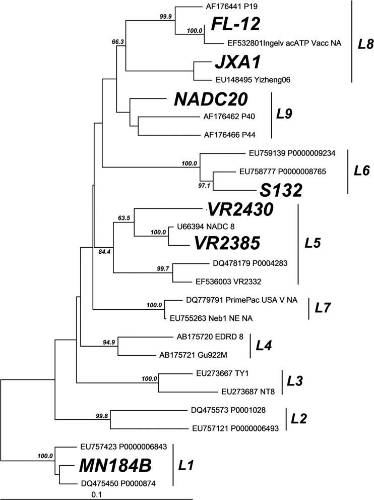
BHK-21 and MARC-145 cells were grown at 37°C in Dulbecco's minimum essential medium (DMEM) supplemented with 10% fetal bovine serum (FBS) and antibiotics. The North American type 2 PRRSV was systematically classified into 9 genetically distinct lineages based on the ORF5 gene sequences of 8,624 PRRSV strains (16). To produce a chimeric virus by molecular breeding, a total of 7 genetically different strains of PRRSV, each representing a distinct genetic lineage or sublineage in the phylogenetic tree (16), i.e., MN184B (lineage 1), VR2385 (lineage 5.1), VR2430 (lineage 5.2), S132 (lineage 6), Chinese highly pathogenic strain JXA1 (lineage 8.7), FL-12 (lineage 8.9), and NADC20 (lineage 9), were selected for DNA shuffling in the study. The genetic relationship of these selected strains of PRRSV used in DNA shuffling is shown in a phylogenetic tree (Fig. 1). The GP5 gene sequences of VR2385 and FL-12 were amplified from the infectious clones pIR-VR2385-CA (12) and pFL-12 (5), respectively. The GP5 gene sequence of strain VR2430 was amplified from viral stock. The GP5 gene sequences of the other 4 PRRSV strains (MN184B, S132, JXA1, and NADC20) were commercially synthesized (Genscript) based on the sequences in the GenBank database.
Fig 1.
Phylogenetic tree based on the GP5 genes of selected PRRSV strains from different genetic lineages of type 2 PRRSV, as reported by Shi et al. (16). The phylogenetic tree was constructed by using the neighbor-joining method with bootstraps in 1,000 replicates. The number above each major branch indicates the bootstrap value. The GP5 sequences of the 7 parental strains selected for shuffling in the study are in boldface italics in the tree. Each lineage corresponding to those reported by Shi et al. (16) is labeled with L and a number beside a vertical line.
DNA shuffling of the GP5 and GP5-M genes.
For GP5 gene shuffling, the GP5 genes from seven strains of PRRSV were mixed in equimolar amounts with a total of 5 μg DNA and diluted in 50 μl of 50 mM Tris-HCl (pH 7.4) and 10 mM MgCl2. The mixture was incubated at 15°C for 2 min with 0.15 U of DNase I (Sigma). DNA fragments 50 to 150 bp in size were purified from 2% agarose gels. The purified DNA fragments were subsequently added to the Pfu PCR mixture consisting of 1× Pfu buffer, 0.4 mM each deoxynucleoside triphosphate (dNTP), and 0.06 U Pfu polymerase. A PCR program without primers (95°C for 4 min; 35 cycles of 95°C for 30 s, 60°C for 30 s, 57°C for 30 s, 54°C for 30 s, 51°C for 30 s, 48°C for 30 s, 45°C for 30 s, 42°C for 30 s, and 72°C for 2 min; and finally, 72°C for 7 min) was performed to reassemble the digested DNA fragments. Subsequently, specific primers flanking the shuffled GP5 gene region, GP5trunc-F (5′-GGGAACAGCAGCTCAAATTTACAG-3′) and GP5trunc-R (5′-AGGGGTAGCCGCGGAACCAT-3′), were used to amplify the assembled shuffling products.
Similar approaches were used to shuffle the GP5-M genes from 6 different strains of PRRSV. Unlike GP5 shuffling, strain S132 was not included in the GP5-M gene shuffling, since the M gene sequence strain S132 was not available. Primers GP5F (5′-ATGTTGGGGAAATGCTTGACCG-3′) and mfu3′R (5′-GCCGCAATCGGATGAAAGCCTG-3′) were used to amplify the assembled shuffling products.
Construction of chimeric PRRSV libraries.
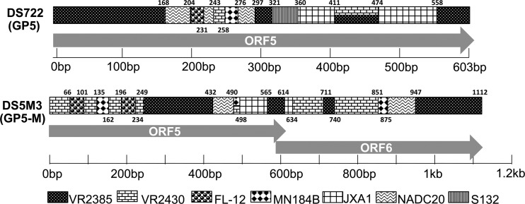
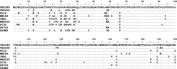
The shuffled product libraries were cloned into a blunt-end vector, pCR-BLUNT, to assess the quality of the DNA shuffling. The recombination efficiency was analyzed by sequencing the shuffled genes from 30 randomly selected clones to delineate crossovers. The nucleotide changes among the parental strains served as markers to delineate the origin of each fragment between two proximate crossover sites incorporated in the shuffled product. The fragment between the crossover sites with the same nucleotide pattern as a particular parental strain was considered to be derived from that parental strain (Fig. 2 and 3). The shuffled products that contained segments derived from all parental viruses and that had a good number of crossovers were selected for the study. The GP5 clone DS722 and the GP5-M clone DS5M3 were ultimately selected from their respective libraries for the construction of chimeric viruses in the backbone of a DNA-launched PRRSV infectious clone, pIR-VR2385-CA (38, 39). For cloning purpose, two flanking fragments amplified from pIR-VR2385-CA containing the naturally occurring unique restriction sites Acl1 and XbaI, respectively, were fused to the corresponding shuffled products, and the fusion fragments were cloned into the DNA-launched infectious clone to produce chimeric viruses containing the shuffled GP5 or GP5-M gene. The amino acid differences in GP5 among the 7 parental virus strains and the two chimeric viruses (DS722 and DS5M3) are presented in Fig. 3.
Fig 2.
Schematic diagram of the shuffled chimeric GP5 gene sequences in two representative chimeras (DS722 and DS5M3). The parental virus sequences of the GP5 gene for the two chimeras are depicted schematically. The exact boundaries of crossovers are indicated with nucleotide position numbers relative to the GP5 gene. Each pattern represents the sequence derived from an individual parental virus strain. If two patterns are displayed in the same region, it indicates that the region contains sequences shared by two different parental strains.
Fig 3.
Alignment of the GP5 amino acid sequences among the seven parental virus strains and the two chimeras (DS722 and DS5M3). The GP5 sequence of the backbone virus, VR2385, is shown at the top. Only differences are indicated for other strains.
In vitro transfection and immunofluorescence assay (IFA).
To rescue the infectious chimeric PRRSV from the recombinant DNA-launched infectious clones, BHK-21 cells at 60% confluence in 6-well plates were transfected with 3 μg of chimeric PRRSV DNA using 8 μl of Lipofectamine LTX (Invitrogen) according to the manufacturer's instructions. At 48 h posttransfection, the cell culture supernatant was harvested and passaged onto MARC-145 cells. Two days later, the cells were washed with 0.05% phosphate-buffered saline (PBS)-Tween and fixed with 80% acetone (Sigma). The fixed cells were incubated with anti-PRRSV N monoclonal antibody SDOW17 (Rural Technologies, Inc.) at 37°C for 1 h. After washing three times, the cells were then incubated with fluorescein isothiocyanate (FITC)-conjugated goat anti-mouse IgG at 37°C for 1 h. The stained cells were visualized with a Nikon Eclipse TE300 fluorescence microscope fitted with a digital camera (Nikon).
Plaque assay.
Confluent monolayers of MARC-145 cells cultured in a 6-well plate were infected with 10-fold serially diluted viruses (10−1, 10−2, 10−3, 10−4, and 10−5). After 1 h of incubation, the inoculum was removed and an agar overlay was applied to the cell monolayer. Plaques were stained with neutral red solution (Sigma) 4 days postinfection at 37°C. The wells containing 10 to 100 plaques in each plate were selected to measure the diameter of each plaque. Plaque morphology and size were compared between the parental virus VR2385 and the two chimeric viruses.
Growth characterization of the chimeric viruses in vitro.
To analyze the growth characteristics of the chimeric viruses in vitro, a growth curve was performed in MARC-145 cells, as well as in porcine alveolar macrophages (PAMs). The PAMs were obtained by lung lavage of a piglet from a PRRSV-free university research herd. Confluent monolayers of MARC-145 or PAMs seeded in 96-well plates were infected with the parental virus VR2385 and two rescued chimeric viruses (DS722 and DS5M3) at the same multiplicity of infection (MOI) of 0.1. Both the infected MARC-145 cells and PAMs were harvested at 6, 12, 24, 36, 48, 60, and 72 h postinfection (p.i.), and an additional time point at 84 h p.i. was added for the infected MARC-145 cells. The titers of virus harvested at different time points were determined by IFA in MARC-145 cells and quantified as 50% tissue culture infective doses (TCID50)/ml. All in vitro experiments were performed in triplicate.
Pathogenicity study of the two chimeric viruses in specific-pathogen-free (SPF) pigs.
To determine and compare the virulences of the two chimeric viruses and the parental virus in pigs, we used a nursery pig respiratory disease model to assess the pathogenicity of PRRSV, since the nursery pig model has been widely used worldwide for evaluating PRRSV virulence and vaccine efficacy (40–44). A total of 24 SPF pigs at 3 weeks of age were divided into 4 groups of 6 each and intramuscularly inoculated with the respective viruses, as shown in Table 1. All six pigs from each group were necropsied at 14 days p.i. At necropsy, the lung tissues were collected from each pig for histological examination and for quantification of viral-RNA loads.
Table 1.
Experimental design for comparative pathogenicity study to determine virulence of shuffled chimeric viruses in pigs
| Group | Inoculum (2 × 10⁵ TCID50/pig) | No. of pigs | Necropsy at 14 days postinoculation (no. of pigs) |
|---|---|---|---|
| A | Parental VR2385 | 6 | 6 |
| B | Chimera DS722 | 6 | 6 |
| C | Chimera DS5M3 | 6 | 6 |
| D | DMEM control | 6 | 6 |
Challenge-and-protection study in SPF pigs vaccinated with chimeric viruses.
A total of 72 SPF pigs at 3 weeks of age were divided into 8 groups of 9 pigs per group and vaccinated as shown in Table 2. At 35 days postvaccination (p.v.), pigs in each group were challenged with either the parental VR2385 virus or a heterologous NADC20 virus (Table 2). At 14 days postchallenge (p.c.), all pigs were necropsied, and gross pathological lung lesions were recorded and scored (35). Lung tissues were also collected for histological examination and viral-RNA load quantification.
Table 2.
PRRSV challenge and protection study in pigs vaccinated with attenuated chimeric viruses or with parental virus
| Group | No. of pigs | Vaccination at day 0 (2 × 10⁵ TCID50/pig) | Challenge virus at 35 days postvaccinationa | Necropsy at 14 days postchallenge (no. of pigs) |
|---|---|---|---|---|
| 1 | 9 | Parental VR2385 | VR2385 | 8b |
| 2 | 9 | Parental VR2385 | NADC20 | 9 |
| 3 | 9 | Chimera DS722 | VR2385 | 9 |
| 4 | 9 | Chimera DS722 | NADC20 | 9 |
| 5 | 9 | Chimera DS5M3 | VR2385 | 9 |
| 6 | 9 | Chimera DS5M3 | NADC20 | 9 |
| 7 | 9 | DMEM control | VR2385 | 9 |
| 8 | 9 | DMEM control | NADC20 | 9 |
The challenge virus dose for all viruses was 2 × 10⁵ TCID50/pig.
One pig died from an unrelated cause before the challenge.
Real-time PCR to quantify viral-RNA loads in sera and lung tissues of pigs.
To quantify viral-RNA loads in lung tissues, samples of lung tissues (500 mg) collected at each necropsy were homogenized in 10% (wt/vol) sterile PBS. The homogenates were centrifuged at 3,000 rpm at 4°C for 15 min, and the supernatants were used for quantification of PRRSV RNA. Total RNAs were extracted from weekly serum samples and homogenates of lung tissues using TRI Reagent (MRC) and used to synthesize cDNA using a Superscript II kit (Invitrogen). PRRSV genomes were quantified using a SYBR green-based quantitative PCR (qPCR). A pair of primers (forward primer, 5′-TTAAATATGCCAAATAACAACGG-3′; reverse primer, 5′-TGCCTCTGGACTGGTT-3′) were designed based on the conserved region in the nucleocapsid gene using Beacon software. The qPCR assay was conducted in a CFX96 real-time (RT) PCR system (Bio-Rad). The reactions were performed in a 20-μl PCR volume containing 10 μl SsoFast EvaGreen Supermix (Bio-Rad), 0.5 μl of each primer (10 μM), 5 μl of the template cDNA, and 4 μl of nuclease-free water. The cycling parameters included an initial denaturation at 95°C for 10 min, followed by 39 cycles of denaturation at 95°C for 10 s and annealing and extension at 58°C for 20 s. Dissociation curve analysis was performed using the instrument's default setting immediately after each PCR run. Each reaction was measured in triplicate.
Necropsy and gross pathology evaluation.
All pigs were humanely euthanized by intravenous pentobarbital overdose (Fatal-Plus; Vortech Pharmaceutical, Ltd., Dearborn, MI). Veterinary pathologists were blinded to the treatment status of the pigs for evaluation of gross lung lesions. The total amount of lung affected by pneumonia (0 to 100% of the lung affected by grossly visible pneumonia) was recorded for each pig at necropsy while blinded to the treatment status, as described previously (35). Briefly, the scoring system is based on the approximate volume that each lung lobe contributes to the entire lung: the right cranial lobe, right middle lobe, cranial part of the left cranial lobe, and caudal part of the left cranial lobe contribute 10% each to the total lung volume; the accessory lobe contributes 5%; and the right and left caudal lobes contribute 27.5% each (35). Five defined sections of lungs (35) were collected, immediately immersed in 10% neutral buffered formalin, and routinely processed for histological examination. In addition, fresh lung tissues were collected separately and immediately stored at −80°C for virological testing.
Histopathology evaluation.
Microscopic lung lesions were evaluated independently by two veterinary pathologists (T.O. and P.G.H.) blinded to the treatment status. Lung sections were scored for the presence and severity of interstitial pneumonia, ranging from 0 to 6 (0, normal; 1, mild multifocal; 2, mild diffuse; 3, moderate multifocal; 4, moderate diffuse; 5, severe multifocal; 6, severe diffuse) (35). If the results obtained by the two pathologists on a certain tissue differed, the mean of the two scores was used.
Statistical analyses.
A two-tailed Student's t test was used to evaluate the differences (P < 0.05) between the samples in the two groups for both in vitro and in vivo studies. The data were analyzed using GraphPad Prism (version 5.0).
Nucleotide sequence accession numbers.
The GP5 sequences of the parental virus strains and the two shuffled chimeras were deposited in the GenBank database under accession numbers JX044140 (VR2385), JX050225 (VR2430), AY545985 (FL-12), DQ176020 (MN184B), EF112445 (JXA1), JX069953 (NADC20), JX069952 (S132), JX044138 (DS722), and JX044139 (DS5M3).
RESULTS
Generation of infectious chimeric viruses containing well-shuffled GP5 or GP5-M genes from 7 and 6 genetically distinct strains of PRRSV, respectively.
To generate GP5-shuffled chimeric viruses with good growth fitness, we excluded 96 nucleotides (nt) from the 5′ end of the GP5 gene, including the signal peptide sequence and the 16-nt region overlapping the M gene, as well as the junction site sequence from 23 to 18 nt upstream of the M gene, for DNA shuffling of the GP5 genes. The resulting 468-nt GP5 genes from seven PRRSV strains, each representing a distinct genetic lineage or sublineage (16) (Fig. 1), were shuffled with DNase I digestion, followed by PCR without primers for reassembly. A PCR product consisting of reassembled shuffled products with an expected size of 468 bp was obtained after a second PCR with specific primers flanking the shuffled region. To generate chimeras containing segments derived from all seven parental viral strains, the shuffling process was iterated by using the shuffled DNA pool from the first-round shuffling as the parents (2, 4).
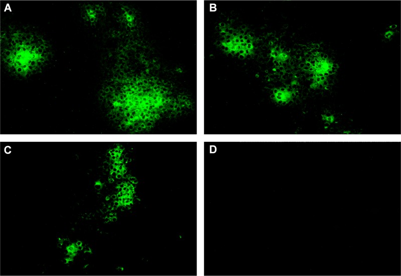
Sequence analyses of 30 representative clones that were randomly selected from the shuffled library revealed that they all contained chimeric GP5 sequences, but only two clones contained sequences from all 7 parental viruses. The numbers of crossovers ranged from 8 to 12 in the shuffled GP5 gene products. The GP5 clone DS722, which contains segments derived from all seven parental viral sequences with 12 crossovers, was selected from the shuffled library (Fig. 2) and cloned into the backbone of a DNA-launched PRRSV infectious clone (39). The resulting chimeric virus, DS722, containing the shuffled GP5 genes from 7 different strains of PRRSV was successfully rescued from transfected cells (Fig. 4B).
Fig 4.
Rescue and in vitro passages of the shuffled chimeric viruses DS722 and DS5M3. (A) IFA confirmation using anti-PRRSV N monoclonal antibody for the rescue of the parental virus, VR2385, in MARC-145 cells infected with the supernatant of BHK-21 cells transfected with a DNA-launched PRRSV infectious clone, pIR-VR2385-CA. (B and C) IFA confirmation of the rescue of the chimeric viruses DS722 (B) and DS5M3 (C) in MARC-145 cells infected with the supernatant of BHK-21 cells transfected with shuffled clones pIR-DS722 and pIR-DS5M3, respectively. (D) There was no fluorescent signal in the MARC-145 cells infected with the supernatant of BHK-21 cells transfected with a plasmid containing a defective PRRSV VR2385 backbone.
Similar approaches were used to shuffle the region spanning the GP5-M genes of 6 distinct strains of PRRSV. Sequence analyses of 10 representative clones that were randomly selected from the shuffled library revealed that all contained the chimeric GP5 sequences, and 6 of the 10 clones contained sequences derived from all 6 parental viruses. The numbers of crossovers ranged from 12 to 22 in the shuffled GP5 gene products. The chimeric GP5-M clone DS5M3, containing segments derived from all 6 parental viral sequences with 18 crossovers (Fig. 2), was selected and cloned into the backbone of the DNA-launched PRRSV infectious clone. The GP5-M chimeric virus DS5M3 was successfully rescued from transfected cells (Fig. 4C). The amino acid differences in GP5 among the parental and shuffled viruses are indicated in Fig. 3.
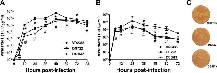
The two chimeric viruses replicated at a lower level, both in MARC-145 cells and in PAMs, and formed smaller plaques in MARC-145 cells than the parental virus.
To characterize and compare the growth characteristics between the two chimeric viruses (DS722 and DS5M3) and the parental virus (VR2385), the growth kinetics of the three viruses were analyzed by infection of MARC-145 cells or PAMs with the respective virus at the same MOI of 0.1. The infectious-virus titers were determined at different times p.i. The results showed that, in MARC-145 cells, the chimeric virus DS722 replicated to significantly lower levels than the parental virus, VR2385, at all time points except 60 and 84 h p.i., whereas the chimeric virus DS5M3 also displayed a significantly lower level of replication than VR2385 (Fig. 5A). In PAMs, the chimeric virus DS722 replicated to significantly lower levels than the parental virus, VR2385, at all time points except 6 and 12 h p.i. The other chimeric virus, DS5M3, displayed a significantly lower level of replication at all time points than VR2385 (Fig. 5B).
Fig 5.
In vitro growth kinetics and plaque morphology of the shuffled chimeric viruses DS722 and DS5M3. (A) Growth kinetics of the parental virus, VR2385, and the two chimeric viruses (DS722 and DS5M3) in MARC-145 cells. The virus infectious titers (TCID50/ml) were determined at the indicated time points postinfection. * and #, statistically significant difference between the chimeric viruses and the parental virus at that time point. The experiments were performed in triplicate, and the error bars indicate standard errors. (B) In vitro growth kinetics of the chimeras DS722 and DS5M3 on PAMs. The virus infectious titers (TCID50/ml) were determined at the indicated time points postinfection. * and #, statistically significant difference between each of the chimeric viruses and the parental virus at that time point. The experiments were performed in triplicate, and the error bars indicate standard errors. (C) Plaque morphologies and diameters of the parental virus VR2385 and chimeric viruses DS722 and DS5M3 on MARC-145 cells.
Both chimeric viruses and the parental virus developed plaques within 4 days p.i. in MARC-145 cells. However, the diameters of the plaques formed by the two chimeric viruses were significantly smaller than that formed by the parental virus (P < 0.001) (Fig. 5C), further indicating that the chimeric viruses had a lower growth capacity in vitro than the parental virus.
Both chimeric viruses (DS722 and DS5M3) were attenuated in pigs.
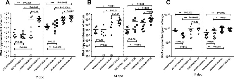
The pathogenicity study revealed that pigs infected with the chimera DS5M3 had significantly lower serum viral-RNA loads than pigs infected with the parental virus, VR2385, at both 7 (P = 0.02) (Fig. 6A) and 14 (Fig. 6B) days p.i. (P = 0.0009). Similarly, the serum samples from pigs infected with chimera DS722 also had lower viral-RNA loads than pigs infected with the parental virus at both 7 and 14 days p.i., and the difference was significant at 7 days p.i. (P = 0.03) (Fig. 6A), but not at 14 days p.i., although most sera from the DS722 group displayed viral-RNA loads lower than those from the VR2385 group at 14 days p.i. (Fig. 6B).
Fig 6.
Viral-RNA loads in serum samples and lung tissues in the comparative-pathogenicity study from pigs infected with the parental virus, VR2385, and chimeric virus DS722 or DS5M3 or inoculated with DMEM (negative control) at 7 and 14 days p.i., respectively. (A) PRRSV viral-RNA loads in serum samples at 7 days p.i. (B) PRRSV viral-RNA loads in serum samples at 14 days p.i. (C) Viral-RNA loads in the lung collected during necropsy. Significant differences are indicated with asterisks (*, P < 0.05; ***, P < 0.001). In all three panels, the numbers within circles along the x axis indicate the numbers of animals in each group that tested negative for viral RNA.
Similarly, pigs infected with the chimera DS5M3 had significantly lower viral-RNA loads (P < 0.0001) in the lung tissues than pigs infected with VR2385. The pigs infected with the chimera DS722 also had a lower viral load in the lung than pigs infected with the parental virus, although the difference was not significant (Fig. 6C).
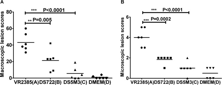
Macroscopic lung lesions were generally absent or mild in pigs inoculated with DMEM and with the two chimeras, DS5M3 and DS722 (Fig. 7A). In pigs infected with the parental VR2385 virus, visible gross lung lesions were more pronounced and affected an average of 42% of the lung surfaces (Fig. 7A). The mean scores of the gross lung lesions in pigs inoculated with the two chimeric viruses, DS722 (P = 0.005) and DS5M3 (P < 0.0001), were significantly lower than that of the pigs inoculated with the parental virus, VR2385 (Fig. 7A). The mean scores of the histological lung lesions in pigs infected with the chimeric viruses DS722 (P = 0.0002) and DS5M3 (P < 0.0001) were significantly lower than that in pigs infected by the parental VR2385 virus (Fig. 7B).
Fig 7.
Macroscopic and microscopic lesions in the lung tissues from pigs experimentally infected with the parental virus, VR2385, and the two chimeric viruses DS722 and DS5M3 during necropsy at 14 days p.i. (A) Macroscopic-lesion scores of lung tissues at 14 days p.i. from the 6 pigs infected with the parental virus, VR2385, and the chimeric viruses DS722 and DS5M3 or inoculated with DMEM (negative control). (B) Microscopic-lesion scores of lung tissues at 14 days p.i. Significant differences are indicated with asterisks (**, P < 0.01; ***, P < 0.001).
Chimera DS722, but not chimera DS5M3, elicited protection in pigs against PRRSV challenge at a level similar to that in the parental virus.
To investigate whether the two attenuated chimeras could still elicit protection against PRRSV, pigs were first vaccinated with the parental virus, VR2385; the chimera DS722 or DS5M3; or DMEM (Table 2). Eight or nine vaccinated pigs each in groups 1, 3, 5, and 7 were then challenged at 35 days p.v. with the parental VR2385 virus (lineage 5). Nine vaccinated pigs each in groups 2, 4, 6, and 8 were challenged at 35 days p.v. with a heterologous NADC20 virus (lineage 9) (Table 2). At the time of challenge at 35 days p.v., viremia was not detected in any of the pigs by RT-PCR. All pigs were necropsied at 14 days p.c.
For the pigs challenged with the parental strain VR2385, at 7 days p.c., the serum viral-RNA loads significantly decreased in pigs vaccinated with VR2385 (P = 0.005) or with the chimera DS722 (P = 0.003), but not in pigs vaccinated with the chimera DS5M3, compared to group 7 control pigs (Fig. 8A). Similarly, at 14 days p.c., the serum viral-RNA loads significantly decreased in pigs that were vaccinated with VR2385 (P = 0.01) or with the chimera DS722 (P = 0.01), but not in pigs vaccinated with the chimera DS5M3, compared to group 7 control pigs (Fig. 8B). The reduction of the serum viral-RNA loads against challenges with parental VR2385 and heterologous NADC20 in pigs vaccinated with the chimera DS722 was similar to that in pigs vaccinated with the parental virus, VR2385, at both 7 and 14 days p.c. (Fig. 8A and B). The viral-RNA loads in the lung tissues at 14 days p.c. were significantly decreased in pigs vaccinated with the parental virus, VR2385 (P = 0.002), or with the chimera DS722 (P < 0.0001), but not in pigs vaccinated with the chimera DS5M3, compared to group 7 control pigs (Fig. 8C). The pigs vaccinated with the chimera DS722 displayed significantly lower viral-RNA loads in the lung tissues than the pigs vaccinated with VR2385 (Fig. 8C).
Fig 8.
Viral-RNA loads in serum samples at 7 and 14 days and in lung tissues at 14 days p.c. from the challenge/protection study in pigs vaccinated with the chimeric viruses (DS722 or DS5M3), followed by challenge. (A) PRRSV viral-RNA loads in serum samples from pigs at 7 days p.c. with a homologous PRRSV, VR2385, or a heterologous PRRSV, NADC20. (B) PRRSV viral-RNA loads in serum samples from pigs at 14 days p.c. with PRRSV VR2385 or NADC20. (C) PRRSV viral-RNA loads in the lung tissues from pigs at 14 days p.c. Significant differences are indicated with asterisks (*, P < 0.05; **, P < 0.01; ***, P < 0.001). In all three panels, the numbers within circles along the x axis indicate the numbers of animals in each group that tested negative for serum viral RNA.
For pigs challenged with a heterologous NADC20 strain, at 7 days p.c., the serum viral-RNA loads were significantly lower in pigs vaccinated with the parental virus (P = 0.0002) or with the two chimeras DS722 (P = 0.0002) and DS5M3 (P = 0.02) than in group 8 controls (Fig. 8A). Similarly, at 14 days p.c., there were significant reductions in the serum viral-RNA loads in pigs vaccinated with the parental virus, VR2385 (P = 0.01), or with two chimeras, DS722 (P = 0.03) and DS5M3 (P = 0.02), compared to group 8 control pigs (Fig. 8B). Also, the viral-RNA loads in the lung tissues at 14 days p.c. were significantly reduced in pigs vaccinated with the parental virus, VR2385 (P = 0.002), and with the chimera DS722 (P = 0.01), but not in pigs vaccinated with the chimera DS5M3, compared to group 8 control pigs (Fig. 8C). The reduction of viral-RNA loads in the lung tissues of pigs vaccinated with the chimera DS722 was similar to that in pigs vaccinated with the parental virus, VR2385, at both 7 and 14 days p.c. (Fig. 8C).
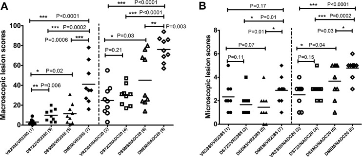
At necropsy, the average scores of both macroscopic and microscopic lung lesions in pigs vaccinated with two chimeras (groups 3, 4, 5, and 6) were significantly lower than those in the control pigs in groups 7 and 8 (Fig. 9A and B). The protection, based on the macroscopic and microscopic lung lesions, was much more effective in the DS722-vaccinated pigs than in DS5M3-vaccinated pigs. The average scores of gross and microscopic lung lesions in DS722-vaccinated pigs were mostly similar to those in VR2385-vaccinated pigs, although the scores in DS5M3-vaccinated pigs were significantly higher than those in VR2385-vaccinated pigs (Fig. 9).
Fig 9.
Macroscopic- and microscopic-lesion scores of lung tissues at necropsy at 14 days following challenge in pigs vaccinated with the chimeric viruses DS722 and DS5M3. (A) Macroscopic-lung-lesion scores from pigs at necropsy 14 days p.c. (B) Microscopic-lesion scores of lung tissues from pigs at necropsy 14 days p.c. Significant differences are indicated with asterisks (*, P < 0.05; ***, P < 0.001).
Both chimeras DS722 and DS5M3 were stable in vivo.
The shuffled genes of chimeric viruses DS722 and DS5M3 recovered from the sera of pigs in the respective groups at 14 days p.i. were amplified by RT-PCR and sequenced. Sequence analyses revealed that the sequences of the recovered viruses were the same as those of the original virus inocula, indicating the genetic stability of these two chimeric viruses in animals.
DISCUSSION
Molecular breeding through DNA shuffling can direct the evolution of viruses in vitro and select new strains with desired traits. To determine if molecular breeding of virus envelope genes that are important virulence determinants can produce an attenuated virus that retains its protective ability against challenge, we bred the GP5 genes of 7 and the GP5-M genes of 6 genetically distinct strains of PRRSV by DNA shuffling and iteration of the shuffling process. The application of iteration of the DNA-shuffling process increased the chances to incorporate all parental viral genes into the small GP5 region (2, 4). Two representative chimeric viruses, a GP5 chimera, DS722, and a GP5-M chimera, DS5M3, were rescued and selected for further studies. Although both chimeras were infectious in vitro, they both displayed a lower level of virus replication in both MARC-145 cells and PAMs. In addition, both chimeric viruses formed smaller plaques in MARC-145 cells than the parental virus, indicating that the two shuffled chimeric viruses exhibited an attenuated phenotype in vitro.
To further determine whether DNA shuffling of the GP5 or GP5-M gene altered virus virulence in vivo, we conducted a pathogenicity study (Table 1) and showed that there was a significant reduction in both the macroscopic- and microscopic-lung-lesion scores in pigs infected with the two chimeras compared to those infected with the parental virus. Significant reductions in viral-RNA loads in sera and lung tissues were also found in pigs infected with the chimera DS5M3. The in vitro growth and the in vivo pathogenicity studies indicated that both chimeras were attenuated. Therefore, rapid attenuation of PRRSV was achieved in this study by shuffling of the virulence determinant GP5 genes from multiple genetically divergent virus strains. It is important to note that GP5 is not the sole gene responsible for PRRSV virulence (37), and thus, DNA shuffling of other PRRSV genes also involved in virulence in the future may further improve virus attenuation. Nevertheless, this unique DNA-shuffling approach to attenuate a virus is more advantageous than many other traditional reverse-genetics system approaches in that DNA shuffling mimics the natural evolution of viruses and does not require an understanding of the functionality of the shuffling regions; rather, the approach relies on functional screening for the desired traits of the shuffled viruses, such as the attenuation phenotype in this study.
Since the two chimeric viruses displayed an attenuated phenotype in vivo, we next evaluated whether the chimeric viruses could still induce protection against PRRSV challenge. Eight groups of pigs were first vaccinated with the parental virus, VR2385; the two chimeras; or DMEM (Table 2). At 35 days p.c., pigs in each group were challenged with a homologous (lineage 5) or a heterologous (lineage 9) PRRSV. The results revealed that the chimera DS722 still elicited solid protection against challenges by both homologous and heterologous PRRSV strains. However, the GP5-M-shuffled chimeric virus DS5M3 did not induce a sufficient level of protection, even though there was a significant reduction in macroscopic- and microscopic-lung-lesion scores. Thus, the GP5-shuffled chimeric virus DS722 still retains its ability to elicit protection against PRRSV, suggesting that the DNA shuffling of the virulence determinant gene attenuated the virus but did not impair the ability of the shuffled virus to elicit protection.
We had initially thought that the GP5-M chimera DS5M3 would also retain its ability to elicit protection, since GP5 and M form heterodimers (45). The observed poor protection of the chimera DS5M3 was likely due to the low replication fitness of the chimera in vivo, since only low levels of chimera DS5M3 viral RNA were detected in both sera and lung tissues in the pathogenicity study. The GP5-M DNA shuffling included some critical regions for virus replication, such as the GP5 signal peptide sequence and the overlapping region, and thus, shuffling of these critical regions likely affected the viral replication efficiency in vivo, leading to overattenuation of the chimera DS5M3 and thus poor protection compared to the GP5 chimera, DS722. In addition, glycosylation of the major envelope protein GP5 is known to play an important role in PRRSV virulence (46). For the chimera DS5M3, it appears that DNA shuffling resulted in the loss of an important glycosylation site at amino acid position 34 of the chimera compared to its parental virus, VR2385 (Fig. 3), and this might contribute to the virus attenuation phenotype of the chimera (46). Although the exact mechanism of attenuation by DNA shuffling remains unknown, the attenuation phenotype of the shuffled viruses may be attributed to the altered growth efficiency of the chimeric viruses. In addition, potential conformational changes of the shuffled GP5 in the chimeras may alter its interactions with other viral proteins or host cells, leading to attenuation (6).
In conclusion, attenuation of a virus by DNA shuffling of its envelope genes was demonstrated for the first time. We successfully produced two chimeric viruses that displayed attenuated phenotypes both in vitro and in vivo by shuffling the GP5 gene containing major virulence determinants and the GP5-M genes. The attenuated shuffled virus DS722 still induced protection similar to that of its parental virus against PRRSV infection. Although development of an improved PRRSV vaccine with better protection was not within the scope of the present study, it is logical to speculate that further shuffling of other structural genes, such as GP3 and GP4, which are relevant for neutralizing activities, in the future may lead to the generation of a more broadly protective PRRSV modified live-attenuated vaccine. Therefore, attenuation of a positive-strand RNA virus by DNA shuffling, as demonstrated in this study, has important implications for potential future vaccine development and thus is of broad general interest to the scientific community, as this approach of rapid virus attenuation can be easily applied to other important human and veterinary viruses.
ACKNOWLEDGMENTS
We thank B. A. Dryman for technical assistance; Z. Zhao for expert support in bioinformatic analyses; Pablo Piñeyro for lung lavage and collection of porcine alveolar macrophages; C. Branstad, P. Gerber, L. Gimenéz-Lirola, M. Hemann, K. O'Neill, X. Wang, and C. Xiao for their assistance with the animal studies; F. A. Osorio and A. Pattnaik of the University of Nebraska—Lincoln for the pFL-12 infectious clone; K. M. Larger of the National Animal Disease Center, Ames, IA, for the NADC20 PRRSV strain; and F. C. Leung of the University of Hong Kong for the genetic lineage data set.
This project was supported in part by Agriculture and Food Research Initiative competitive grant no. 2011-67015-30165 from the USDA National Institute of Food and Agriculture and by a PRRS CAP grant (2008-55620-19132).
Footnotes
Published ahead of print 17 October 2012
REFERENCES
- 1. Locher CP, Paidhungat M, Whalen RG, Punnonen J. 2005. DNA shuffling and screening strategies for improving vaccine efficacy. DNA Cell Biol. 24:256–263 [DOI] [PubMed] [Google Scholar]
- 2. Chang CC, Chen TT, Cox BW, Dawes GN, Stemmer WP, Punnonen J, Patten PA. 1999. Evolution of a cytokine using DNA family shuffling. Nat. Biotechnol. 17:793–797 [DOI] [PubMed] [Google Scholar]
- 3. Stemmer WP. 1994. DNA shuffling by random fragmentation and reassembly: in vitro recombination for molecular evolution. Proc. Natl. Acad. Sci. U. S. A. 91:10747–10751 [DOI] [PMC free article] [PubMed] [Google Scholar]
- 4. Zhang JH, Dawes G, Stemmer WP. 1997. Directed evolution of a fucosidase from a galactosidase by DNA shuffling and screening. Proc. Natl. Acad. Sci. U. S. A. 94:4504–4509 [DOI] [PMC free article] [PubMed] [Google Scholar]
- 5. Apt D, Raviprakash K, Brinkman A, Semyonov A, Yang S, Skinner C, Diehl L, Lyons R, Porter K, Punnonen J. 2006. Tetravalent neutralizing antibody response against four dengue serotypes by a single chimeric dengue envelope antigen. Vaccine 24:335–344 [DOI] [PubMed] [Google Scholar]
- 6. Soong NW, Nomura L, Pekrun K, Reed M, Sheppard L, Dawes G, Stemmer WP. 2000. Molecular breeding of viruses. Nat. Genet. 25:436–439 [DOI] [PubMed] [Google Scholar]
- 7. Yang L, Jiang J, Drouin LM, Agbandje-McKenna M, Chen C, Qiao C, Pu D, Hu X, Wang DZ, Li J, Xiao X. 2009. A myocardium tropic adeno-associated virus (AAV) evolved by DNA shuffling and in vivo selection. Proc. Natl. Acad. Sci. U. S. A. 106:3946–3951 [DOI] [PMC free article] [PubMed] [Google Scholar]
- 8. Kenney JL, Volk SM, Pandya J, Wang E, Liang X, Weaver SC. 2011. Stability of RNA virus attenuation approaches. Vaccine 29:2230–2234 [DOI] [PMC free article] [PubMed] [Google Scholar]
- 9. Lunney JK, Benfield DA, Rowland RR. 2010. Porcine reproductive and respiratory syndrome virus: an update on an emerging and re-emerging viral disease of swine. Virus Res. 154:1–6 [DOI] [PMC free article] [PubMed] [Google Scholar]
- 10. Nieuwenhuis N, Duinhof TF, van Nes A. 2012. Economic analysis of outbreaks of porcine reproductive and respiratory syndrome virus in nine sow herds. Vet. Rec. 170:225. [DOI] [PubMed] [Google Scholar]
- 11. Neumann EJ, Kliebenstein JB, Johnson CD, Mabry JW, Bush EJ, Seitzinger AH, Green AL, Zimmerman JJ. 2005. Assessment of the economic impact of porcine reproductive and respiratory syndrome on swine production in the United States. J. Am. Vet. Med. Assoc. 227:385–392 [DOI] [PubMed] [Google Scholar]
- 12. An TQ, Tian ZJ, Leng CL, Peng JM, Tong GZ. 2011. Highly pathogenic porcine reproductive and respiratory syndrome virus, Asia. Emerg. Infect. Dis. 17:1782–1784 [DOI] [PMC free article] [PubMed] [Google Scholar]
- 13. Tian K, Yu X, Zhao T, Feng Y, Cao Z, Wang C, Hu Y, Chen X, Hu D, Tian X, Liu D, Zhang S, Deng X, Ding Y, Yang L, Zhang Y, Xiao H, Qiao M, Wang B, Hou L, Wang X, Yang X, Kang L, Sun M, Jin P, Wang S, Kitamura Y, Yan J, Gao GF. 2007. Emergence of fatal PRRSV variants: unparalleled outbreaks of atypical PRRS in China and molecular dissection of the unique hallmark. PLoS One 2:e526 doi:10.1371/journal.pone.0000526 [DOI] [PMC free article] [PubMed] [Google Scholar]
- 14. Benfield DA, Nelson E, Collins JE, Harris L, Goyal SM, Robison D, Christianson WT, Morrison RB, Gorcyca D, Chladek D. 1992. Characterization of swine infertility and respiratory syndrome (SIRS) virus (isolate ATCC VR-2332). J. Vet. Diagn. Invest. 4:127–133 [DOI] [PubMed] [Google Scholar]
- 15. Nelsen CJ, Murtaugh MP, Faaberg KS. 1999. Porcine reproductive and respiratory syndrome virus comparison: divergent evolution on two continents. J. Virol. 73:270–280 [DOI] [PMC free article] [PubMed] [Google Scholar]
- 16. Shi M, Lam TT, Hon CC, Murtaugh MP, Davies PR, Hui RK, Li J, Wong LT, Yip CW, Jiang JW, Leung FC. 2010. Phylogeny-based evolutionary, demographical, and geographical dissection of North American type 2 porcine reproductive and respiratory syndrome viruses. J. Virol. 84:8700–8711 [DOI] [PMC free article] [PubMed] [Google Scholar]
- 17. Fang Y, Snijder EJ. 2010. The PRRSV replicase: exploring the multifunctionality of an intriguing set of nonstructural proteins. Virus Res. 154:61–76 [DOI] [PMC free article] [PubMed] [Google Scholar]
- 18. Meng XJ, Paul PS, Halbur PG. 1994. Molecular cloning and nucleotide sequencing of the 3′-terminal genomic RNA of the porcine reproductive and respiratory syndrome virus. J. Gen. Virol. 75:1795–1801 [DOI] [PubMed] [Google Scholar]
- 19. Dokland T. 2010. The structural biology of PRRSV. Virus Res. 154:86–97 [DOI] [PMC free article] [PubMed] [Google Scholar]
- 20. Meng XJ, Paul PS, Morozov I, Halbur PG. 1996. A nested set of six or seven subgenomic mRNAs is formed in cells infected with different isolates of porcine reproductive and respiratory syndrome virus. J. Gen. Virol. 77:1265–1270 [DOI] [PubMed] [Google Scholar]
- 21. Wissink EH, Kroese MV, van Wijk HA, Rijsewijk FA, Meulenberg JJ, Rottier PJ. 2005. Envelope protein requirements for the assembly of infectious virions of porcine reproductive and respiratory syndrome virus. J. Virol. 79:12495–12506 [DOI] [PMC free article] [PubMed] [Google Scholar]
- 22. Delputte PL, Costers S, Nauwynck HJ. 2005. Analysis of porcine reproductive and respiratory syndrome virus attachment and internalization: distinctive roles for heparan sulphate and sialoadhesin. J. Gen. Virol. 86:1441–1445 [DOI] [PubMed] [Google Scholar]
- 23. Faaberg KS, Even C, Palmer GA, Plagemann PG. 1995. Disulfide bonds between two envelope proteins of lactate dehydrogenase-elevating virus are essential for viral infectivity. J. Virol. 69:613–617 [DOI] [PMC free article] [PubMed] [Google Scholar]
- 24. Snijder EJ, Dobbe JC, Spaan WJ. 2003. Heterodimerization of the two major envelope proteins is essential for arterivirus infectivity. J. Virol. 77:97–104 [DOI] [PMC free article] [PubMed] [Google Scholar]
- 25. Hansen W, Loser K, Westendorf AM, Bruder D, Pfoertner S, Siewert C, Huehn J, Beissert S, Buer J. 2006. G protein-coupled receptor 83 overexpression in naive CD4+CD25- T cells leads to the induction of Foxp3+ regulatory T cells in vivo. J. Immunol. 177:209–215 [DOI] [PubMed] [Google Scholar]
- 26. Ansari IH, Kwon B, Osorio FA, Pattnaik AK. 2006. Influence of N-linked glycosylation of porcine reproductive and respiratory syndrome virus GP5 on virus infectivity, antigenicity, and ability to induce neutralizing antibodies. J. Virol. 80:3994–4004 [DOI] [PMC free article] [PubMed] [Google Scholar]
- 27. Faaberg KS, Hocker JD, Erdman MM, Harris DL, Nelson EA, Torremorell M, Plagemann PG. 2006. Neutralizing antibody responses of pigs infected with natural GP5 N-glycan mutants of porcine reproductive and respiratory syndrome virus. Viral Immunol. 19:294–304 [DOI] [PubMed] [Google Scholar]
- 28. Ostrowski M, Galeota JA, Jar AM, Platt KB, Osorio FA, Lopez OJ. 2002. Identification of neutralizing and nonneutralizing epitopes in the porcine reproductive and respiratory syndrome virus GP5 ectodomain. J. Virol. 76:4241–4250 [DOI] [PMC free article] [PubMed] [Google Scholar]
- 29. Plagemann PG, Rowland RR, Faaberg KS. 2002. The primary neutralization epitope of porcine respiratory and reproductive syndrome virus strain VR-2332 is located in the middle of the GP5 ectodomain. Arch. Virol. 147:2327–2347 [DOI] [PubMed] [Google Scholar]
- 30. Vu HL, Kwon B, Yoon KJ, Laegreid WW, Pattnaik AK, Osorio FA. 2011. Immune evasion of porcine reproductive and respiratory syndrome virus through glycan shielding involves both glycoprotein 5 as well as glycoprotein 3. J. Virol. 85:5555–5564 [DOI] [PMC free article] [PubMed] [Google Scholar]
- 31. Vanhee M, Van Breedam W, Costers S, Geldhof M, Noppe Y, Nauwynck H. 2011. Characterization of antigenic regions in the porcine reproductive and respiratory syndrome virus by the use of peptide-specific serum antibodies. Vaccine 29:4794–4804 [DOI] [PubMed] [Google Scholar]
- 32. Wissink EH, Kroese MV, Maneschijn-Bonsing JG, Meulenberg JJ, van Rijn PA, Rijsewijk FA, Rottier PJ. 2004. Significance of the oligosaccharides of the porcine reproductive and respiratory syndrome virus glycoproteins GP2a and GP5 for infectious virus production. J. Gen. Virol. 85:3715–3723 [DOI] [PubMed] [Google Scholar]
- 33. Johnson CR, Griggs TF, Gnanandarajah J, Murtaugh MP. 2011. Novel structural protein in porcine reproductive and respiratory syndrome virus encoded by an alternative ORF5 present in all arteriviruses. J. Gen. Virol. 92:1107–1116 [DOI] [PMC free article] [PubMed] [Google Scholar]
- 34. Music N, Gagnon CA. 2010. The role of porcine reproductive and respiratory syndrome (PRRS) virus structural and non-structural proteins in virus pathogenesis. Anim. Health Res. Rev. 11:135–163 [DOI] [PubMed] [Google Scholar]
- 35. Halbur PG, Paul PS, Frey ML, Landgraf J, Eernisse K, Meng XJ, Lum MA, Andrews JJ, Rathje JA. 1995. Comparison of the pathogenicity of two US porcine reproductive and respiratory syndrome virus isolates with that of the Lelystad virus. Vet. Pathol. 32:648–660 [DOI] [PubMed] [Google Scholar]
- 36. Kapur V, Elam MR, Pawlovich TM, Murtaugh MP. 1996. Genetic variation in porcine reproductive and respiratory syndrome virus isolates in the midwestern United States. J. Gen. Virol. 77:1271–1276 [DOI] [PubMed] [Google Scholar]
- 37. Kwon B, Ansari IH, Pattnaik AK, Osorio FA. 2008. Identification of virulence determinants of porcine reproductive and respiratory syndrome virus through construction of chimeric clones. Virology 380:371–378 [DOI] [PubMed] [Google Scholar]
- 38. Huang YW, Fang Y, Meng XJ. 2009. Identification and characterization of a porcine monocytic cell line supporting porcine reproductive and respiratory syndrome virus (PRRSV) replication and progeny virion production by using an improved DNA-launched PRRSV reverse genetics system. Virus Res. 145:1–8 [DOI] [PubMed] [Google Scholar]
- 39. Ni YY, Huang YW, Cao D, Opriessnig T, Meng XJ. 2011. Establishment of a DNA-launched infectious clone for a highly pneumovirulent strain of type 2 porcine reproductive and respiratory syndrome virus: identification and in vitro and in vivo characterization of a large spontaneous deletion in the nsp2 region. Virus Res. 160:264–273 [DOI] [PubMed] [Google Scholar]
- 40. Cano JP, Dee SA, Murtaugh MP, Pijoan C. 2007. Impact of a modified-live porcine reproductive and respiratory syndrome virus vaccine intervention on a population of pigs infected with a heterologous isolate. Vaccine 25:4382–4391 [DOI] [PubMed] [Google Scholar]
- 41. Fang Y, Christopher-Hennings J, Brown E, Liu H, Chen Z, Lawson SR, Breen R, Clement T, Gao X, Bao J, Knudsen D, Daly R, Nelson E. 2008. Development of genetic markers in the non-structural protein 2 region of a US type 1 porcine reproductive and respiratory syndrome virus: implications for future recombinant marker vaccine development. J. Gen. Virol. 89:3086–3096 [DOI] [PubMed] [Google Scholar]
- 42. Halbur PG, Paul PS, Meng XJ, Lum MA, Andrews JJ, Rathje JA. 1996. Comparative pathogenicity of nine US porcine reproductive and respiratory syndrome virus (PRRSV) isolates in a five-week-old cesarean-derived, colostrum-deprived pig model. J. Vet. Diagn. Invest. 8:11–20 [DOI] [PubMed] [Google Scholar]
- 43. Johnson W, Roof M, Vaughn E, Christopher-Hennings J, Johnson CR, Murtaugh MP. 2004. Pathogenic and humoral immune responses to porcine reproductive and respiratory syndrome virus (PRRSV) are related to viral load in acute infection. Vet. Immunol. Immunopathol. 102:233–247 [DOI] [PubMed] [Google Scholar]
- 44. Mengeling WL, Lager KM, Vorwald AC, Clouser DF. 2003. Comparative safety and efficacy of attenuated single-strain and multi-strain vaccines for porcine reproductive and respiratory syndrome. Vet. Microbiol. 93:25–38 [DOI] [PubMed] [Google Scholar]
- 45. Osorio FA, Galeota JA, Nelson E, Brodersen B, Doster A, Wills R, Zuckermann F, Laegreid WW. 2002. Passive transfer of virus-specific antibodies confers protection against reproductive failure induced by a virulent strain of porcine reproductive and respiratory syndrome virus and establishes sterilizing immunity. Virology 302:9–20 [DOI] [PubMed] [Google Scholar]
- 46. Wei Z, Lin T, Sun L, Li Y, Wang X, Gao F, Liu R, Chen C, Tong G, Yuan S. 2012. N-linked glycosylation of GP5 of porcine reproductive and respiratory syndrome virus is critically important for virus replication in vivo. J. Virol. 86:9941–9951 [DOI] [PMC free article] [PubMed] [Google Scholar]


