Abstract
Axons of various hippocampal neurons are myelinated mainly postnatally, which is important for the proper function of neural circuits. Demyelination in the hippocampus has been observed in patients with multiple sclerosis, Alzheimer’s disease or temporal lobe epilepsy. However, very little is known about the mechanisms and exact functions of the interaction between the myelin-making oligodendrocytes and the axons within the hippocampus. This is mainly attributable to the lack of a system suitable for molecular studies. We recently established a new myelin coculture from embryonic day (E) 18 rat embryos consisting of hippocampal neurons and oligodendrocytes, with which we identified a novel intra-axonal signaling pathway regulating the juxtaparanodal clustering of Kv1.2 channels. Here we describe the detailed protocol for this new coculture. It takes about 5 weeks to set up and use the system. This coculture is particularly useful for studying myelin-mediated regulation of ion channel trafficking and for understanding how neuronal excitability and synaptic transmission are regulated by myelination.
INTRODUCTION
The hippocampus, a major component of the mammalian brain, belongs to the limbic system. It is a paired structure with mirror-image halves in the left and right sides of the brain. In humans, the hippocampus is located inside the medial temporal lobe beneath the cortical surface. The hippocampus plays important roles in higher brain functions, most notably the consolidation of short-term memories to long-term memories. The hippocampus directly contacts the amygdala, allowing emotional responses to memory. In addition, the hippocampus is involved in spatial recognition and navigation. It helps us remember where we are and how to get there. It is believed that the long-term potentiation of synaptic transmission between neurons is a major underlying mechanism of neural plasticity, which is critical for these important functions of the hippocampus.
Hippocampal neuron cultures have been widely used as an important model system for cell biological studies of the central nervous system (CNS)1–3. One reason for its popularity may be its feasibility. Dissociated hippocampal neurons can be cultured in multiple different ways in chemically defined medium. Unlike some other types of neurons, such as cerebellar Purkinje neurons, hippocampal neurons can survive by themselves in a culture dish in the absence of other types of cells. This may reflect the fact that neurons in the hippocampus are densely packed in vivo, leaving limited space for other types of cells. Hippocampal neuron cultures are used for studying the following: neuronal survival and polarity; dendrite and axon morphogenesis; trafficking of receptors and ion channels; and synapse formation4–14.
The hippocampus contains many myelinated axons not only including those projecting into or out of the hippocampus, but also those within the hippocampus15,16. As it is widely accepted that myelin enhances the speed and efficacy of axon conduction, myelination of hippocampal axons may profoundly affect learning and memory via regulation of the information processing in neural circuits. Demyelination in hippocampal lesions, observed in patients with multiple sclerosis, Alzheimer’s disease, temporal lobe epilepsy or psychotic disorders, may play an important role in pathogenic processes17–20. However, very little is known about the mechanism of hippocampal myelination mainly because of the lack of a suitable system for molecular studies.
Myelination coculture systems
Several different myelin culture systems have already been described. The most sophisticated ones are the myelin cocultures composed of purified sensory neurons (dorsal root ganglion neurons or retinal ganglion neurons) and oligodendrocyte precursor cells (OPCs)21,22. A major advantage of these cocultures is that the cells involved, including neurons and OPCs, are close to homogenous, which allows for analysis of gene expression profiles by micro-arrays. However, these cocultures involve sophisticated purification and maintenance techniques for multiple types of cells. These procedures use expensive reagents (such as growth factors) and are labor intensive. The myelin coculture of spinal cord neurons and oligodendrocytes includes the culture of an astrocyte feeding layer and subsequently involves seeding embryonic cells dissociated from rodent spinal cord23,24. This method provides a valuable tool to investigate myelination/demyelination of spinal cord motor neurons, but the culture also contains other types of cells. Finally, the mixed myelin culture containing cortical neurons and oligodendrocytes was described25. This method uses the cortical tissue of rodent embryos at E15, in which different types of cells are all mixed together. Here we describe a relatively simple method of CNS myelin coculture, which is composed of hippocampal neurons and oligodendrocytes. This coculture does not require cell purification via immunopanning, but allows the examination of the different roles of neurons and oligodendrocytes in myelination.
Experimental design
The culture protocol detailed here has three major sections: hippo-campal neuron culture, coculture of hippocampal neurons and oligodendrocytes, and analysis of the neuronal function and the effect of myelination.
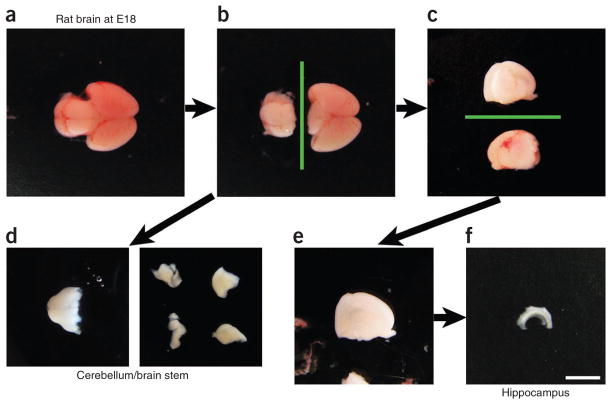
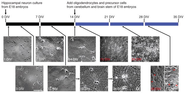
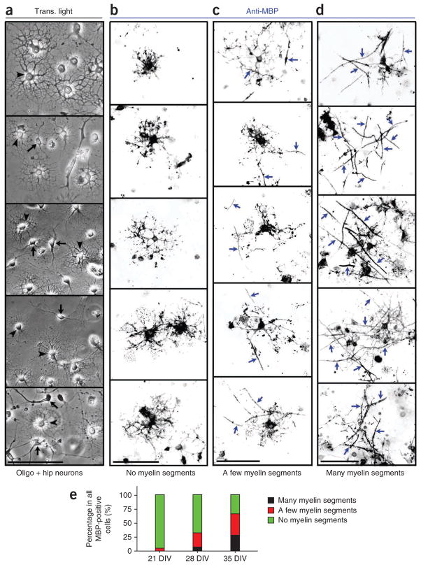
In the first section, we use perhaps the simplest neuron culture currently available, which we have been using successfully for more than 10 years7,9,10,26–28. This culture involves dissecting rat E18 embryos (Fig. 1) and culturing the neurons for up to 6 weeks in a chemically defined medium (Fig. 2). In contrast with the classic Banker culture29, an astrocyte feeding layer is not used in this protocol. In the next section of the protocol, after the hippo-campal neurons reach 14 days in vitro (14 DIV), oligodendrocytes and precursor cells dissociated from cerebellum and brain stem of rat E18 embryos (Fig. 1) are seeded onto the culture (Fig. 2). Initially, individual hippocampal neurons (>14 DIV) and developing oligodendrocytes with multiple processes can be clearly identified (Figs. 2 and 3a, respectively). At later stages, all of the cells are densely mingled together. Although it becomes more difficult to do so, hippocampal neurons can still be identified because of their large soma and proximal dendrites (Fig. 2). The unequivocal identification of individual neurons and oligodendrocytes often requires immunostaining. Myelination starts between 1 and 2 weeks of coculture (21–28 DIV) and peaks around 35 DIV. In this period, the myelin coculture is ready for use, although not all oligodendro-cytes become fully mature (Fig. 3). Even after 3 weeks of coculture, about one-third of myelin basic protein (MBP)-positive cells have multiple myelin segments, and one-third have one or two segments (Fig. 3)30. Axons of hippocampal neurons transfected with YFP before coculture can be myelinated (Fig. 4). Those mature oligodendrocytes with multiple myelin segments can be revealed by the fluorescence of viral (adeno-associated virus serotype-9)-mediated expression of green fluorescence protein (GFP) (AAV9-GFP) in these cells and the immunostaining for endogenous MBP (Fig. 5a,b). Each mature oligodendrocyte has multiple myelin segments that are both GFP and MBP positive (Fig. 5a,b). In contrast, the soma of these cells only expresses GFP but not MBP (Fig. 5a,b). This is consistent with the MBP distribution pattern in vivo, in which MBP is only present in the processes but not in the soma of myelinating oligodendrocytes. Images of transmission electron microscopy further show myelin membranes wrapping around axons with varied degrees of thickness and compactness in vitro (Fig. 5c). The variation may be due to in vitro unsynchronized development of neuron and myelin in culture, and possible intrinsic variability of gray matter myelination. Furthermore, myelin clusters the endogenous axonal Kv1.2 channels in hippocampal neurons30. Nodes of Ranvier and heminodes can also be observed30.
Figure 1.
Dissection procedure of rat brain from the E18 embryos for making the myelin coculture. (a) The top view of a whole brain of rat embryo at day 18. The frontal lobe is on the right and the cerebellum/brain stem is on the left. (b) The cerebellum and brain stem are cut away. Green line, cutting position. (c) The two hemispheres are split. One with the meninges removed (top) and the other with meninges remaining (bottom). Green line, cutting position. (d) The cerebellum and brain stem with the meninges removed (left) and after being cut into four pieces (right). (e) A cortical hemisphere without the hippocampus. (f) A dissected hippocampus. The dissection of hippocampus follows panels a–c, then e,f, whereas the dissection of cerebellum/brain stem follows panels a,b, then d. Scale bar, 3 mm. Black arrows show the sequence of dissection. All animal experiments have been conducted in accordance with the National Institutes of Health Animal Use Guidelines and approved by the Institutional Animal Care and Use Committee at The Ohio State University.
Figure 2.
The timeline for making hippocampal neuron culture and coculture. Hippocampal neurons from E18 rat embryos are dissociated and cultured for 14 d in the maintenance medium. Dividing glial cells are eliminated by the Ara-C treatment from 3 DIV to 5 DIV. Oligodendrocytes and precursor cells are dissociated from the cerebellum and brain stem of E18 rat embryos and added to the hippocampal neuron culture at 14 DIV. The maintenance medium is replaced with the myelin medium at 15 DIV. Then the culture medium is refreshed every 3 d. Myelination commences after 1–2 weeks of coculture. The example differential interference contrast microscopy images from hippocampal neuron culture (time labeled in white from 1 DIV to 26 DIV) and coculture (time labeled in red from 15 DIV to 29 DIV) at different stages are shown under the timeline. Red arrowheads, myelin segments. All images are at the same scale. Scale bar, 150 μm. Modified with permission from ref. 30. All animal experiments have been conducted in accordance with the National Institutes of Health Animal Use Guidelines and approved by the Institutional Animal Care and Use Committee at the Ohio State University.
Figure 3.
Maturation of oligodendrocytes cocultured with hippocampal neurons. (a) Light microscopic images obtained 2 d after oligodendrocytes (oligo) were added to 14 DIV hippocampal (hip) neurons. Black arrows, hippocampal neurons; black arrowheads, differentiating oligodendrocytes. (b–d) Oligo-dendrocytes in the coculture were stained for MBP, a marker of mature myelin. The coculture was fixed with 4% (wt/vol) formaldehyde, permeabilized with 0.2% (vol/vol) Triton X-100 and stained with a rat monoclonal anti-MBP antibody (Chemicon) at a 1:200 dilution. Signals are inverted. After coculture for 21 d, MBP-positive cells were categorized into three major groups: cells without myelin segments (b), cells with only one or two myelin segments (c), and cells with multiple myelin segments and reduced MBP staining in the soma (d). (e) Percentage of the three major groups of MBP-positive oligodendrocytes in the coculture at 21, 28 and 35 DIV (n = 50). Blue arrows, MBP-positive myelin segments. Scale bars, 100 μm. Trans., transmitted.
Figure 4.
Myelination of hippocampal neurons expressing YFP. (a) A YFP-expressing hippocampal neuron at 21 DIV in regular culture. The neuron was transfected with YFP (green; left) at 18 DIV, using the Ca2+-phosphate method32. The transmitted light image is on the right. (b–d) The soma (white asterisk) and proximal axon (white arrowheads) (b) of a YFP-expressing hippocampal neuron in myelin coculture at 30 DIV; the hippocampal neurons were transfected at 7 DIV using Lipofectamine 2000, before oligodendrocytes were seeded (b–d). A myelin segment, revealed by the rat monoclonal anti-myelin basic protein (MBP) antibody (1:200 dilution; Chemicon) (red), formed along the proximal axon. YFP is in green. (c,d) YFP-positive axons of transfected hippocampal neurons were myelinated revealed by MBP staining (white arrows)30. Scale bars: a, 100 μm; b, 50 μm; c,d, 25 μm.
Figure 5.
Myelin segments of mature oligodendrocytes in the coculture. Myelin coculture was infected with AAV9-GFP at 16 DIV, 2 d after oligodendrocytes/precursor cells were seeded. The coculture was fixed and stained after 28 DIV. Some mature oligodendrocytes expressed GFP. (a,b) Two examples of GFP-positive and mature oligodendrocytes (green) developed multiple myelin basic protein (MBP)-positive (red) myelin segments. White arrowheads, GFP- and MBP-positive myelin segments. (c) Two example images of transmission electron microscopy show the ultrastructure of myelin membranes. Red arrows, outer membranes. Blue arrowheads, inner membranes. Scale bars: a,b, 100 μm; c, 200 nm.
Applications of the protocol
This system can be useful for many different research projects. One example is the study of myelin-mediated regulation of trafficking and targeting of axonal ion channels, as shown in our previous published works30,31. This system can also be used to determine the factor(s) involved in regulating the interaction between oligodendrocytes and axons, and the efficacy of myelination, comparing the results from other myelin cocultures. Molecular mechanisms underlying the potential myelin-mediated regulation of intra-axonal trafficking of cellular organelles, such as mitochondria, and the targeting of other axonal proteins, can also be pursued with the system. Furthermore, the impact of myelin on neuronal function can be examined by using patch-clamp recordings or imaging techniques, which are currently used with the regular neuron culture10,28. Finally, this coculture system can be further developed to perform large-scale high-throughput drug screens.
Limitations of the protocol
Despite important potential applications of this system, there are still some limitations. To our knowledge, this protocol is already the simplest among myelin cocultures. There is no astrocyte feeding layer for hippocampal neurons and no purified OPCs (via immunopanning) are required. Nonetheless, making this coculture is still a lengthy process. Myelin segments can only be observed after at least 1 week of coculture. In the end, many axons in the coculture remain unmyelinated and many oligodendrocytes still do not fully mature (Fig. 2)30. Finally, this coculture includes a lengthy procedure and is certainly not the easiest one, but a technician with reasonable cell culture background and some patience can still successfully set up the whole system within a relatively short period of time.
MATERIALS
REAGENTS
Poly-D-lysine (PDL; Sigma-Aldrich, cat. no. P6407)
Rat tail collagen (Roche, cat. no. 11179179001)
Ultra Pure 10× PBS (National Diagnostics, cat. no. CL-253)
Acetic acid, glacial (Fisher Scientific, cat. no. A38-212)
NaOH (Fisher Scientific, cat. no. SS255-1)
Nitric acid (Fisher Scientific, cat. no. A200-500) ! CAUTION This is a highly corrosive and toxic strong mineral acid.
Na2SO4 (Fisher Scientific, cat. no. S421)
K2SO4 (Fisher Scientific, cat. no. P304)
HEPES (Fisher Scientific, cat. no. BP410)
D-Glucose (Fisher Scientific, cat. no. D16)
MgCl2 (Fisher Scientific, cat. no. BP214)
Proteinase 23 (Sigma-Aldrich, cat. no. P-4032)
FBS (Invitrogen, cat. no. 26140)
Sodium pyruvate, 100× (100 mM; Invitrogen, cat. no. 11360-070)
Glutamine, 100× (200 mM; Invitrogen, cat. no. 25030-81)
Penicillin-streptomycin (100×; Invitrogen, cat. no. 15140-122). This solution contains 10,000 units of penicillin and 10,000 μg of streptomycin per ml
MEM (with Earle’s salts with no L-glutamine; Invitrogen, cat. no. 11090-81)
Neurobasal medium (Invitrogen, cat. no. 21103-049)
B-27 supplement, 50× (Invitrogen, cat. no. 17504-044)
Cytosine arabinose (Ara-C; Sigma-Aldrich, cat. no. C1768-1G)
(Two) timed pregnant Sprague-Dawley rats at E18, with pregnancies spaced 14 d apart (Charles River Laboratory), one for the initial hippocampal neuron culture and the other for obtaining oligodendrocytes and precursor cells 2 weeks later ! CAUTION Experiments involving rats must conform to all relevant governmental and institutional ethics regulations.
Compressed CO2 (Ohio State University Stores)
Hydrochloric acid (Fisher Scientific, cat. no. A144) ! CAUTION This is a highly corrosive, strong mineral acid.
Transferrin (Sigma-Aldrich, cat. no. T-1147)
BSA (Sigma-Aldrich, cat. no. A-4161)
Progesterone (Sigma-Aldrich, cat. no. P8783)
Putrescine (Sigma-Aldrich, cat. no. P5780)
Sodium selenite (Sigma-Aldrich, cat. no. S5261)
Triiodo-thyronine (Sigma-Aldrich, cat. no. T6397)
Dulbecco’s phosphate-buffered saline (DPBS; Invitrogen, cat. no. 14040117)
Deionized water
N-acetyl cysteine (NAc; Sigma-Aldrich, cat. no. A8199)
Gibco Cellgro Trace Elements B (Invitrogen, cat. no. MT99-175-C1)
Biotin (Sigma-Aldrich, cat. no. B4639)
Insulin (Sigma-Aldrich, cat. no. I6634)
DMEM (high glucose; Invitrogen, cat. no. 11960-040)
EQUIPMENT
Pipetman, 1 ml (Molecular Technology)
Cell culture plates, 24 well (BD Falcon, cat. no. 353047)
Cell culture plates, six well (BD Falcon, cat. no. 353046)
Cell culture dishes, 10 cm (BD Falcon, cat. no. 353003)
Parafilm M (Pechiney Plastic Packaging, cat. no. PM-996)
Small (12 mm diameter) coverslips (Fisher Scientific, cat. no. 12-545-80)
Large (25 mm diameter) coverslips (Fisher Scientific, cat. no. 12-545-102)
Small forceps (FST by Dumont)
Large forceps (FST, cat. no. 11021-14)
Hemocytometer (Sigma-Aldrich, cat. no. Z359629)
Euthanasia box
Large scissors (FST, cat. no. 14130-17)
Spatula
Large ice bucket
Plastic bags (for animal carcass)
Paper towels
Dissection microscope (Nikon, cat. no. SMZ645)
Stericup filter unit (0.22 μm; Millipore, cat. no. SCGPTO5RE)
Syringe filter (0.22-μm pore size; Fisher Scientific 09-719C)
Syringe filter (0.45-μm pore size; Fisher Scientific 09-719D)
Microcentrifuge tubes (Fisher Scientific, cat. no. 02-682-550)
Incubator at 5% CO2 (NapCo Scientific Company)
Tissue culture fume hood (The Baker Company)
IEC HN-SII general purpose centrifuge (International Equipment Company)
REAGENT SETUP
Coverslip coating solution
Add 20 μl of rat tail collagen (3 mg ml−1 stock) and 200 μl PDL (0.5 mg ml−1 stock) to 780 μl of acetic acid (17 mM stock). Freshly prepare before use.
Poly-D-lysine (100×)
Add 10 ml of dH2O to 5 mg of PDL. Filter-sterilize PDL through a 0.22-μm filter and make 200-μl aliquots. ▲ CRITICAL Store at −80 °C for up to 12 months.
Slice dissection solution (SLDS)
Mix deionized water with 82 mM Na2SO4, 30 mM K2SO4, 10 mM HEPES (wt/vol), 10 mM glucose and 5 mM MgCl2; adjust the pH to 7.4 and filter-sterilize the solution through a 0.22-μm filter. ▲ CRITICAL Make a 5× stock without MgCl2 and store it at 4 °C. When you are ready to use it, dilute the stock to 1× and add 1 M MgCl2 to obtain a final concentration of 5 mM. The 1× solution can also be stored at 4 °C for up to 6 months.
Enzyme digestion solution
Mix 1× SLDS with 3 mg ml−1 proteinase 23. ▲ CRITICAL Make 3-ml aliquots and store them at −20 °C for up to 6 months.
Plating medium
Mix MEM with 10% (vol/vol) FBS, 0.45% (vol/vol) glucose, 1 mM sodium pyruvate, 25 μM glutamine and 1× penicillin-streptomycin. Filter-sterilize the medium through a 0.22-μm filter and store it at 4 °C for up to 3 months.
Maintenance medium
Combine neurobasal medium with 2% (vol/vol) 50× B-27 supplement, 0.5 mM glutamine and 1× penicillin-streptomycin. Filter-sterilize the medium through a 0.22-μm filter and store it at 4 °C for up to 3 months.
T3 (100×)
Dissolve 3.2 mg of triiodo-thyronine in 400 μl of 0.1 N NaOH. Add 10 μl of this mixture to 20 ml of DPBS. Filter-sterilize the solution through a 0.22-μm filter and store it at −20 °C in 200-μl aliquots for up to 6 months.
BSA, 4% (wt/vol, 20×)
Dissolve 2 g of BSA into 50 ml of DPBS at 37 °C. Adjust the pH to 7.4 and filter-sterilize the solution first through a 0.45-μm filter and then through a 0.22-μm filter. ▲ CRITICAL Store at −20 °C in 500-μl aliquots for up to 12 months.
Insulin (0.5 mg ml−1, 100×)
Dissolve 10 mg of insulin into 20 ml of dH2O. Add 100 μl of 1.0 N HCl and mix well. Filter-sterilize the solution through a 0.22-μm filter. ▲ CRITICAL Store insulin at −30 °C for up to 6 months. Discard any media (stored at 4 °C) containing insulin after 6 weeks.
NAc (5 mg ml−1, 1,000×)
Dissolve 50 mg of NAc in 10 ml of Neurobasal medium and aliquot. Store at −20 °C for up to 6 months.
Sato (100×)
Combine Neurobasal medium with 1% (wt/vol) transferrin, 1% (wt/vol) BSA, 2 μM progesterone, 0.16% (wt/vol) putrescine and 1% (wt/vol) sodium selenite (4.0 mg per 100 μl of 0.1 N NaOH in 10 ml of Neurobasal medium). Filter-sterlize the solution through a 0.22-μm filter and store it at −20 °C for up to 6 months. ▲ CRITICAL Do not reuse progesterone and sodium selenite stocks; freshly prepare them each time.
Myelin coculture medium (~44 ml)
Mix 19.5 ml of neurobasal medium with 19.5 ml of DMEM-high glucose. Thereafter, add the following (400 μl each): 200 mM glutamine, 100× penicillin-streptomycin, 100× sodium pyruvate (100 mM), insulin (0.5 mg ml−1), 100× Sato and 100× T3. (As these stock solutions are all at 100×, adding 400 μl each to the 40-ml final volume will give each 1× concentration.) Add 800 μl of 50× B-27 supplement. Next, add 40 μl of 1,000× NAc (5 mg ml−1), 40 μl of 1,000× biotin (10 μg ml−1) and 40 μl of 1,000× Cellgro Trace Elements B. Filter-sterilize the solution through a 0.22-μm filter and store it at 4 °C for up to 6 weeks.
PROCEDURE
Preparing coverslips ● TIMING ~3 d
-
1|
Place coverslips in 50 ml of nitric acid overnight.
-
2|
Wash treated coverslips with distilled water in a 1-liter beaker while shaking for 1 h. Repeat 8 times.
-
3|
Bake the washed coverslips at 250 °C for 6 h.
-
4|
Place one small or large coverslip in each well of a 24-well or 6-well plate, respectively.
▲ CRITICAL STEP Perform all of the following steps in a tissue culture hood.
-
5|
Add 30 μl of coating solution onto each coverslip in the 24-well plate, and 100 μl onto each coverslip in the 6-well plate, and swirl to coat the coverslips.
-
6|
Wait for ~10 min, add 1 ml of sterile PBS and then incubate at 4 °C overnight.
▲ CRITICAL STEP Coating will not be optimal if the PBS incubation is less than 10 h or longer than 2 d.
■ PAUSE POINT If coverslips are not used immediately after overnight PBS incubation, aspirate the PBS/coating solution, allow the cover slips to air-dry in the hood with the UV light on (~2 h), seal the plates with Parafilm and store them at 4 °C for up to 1 week.
Dissociating and culturing hippocampal neurons ● TIMING 2–3 h
-
7|
Remove PBS from plates and allow them to air-dry in the hood with the UV light on.
-
8|
Thaw the enzyme digestion solution and warm the plating medium to 37 °C.
-
9|
Place a few layers of paper towels in a plastic bag (for the carcass) and arrange surgical items on a clean surface near the dissection microscope.
-
10|
Take a pregnant female rat (E18) out of the animal facility, euthanize it by CO2 inhalation (~15 min) followed by cervical dislocation, and then use the large scissors to make an incision in the lower abdomen of the rat. Use the large scissors to cut open the abdomen and the large forceps to pull out embryos (6–12 embryos is normal). Cut the connecting tissue away and place the embryos in an empty Petri dish on the countertop, and dispose of the carcass.
-
11|
Add SLDS to two Petri dishes, one on ice and the other on the microscope.
-
12|
By using small forceps, open the embryonic sac and hold the first embryo at the neck. Use the other forceps to peel back the skin and reveal the skull.
-
13|
By following the midline of the brain, use forceps to impale the skull and peel it back.
▲ CRITICAL STEP Try not to injure the brain tissue.
-
14|
Use the spatula (treated with SLDS solution) to carefully remove the brain (both cerebrum and cerebellum) and place it in the SLDS buffer in a Petri dish on ice. Repeat Steps 12–14 until 8–10 brains are collected.
-
15|
Transfer one brain to the SLDS solution in the Petri dish on the dissection scope and use the forceps to remove the cerebellum. Cut the cerebrum in half and remove the meninges from around the brain. At this point, the brain should go from a red/pink color to a white color (Fig. 1c,d).
-
16|
Next, flip the cortical hemisphere onto its side with the inner surface facing up, and then use forceps to cut away the large inner area of the brain (Fig. 1d,e).
-
17|
Once the cortex has been cleaned, a C-shaped area should be revealed. This is the hippocampus. By using forceps, hold the cortex steady, remove the hippocampus and carefully transfer the hippocampus into a new Petri dish filled with SLDS on ice. Repeat Steps 15–17 to remove hippocampi from the other brains. Place all hippocampi in a single SLDS-filled Petri dish.
-
18|
By using a 1-ml pipette, suck up the hippocampi and place them in a 15-ml conical tube. Let the hippocampi sink to the bottom of the tube and remove extra SLDS solution with a pipette.
-
19|
Add 3 ml of enzyme digestion solution (30 hippocampi per 3 ml of enzyme digestion solution) to the tube and place it in a 37 °C incubator for 15 min.
-
20|
Under the culture hood, add 10 ml of plating medium to the 15-ml tube containing 3 ml of enzyme digestion solution and hippocampi sitting at the bottom of the tube. Wait until the hippocampi settle to aspirate and discard the wash, and then add another 10 ml of plating medium to the tube. Aspirate and discard the second wash and add another 10 ml of plating medium. The hippocampi will be dissociated in the final 10 ml.
-
21|
By using a glass pipette and rubber bulb (balloon), suck up hippocampi and forcefully expel them. Do this approximately 40 times until the solution is cloudy.
▲ CRITICAL STEP To prevent neuron death, do not do this more than 50 times. Avoid creating air bubbles.
-
22|
Count cells using a hemocytometer.
-
23|
Dilute the dissociated neurons in plating medium as follows: 1 ml per well of the 24-well plate (~105 cells per well), or 3 ml per well of the six-well plate (approximately 3 × 10⁵ cells per well). We estimate that three or four hippocampi are needed for each 24-well plate. A cell density of 3 × 10⁴ cells per well is used for the low-density culture, but it is normally not used for the myelin coculture. Incubate cells in the plating medium for 2–4 h at 37 °C. Then replace the medium with prewarmed maintenance medium and continue to culture at 37 °C for 2 d.
Maintaining hippocampal neuron culture ● TIMING ~14 d
-
24|
After 2 d of culture, replace half of the maintenance medium with 2 μM Ara-C (Ara-C inhibits the growth of fibroblasts, endothelial cells and glial cells), so that the final concentration of Ara-C is 1 μM. As neurons are postmitotic cells, Ara-C is used to eliminate or inhibit proliferating cells in the dish, such as fibroblasts and glial cells.
? TROUBLESHOOTING
-
25|
After 2 d of culture with Ara-C, replace the medium with fresh maintenance medium.
-
26|
Continue to culture the neurons, replacing medium every 3 d. As the neuron culture ages, to retain optimal growth the volume of replaced medium should change. In our experience, before 10 DIV, neurons can survive 100% medium replacement. Between 10 DIV and 20 DIV, we replace 50% of the culture medium. To make the myelin coculture, culture neurons at 12 DIV and proceed to Step 27. If you wish to continue culture of hippocampal neurons in isolation, continue to replace the medium every 3 d. Hippocampal neurons can be maintained for up to 5–8 weeks; however, the tolerance to medium replacement continues to diminish. Thus, after 20 DIV, we replace only 25–40% medium at each medium replacement.
? TROUBLESHOOTING
Seeding with cells dissociated from the cerebellum and brain stem onto cultured hippocampal neurons ● TIMING ~2 h
-
27|
Two days before seeding cells from cerebellum/brain stem onto 14 DIV hippocampal neurons, change half of the maintenance medium in the neuron culture.
-
28|
When the previously cultured hippocampal neurons are at 14 DIV, repeat Steps 8–14 with the second pregnant rat.
-
29|
Transfer one brain to the SDLS solution in the Petri dish on the dissection scope and use the forceps to remove the meninges from the cerebellum/brain stem, separate the halves (Fig. 1b,f) and place them into 3 ml of the enzyme digestion solution; thereafter, incubate at 37 °C for 15 min. We use 1.5 cerebellums or brain stems for one 24-well plate.
-
30|
Similarly to Step 20, under the culture hood, add 10 ml of plating medium to a 15-ml conical tube, aspirate the washes, add another 10 ml of plating medium to the tube, aspirate and add a further 10 ml of plating medium. The cerebellum will be dissociated in the last 10 ml.
-
31|
By using a glass pipette and a rubber bulb, pipette the cerebellum and enzyme solution ~40 times until the medium is cloudy.
▲ CRITICAL STEP Ensure that cells are completely dissociated but not lysed.
-
32|
Spin down cells at 100g with an IEC HN-SII centrifuge for 5 min at room temperature (20–25 °C) and resuspend them in 10 ml of maintenance medium.
-
33|
Count the cells and dilute the suspension to 105 cells per 300 μl.
-
34|
Add the cell suspension to 14 DIV hippocampal neurons (300 μl per well) dropwise with a 1-ml pipette. Incubate the coculture for 24 h. At this step, we do not purify oligodendrocytes or precursor cells from other cell types in these brain regions, such as astrocytes and neurons. Therefore, this part of the brain that is usually discarded during dissection in the hippocampal neuron culture is used here for making the myelin coculture.
Maintaining the myelin coculture ● TIMING 2–3 weeks
-
35|
After the 24-h incubation, remove 700 μl of medium (to account for the added 300 μl of added cells and 400 μl of the medium normally needed to change) and add 400 μl of the myelin medium.
? TROUBLESHOOTING
-
36|
Continue to culture cells and to change myelin medium every 3 d by replacing 250–400 μl of the medium in the wells with fresh myelin medium. In the first week of coculture, we replace 400 μl of medium. In the second week, we replace 300 μl of medium. In the third week, we replace 250 μl of medium.
▲ CRITICAL STEP It is essential to change the myelin medium on time to ensure maximal myelination or full maturation of oligodendrocytes.
? TROUBLESHOOTING
? TROUBLESHOOTING
Troubleshooting advice can be found in Table 1.
TABLE 1.
Troubleshooting table.
| Step | Problem | Possible reason | Solution |
|---|---|---|---|
| 24 | Neurons aggregate and detach from coverslips | Coverslip glass or coating procedure | Glass materials from different companies may differ. We have been using coverslips from the same company over the years. Double-check the cleaning procedure of coverslips (with nitric acid) and check or remake the coating solution |
| 26 | Neuronal cell death or slow development before myelination | Something is wrong with the culture conditions associated with the neuron culture medium | Double-check the CO2and water levels in the incubator. Make fresh neuron culture medium and check the pH of the medium in the incubator as indicated by the color (pH too low—yellow; pH too high—bright pink) |
| 35 | Neuronal cell death or unhealthy neurons after seeding the cells dissociated from cerebellum and brain stem | The medium containing the dissociated cells may damage the hippocampal neurons in culture | Double-check the components and pH of the neuron culture medium used for seeding cerebellar cells. Wash away the proteinase during the digestion. Seed less cells, for instance at the density 5 × 10⁴ cells per well of a 24-well plate |
| 36 | Death or low density of seeded cells | The culture medium conditioned by the 14-DIV hippocampal neurons may not be optimal for newly dissociated cells | Change half of the culture medium of hippocampal neurons around 2 d before the seeding. Double-check the new medium containing the newly dissociated cells |
| Both neurons and seeded cells seem to be alive, but no or little myelin forms | The culture conditions are not optimal for oligodendrocyte maturation and differentiation | Increase the coculture time to 3 weeks. The coculture must be fed on time, once every 3 d. Any variation in feeding can markedly decrease the efficiency of myelin formation without apparent cell death |
● TIMING
Steps 1–6, preparing coverslips: ~3 d
Steps 7–23, dissociating and culturing hippocampal neurons: 2–3 h
Steps 24–26, maintaining the hippocampal neuron culture: ~14 d
Steps 27–34, seeding cells dissociated from the cerebellum and brain stem onto cultured hippocampal neurons: ~2 h
Steps 35 and 36, maintaining the myelin coculture: 2–3 weeks
ANTICIPATED RESULTS
Extensive studies have been done on the development of rodent hippocampal neurons in dissociated culture2,29. The development of hippocampal neurons under our culture conditions is largely consistent with what has been previously published. When initially plated, hippocampal neurons appear like scattered small spheres. About a few hours to half a day after plating, the neurons extend lamella-like short processes around the cell body, called stage 1 (ref. 1). At stage 2 (within 2 DIV), neurites with equal length and large growth cones can be observed. At stage 3 (around 2–3 DIV), one neurite grows much longer than the rest and then becomes the axon. At stage 4 (around 3–7 DIV), dendrites start to grow and branch, whereas the axon continues to grow. At stage 5 (>7 DIV), neurons become mature, synapses start to form and dendritic spines on some neurons can be observed.
Our protocol provides an important advancement to this widely used culture technique. This protocol describes how to induce myelin formation along axons of cultured hippocampal neurons. Oligodendrocytes and precursor cells mixed with other cell types dissociated from cerebellum/brain stem are seeded on hippocampal neurons at 14 DIV, when the neurons are considered mature. Myelin starts to form between 1 and 2 weeks after coculture and gradually increases afterward. Myelin segments shine under the microscope with the transmitted light and are easy to detect. Under our conditions, we have found that myelination reaches its peak after a 3-week coculture. Prolonged incubation does not seem to further increase the formation of myelin segments. However, because our normal experimental time window is within 5–6 weeks, we have not yet extensively tried to optimize the conditions to maintain older cultures. It is possible that with some modification of the protocol these neurons and myelin can survive much longer.
This coculture system is ideal for molecular studies, as both loss-of-function and gain-of-function experiments for hip-pocampal neurons or oligodendrocytes can be performed. This can be done through transient transfection of vector-based RNAi probes or the coding sequence of a gene of interest30, or via viral-mediated infection (Figs. 4 and 5). The best time for Lipofectamine 2000–mediated transfection to neurons is around 5–7 DIV. Efficiency drops and cell death occurs if neurons older than 10 DIV are transfected. Some constructs can be transfected into young neurons (~7 DIV) using Lipofectamine 2000, and their expressed proteins can persist up to 3 weeks (~28 DIV) until myelination occurs in the coculture, allowing assays to be performed. However, the expression level of some other proteins decreases 10 d after transfection. Therefore, a different approach has to be used. To transfect older neurons (from 10 DIV to 21 DIV), we use the Ca2+-phosphate method32. When we transfect constructs at 14 DIV, they are likely to persist through the myelination process and become useful for the assay. In addition, the coculture can be used to perform viral-mediated expression or knockdown. So far, AAV has worked the best. In particular, AAV9-GFP seems to efficiently infect maturating oligodendrocytes and expresses GFP in both cell bodies and myelin processes (Fig. 5a,b).
This coculture system is highly accessible by a variety of different techniques, such as immunostaining, live cell imaging, patch clamp recording, calcium imaging and so on10,28,30. A major advantage of this system over cultured slices is the easy manipulation of either neurons or myelin-making oligodendrocytes. However, we are continuing to develop this system. It should be a very useful tool to study basic neuroscience questions, such as neuron-glia interaction, as well as to investigate pathogenic mechanisms underlying neurological diseases involving hippocampal myelination and to devise better strategies to treat these diseases.
Acknowledgments
We thank J. Barry and Y. Gu for technical assistance, and the Campus Microscopy & Imaging facility at the Ohio State University and Research Core Services at Lerner Research Institute of Cleveland Clinic Foundation for technical assistance in transmission electron microscopy. This work was supported by a career transition fellowship award from the US National Multiple Sclerosis Society (grant TA3012A1) and a grant from the US National Institute of Neurological Disorders and Stroke/National Institutes of Health (R01NS062720) to C.G. All animal experiments have been conducted in accordance with the US National Institutes of Health Animal Use Guidelines.
Footnotes
AUTHOR CONTRIBUTIONS A.G. and C.G. designed and performed the experiments. A.G., P.J. and C.G. wrote the manuscript. C.G. supervised the project.
COMPETING FINANCIAL INTERESTS The authors declare no competing financial interests.
Reprints and permissions information is available online at http://www.nature.com/reprints/index.html.
References
- 1.Dotti CG, Sullivan CA, Banker GA. The establishment of polarity by hippocampal neurons in culture. J Neurosci. 1988;8:1454–1468. doi: 10.1523/JNEUROSCI.08-04-01454.1988. [DOI] [PMC free article] [PubMed] [Google Scholar]
- 2.Craig AM, Banker G. Neuronal polarity. Annu Rev Neurosci. 1994;17:267–310. doi: 10.1146/annurev.ne.17.030194.001411. [DOI] [PubMed] [Google Scholar]
- 3.Horton AC, Ehlers MD. Neuronal polarity and trafficking. Neuron. 2003;40:277–295. doi: 10.1016/s0896-6273(03)00629-9. [DOI] [PubMed] [Google Scholar]
- 4.Dotti CG, Banker GA. Experimentally induced alteration in the polarity of developing neurons. Nature. 1987;330:254–256. doi: 10.1038/330254a0. [DOI] [PubMed] [Google Scholar]
- 5.Sampo B, Kaech S, Kunz S, Banker G. Two distinct mechanisms target membrane proteins to the axonal surface. Neuron. 2003;37:611–624. doi: 10.1016/s0896-6273(03)00058-8. [DOI] [PubMed] [Google Scholar]
- 6.Wisco D, et al. Uncovering multiple axonal targeting pathways in hippocampal neurons. J Cell Biol. 2003;162:1317–1328. doi: 10.1083/jcb.200307069. [DOI] [PMC free article] [PubMed] [Google Scholar]
- 7.Gu C, Jan YN, Jan LY. A conserved domain in axonal targeting of Kv1 (Shaker) voltage-gated potassium channels. Science. 2003;301:646–649. doi: 10.1126/science.1086998. [DOI] [PubMed] [Google Scholar]
- 8.Gu C, et al. The microtubule plus-end tracking protein EB1 is required for Kv1 voltage-gated K+ channel axonal targeting. Neuron. 2006;52:803–816. doi: 10.1016/j.neuron.2006.10.022. [DOI] [PubMed] [Google Scholar]
- 9.Barry J, Gu Y, Gu C. Polarized targeting of L1-CAM regulates axonal and dendritic bundling in vitro. Eur J Neurosci. 2010;32:1618–1631. doi: 10.1111/j.1460-9568.2010.07447.x. [DOI] [PMC free article] [PubMed] [Google Scholar]
- 10.Gu Y, Gu C. Dynamics of Kv1 channel transport in axons. PLoS ONE. 2010;5:e11931. doi: 10.1371/journal.pone.0011931. [DOI] [PMC free article] [PubMed] [Google Scholar]
- 11.Horton AC, et al. Polarized secretory trafficking directs cargo for asymmetric dendrite growth and morphogenesis. Neuron. 2005;48:757–771. doi: 10.1016/j.neuron.2005.11.005. [DOI] [PubMed] [Google Scholar]
- 12.Hayashi T, Thomas GM, Huganir RL. Dual palmitoylation of NR2 subunits regulates NMDA receptor trafficking. Neuron. 2009;64:213–226. doi: 10.1016/j.neuron.2009.08.017. [DOI] [PMC free article] [PubMed] [Google Scholar]
- 13.Ryu J, et al. A critical role for myosin IIb in dendritic spine morphology and synaptic function. Neuron. 2006;49:175–182. doi: 10.1016/j.neuron.2005.12.017. [DOI] [PubMed] [Google Scholar]
- 14.Williams ME, et al. Cadherin-9 regulates synapse-specific differentiation in the developing hippocampus. Neuron. 2011;71:640–655. doi: 10.1016/j.neuron.2011.06.019. [DOI] [PMC free article] [PubMed] [Google Scholar]
- 15.Arnold SE, Trojanowski JQ. Human fetal hippocampal development: I. Cytoarchitecture, myeloarchitecture, and neuronal morphologic features. J Comp Neurol. 1996;367:274–292. doi: 10.1002/(SICI)1096-9861(19960401)367:2<274::AID-CNE9>3.0.CO;2-2. [DOI] [PubMed] [Google Scholar]
- 16.Haber M, Vautrin S, Fry EJ, Murai KK. Subtype-specific oligodendrocyte dynamics in organotypic culture. Glia. 2009;57:1000–1013. doi: 10.1002/glia.20824. [DOI] [PubMed] [Google Scholar]
- 17.Dutta R, et al. Demyelination causes synaptic alterations in hippocampi from multiple sclerosis patients. Ann Neurol. 2011;69:445–454. doi: 10.1002/ana.22337. [DOI] [PMC free article] [PubMed] [Google Scholar]
- 18.Noble M. The possible role of myelin destruction as a precipitating event in Alzheimer’s disease. Neurobiol Aging. 2004;25:25–31. doi: 10.1016/j.neurobiolaging.2003.07.001. [DOI] [PubMed] [Google Scholar]
- 19.Dawodu S, Thom M. Quantitative neuropathology of the entorhinal cortex region in patients with hippocampal sclerosis and temporal lobe epilepsy. Epilepsia. 2005;46:23–30. doi: 10.1111/j.0013-9580.2005.21804.x. [DOI] [PubMed] [Google Scholar]
- 20.Chambers JS, Perrone-Bizzozero NI. Altered myelination of the hippocampal formation in subjects with schizophrenia and bipolar disorder. Neurochem Res. 2004;29:2293–2302. doi: 10.1007/s11064-004-7039-x. [DOI] [PubMed] [Google Scholar]
- 21.Chan JR, et al. NGF controls axonal receptivity to myelination by Schwann cells or oligodendrocytes. Neuron. 2004;43:183–191. doi: 10.1016/j.neuron.2004.06.024. [DOI] [PMC free article] [PubMed] [Google Scholar]
- 22.Watkins TA, Emery B, Mulinyawe S, Barres BA. Distinct stages of myelination regulated by gamma-secretase and astrocytes in a rapidly myelinating CNS coculture system. Neuron. 2008;60:555–569. doi: 10.1016/j.neuron.2008.09.011. [DOI] [PMC free article] [PubMed] [Google Scholar]
- 23.Nash B, et al. Functional duality of astrocytes in myelination. J Neurosci. 2011;31:13028–13038. doi: 10.1523/JNEUROSCI.1449-11.2011. [DOI] [PMC free article] [PubMed] [Google Scholar]
- 24.Thomson CE, et al. Myelinated, synapsing cultures of murine spinal cord—validation as an in vitro model of the central nervous system. Eur J Neurosci. 2008;28:1518–1535. doi: 10.1111/j.1460-9568.2008.06415.x. [DOI] [PMC free article] [PubMed] [Google Scholar]
- 25.Lubetzki C, et al. Even in culture, oligodendrocytes myelinate solely axons. Proc Natl Acad Sci USA. 1993;90:6820–6824. doi: 10.1073/pnas.90.14.6820. [DOI] [PMC free article] [PubMed] [Google Scholar]
- 26.Xu M, Cao R, Xiao R, Zhu MX, Gu C. The axon-dendrite targeting of Kv3 (Shaw) channels is determined by a targeting motif that associates with the T1 domain and ankyrin G. J Neurosci. 2007;27:14158–14170. doi: 10.1523/JNEUROSCI.3675-07.2007. [DOI] [PMC free article] [PubMed] [Google Scholar]
- 27.Xu M, Gu Y, Barry J, Gu C. Kinesin I transports tetramerized Kv3 channels through the axon initial segment via direct binding. J Neurosci. 2010;30:15987–16001. doi: 10.1523/JNEUROSCI.3565-10.2010. [DOI] [PMC free article] [PubMed] [Google Scholar]
- 28.Gu Y, Barry J, McDougel R, Terman D, Gu C. Alternative splicing regulates kv3.1 polarized targeting to adjust maximal spiking frequency. J Biol Chem. 2012;287:1755–1769. doi: 10.1074/jbc.M111.299305. [DOI] [PMC free article] [PubMed] [Google Scholar]
- 29.Kaech S, Banker G. Culturing hippocampal neurons. Nat Protoc. 2006;1:2406–2415. doi: 10.1038/nprot.2006.356. [DOI] [PubMed] [Google Scholar]
- 30.Gu C, Gu Y. Clustering and activity tuning of Kv1 channels in myelinated hippocampal axons. J Biol Chem. 2011;286:25835–25847. doi: 10.1074/jbc.M111.219113. [DOI] [PMC free article] [PubMed] [Google Scholar]
- 31.Gu C, Barry J. Function and mechanism of axonal targeting of voltage-sensitive potassium channels. Prog Neurobiol. 2011;94:115–132. doi: 10.1016/j.pneurobio.2011.04.009. [DOI] [PMC free article] [PubMed] [Google Scholar]
- 32.Jiang M, Chen G. High Ca2+-phosphate transfection efficiency in low-density neuronal cultures. Nat Protoc. 2006;1:695–700. doi: 10.1038/nprot.2006.86. [DOI] [PubMed] [Google Scholar]


