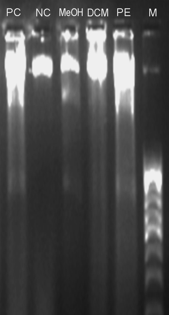
Fig. 6.

The gel electrophoresis image obtained after DNA fragmentation assay for apoptosis detection. From left lanes are: positive control/Curcumin treated (PC), negative control/no treatment (NC), MeOH, DCM and PE extracts of SBOI treated cells and marker/100 bp DNA ladder (M)
