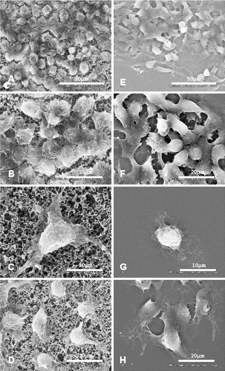
Fig. 3.
SEM images of HepG2 cells. Cells cultured on nitrocellulose membranes at 2 days (d, ×2,000) and 4 days (a, ×1,000; b, ×2,000) after seeding. Various pseudopod-like structures formed on the side of the membrane by which cells had formed a tight bond with the porous membrane (c, ×4,000). Cells cultured on coverslips at the same time as the cells cultured on membranes after seeding as controls: 2 days (h, ×2,000) and 4 days (e, ×1,000; f, ×2,000) after seeding. No obvious pseudopod -like structures formed between cells and coverslips (g, ×4,000)

