Abstract
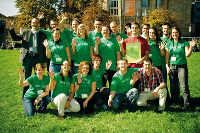
The seventeenth European Bioenergetics Conference (EBEC) took place in September 2012 at the Albert-Ludwigs-University in Freiburg, Germany, and was hosted by Thorsten Friedrich. The conference is a biannual event that brings together researchers from across the globe to discuss progress in this diverse and challenging field.
The EBEC is a major international event in ‘Bioenergetics’, the discipline that studies energy transformation in biological systems. The series began in 1980, shortly after the Nobel Prize in Chemistry was awarded to Peter Mitchell for his work on the chemiosmotic mechanism of energy conservation. The Mitchell theory still provides the framework for understanding how respiratory complexes, photosynthetic complexes and ATP synthases work, and how the transport of ions and metabolites through energy-conserving membranes integrates metabolism and cell respiration within the context of cellular signalling. This perfect match allows the production of ATP to be adapted to the changing energy requirements of the cell, and its derangements often provide the key to understanding disease pathogenesis. Since 1994, in recognition of Mitchell's outstanding contributions to the discipline, the meeting has begun with a plenary lecture by the recipient of the Peter Mitchell Medal, which in two cases preceded the award of a Nobel Prize. This year's Medal was awarded to H. Ronald Kaback (U. California, Los Angeles, USA), who presented a lecture on ‘Lactose permease: a beautiful chemiosmotic machine’. The meeting then unfolded through plenary lectures, parallel symposia and poster presentations. It is impossible to cover all presentations, so we have selected a set of topics and apologize to those who are not featured here.
F-ATPases: much more to be learned
F-ATPases of mitochondria, chloroplasts and bacteria are multi-subunit enzymes that use the proton electrochemical gradient generated by respiration or photosynthesis to synthesize ATP under aerobic conditions. Under anaerobic conditions, ATP might instead be hydrolysed by F-ATPases, if they are not inhibited by the protein IF1. However, not all F-ATPases can hydrolyse ATP. If you had thought that solving the crystal structure of the mitochondrial F-ATPase would have revealed the mechanism by which the enzyme works, you would have been surprised by the opening lecture by Sir John Walker (MRC, Cambridge, UK). The picture is actually far more complex. When 48 ATPase structures were compared for rotation, Walker found that whilst the α- and β-subunits—which form the catalytic core of F1 and carry out ATP synthesis—are similar, the structures of the foot and central stalk—the ‘transmission shaft’—differ. Fortunately, the regulatory features of the F-ATPase should allow us to manipulate the enzyme to solve both structure and rotation through the use of IF1 to block the catalytic cycle at different points. Solving the structures will allow us to remodel the catalytic cycle. Walker also pointed out that the number of c-subunits in the rotating membrane Fo—eight in mammals and invertebrates, ten in yeast and 11–15 in prokaryotes—is linked to the magnitude of the proton electrochemical gradient. Mammals push a smaller ‘gear’ thanks to the high and constant gradient that allows ATP production to be maximized.
Photo credit: Dr Oliver Einsle
Thomas Meier (MPI of Biophysics, Frankfurt, Germany) specifically addressed the problem of coupling ion flux and Fo rotation. Site-directed mutagenesis of a conserved stretch of glycines, of the c11 Na+-motive ATPase of Ilyobacter tartaricus, generated functionally assembled complexes with various numbers of c-subunits and, thus, c-ring sizes. The mutant enzyme harbouring the larger c12 was functional at lower ion-motive force, reflecting an ability to adapt to specific bioenergetic demands. In spite of a generally conserved mechanism that couples ion translocation to ATP synthesis, F-type ATP synthases differ enough for the design of specific drugs, and this is good news. Indeed, Meier recalled that diarylquinolines have proven to be effective anti-tuberculosis agents by selectively inhibiting the F-ATPase of mycobacteria.
Wayne D. Frasch (Arizona State U., Tempe, USA) spoke about the mechanism of F1-ATPase γ-subunit rotation in Escherichia coli. Frasch presented single-molecule measurements obtained by monitoring light scattered from a 75 nm × 35 nm gold nanorod attached to the γ-subunit as a probe of rotation during catalysis. This technique provides the ability to resolve the rotational velocity of the enzyme and observe substeps of rotation that are too short to be observed by other approaches. Effects of treatments on the velocity of rotation were reported that provided new insight into the molecular mechanism of ATPase-driven rotation.
F-type ATP synthases differ enough for the design of specific drugs, and this is good news
Masamitsu Futai (Iwate Medical U., Japan) presented a similar study in which he carried out a mutational analysis of γ-subunit M23 and β-subunit E381, which interact during catalysis in E. coli. A γ-subunit M23K mutant had lower rotation speed due to increased catalytic dwell time, in spite of similar 120° stepping rate, and wild-type behaviour was reinstated by a β E381D mutation. He also studied the effects of polyphenols on rotational catalysis.
F-ATPases are involved not only in ATP synthesis, but also in the organization of mitochondrial cristae. They form dimers that constitute the building blocks of long rows of ATPase oligomers. The organization of F-ATPase into dimers has been observed in fungi, plants and vertebrates and seems essential for the maintenance of normal morphology in all species. Karen Davies (MPI of Biophysics, Frankfurt, Germany) showed that the yeast dimer reveals a twofold symmetrical V-shape structure, with an angle of 86° between monomers, as seen at 3.7 nm resolution. Werner Kühlbrandt (MPI of Biophysics, Frankfurt) showed that a single F-ATPase dimer causes a local deformation of the lipid bilayer that is sufficient to start cristae curvature, and suggested that regions with high membrane curvature might maintain a lower local pH, and hence provide a larger contribution of ΔpH to the protonmotive force. Marie-France Giraud (U. Bordeaux, France) presented organization models of membrane regions of different F-ATPase oligomeric species. Subunits 4 and 6 are located at the dimerization interface; subunits e and g are essential for dimer stabilization and are located at the oligomerization interface. An unexpected finding was presented by Paolo Bernardi's laboratory (U. Padova, Italy), indicating that F-ATPase dimers might form the mysterious permeability transition pore (PTP), a high-conductance inner membrane channel that requires adequate loads of matrix Ca2+ and is favoured by oxidative stress. When reconstituted in lipid bilayers, F-ATPase dimers but not monomers gave rise to currents with the expected features of the PTP, suggesting a new function for the mitochondrial F-ATPase that would directly link the enzyme to the effector pathways of cell death.
Respiratory complexes
Complex (C) I is the largest protein complex of bacterial and mitochondrial respiratory chains. Uli Brandt (U. Frankfurt, Germany) reminded us that 90% of energy conversion takes place here, with a daily production of some 85 kg of NADH, which corresponds to 3,000 l of H2. The first three-dimensional structure of the bacterial complex has been determined, and its overall architecture characterized by X-ray crystallography, yet the mechanism of redox-driven proton pumping remains under study, as does the binding site for ubiquinone. Brandt showed that the quinone head group is mainly stabilized by hydrogen bonds to the conserved Tyr 144 of subunit 49 kDa, whilst the hydrophobic tail interacts at the interface of the 49 kDa and PSST subunits. Leonid Sazanov (MRC, Cambridge, UK) further defined the quinone-binding site in his description of the studies on the structure of the entire Thermus thermophilus CI. More on CI was reported by Ilka Wittig (U. Frankfurt, Germany) who introduced complexome profiling as a powerful method to identify protein–protein interactions and that was used for the identification of TMEM126B as a component of the mitochondrial CI assembly complex.
…a single F-ATPase dimer causes a local deformation of the lipid bilayer that is sufficient to start cristae curvature […] regions with high membrane curvature might maintain a lower local pH
Luca Scorrano (U. Geneva, Switzerland) showed data on the relationship between respiratory chain supercomplexes (RCSs) and the shape of the cristae. RCSs are quaternary structures of the respiratory chain complexes, the best known of which is the so-called ‘respirosome’, which includes CI, CIII and CIV and is capable of electron transfer from NADH to molecular oxygen. Although RCSs are thought to be crucial for efficient function of the respiratory chain, the requirements for their formation, as well as their role in cell and mitochondrial physiology are still a matter of investigation. Scorrano showed that ablation of the master cristae biogenetic molecule optic atrophy 1 (Opa1) impairs the assembly of the quaternary structures of the RCSs. Conversely, during apoptosis, when cristae remodel, the RCSs disassemble and ultimately slow down cell growth. Thus, Opa1-dependent cristae remodelling determines how efficiently mitochondria use substrates by defining the assembly and the stability of the RCSs. Leo Nijtmans (U. Nijmegen, the Netherlands) presented a mouse model lacking the NDUSF4 nuclear gene, which is required for CI assembly. Absence of the gene caused the loss of the electron influx part (N-module), which could be diminished by CI+CIII RCS formation. But how do RCSs assemble, and why are they important? Cristina Ugalde (Hospital U. 12 de Octubre, Madrid) suggested that in human cells RCSs do not originate from the association of fully assembled individual holo-enzymes, but rather from CI intermediates that act as a scaffold for incorporation of free subunits and of subcomplexes from CIII and CIV. On the issue of RCS importance, Maria Luisa Genova (U. Bologna, Italy) showed that the formation of CI–CIII supercomplexes provides the advantage of facilitating electron transfer in the CoQ region, also decreasing the production of reactive oxygen species by CI. Walter Neupert (MPI of Biochemistry, Martinsried, Germany) discussed the components required for the formation of cristae junctions and rims, that is, Fcj1 and subunits e and g of the F-ATPase. He reported the identification of a new supermolecular complex that consists of at least six different proteins including Fcj1. These proteins preferentially locate to cristae junctions and interact with the outer membrane TOM–SAM and Ugo1–Fzo1 complexes, which are essential for biogenesis of β-barrel proteins and fusion, respectively.
Mitochondria and diseases
With an estimated 1017 mitochondria in the body—as compared with 1014 bacteria in the intestine—mitochondrial (mt)DNA is by far the most represented genome in the human body, as Douglas C. Wallace (U. Pennsylvania, USA) noted in his lecture on the mitochondrial aetiology of complex diseases. The main philosophical question Wallace raised was whether anatomical (organ-specific) and classical Mendelian paradigms are still adequate to describe diseases, or whether we should rather consider a ‘bioenergetic threshold’ as the key, which would also involve mitochondrial pathogenesis for diseases that are not caused by mtDNA mutations. The evolution of mtDNA haplotypes—that is, of genetic variants—has allowed us to track the colonization of the world by humanity. Wallace put forward the hypothesis that the different mtDNA haplotypes represent an adaptation to climate and high altitude, which would allow mitochondrial performance to be optimized—higher or lower coupling—to the needs of the environment. In turn, mtDNA background would then dictate whether specific mutations result in disease. For example, there is a striking correlation between life at high altitude, the M9 haplogroup and the T3394C mutation, which causes CI deficiency in N haplogroups B4 and F1, but not in M9.
The maintenance of healthy mitochondria is also mediated by efficient mechanisms that remove damaged organelles. An emerging concept is that depolarization is the trigger for autophagy, and Richard Youle (NIH, Bethesda, USA) discussed the role of PINK1 (a kinase anchored to the mitochondrial surface) and Parkin (a cytosolic E3 ubiquitin ligase) in the process, and in the pathogenesis of Parkinson disease. In healthy mitochondria (high proton gradient and ATP production), PINK1 is degraded by the PARL protease, whilst in depolarized mitochondria (no proton gradient, no ATP production), PINK1 gets stuck in the outer membrane—because of the clogging of its removal process—allowing for Parkin binding and mitochondrial removal. In the discussion, an important point was raised that the mitochondrial membrane potential must be close to zero, which would guarantee that phosphorylating mitochondria—which have a lower membrane potential than resting mitochondria—are not removed. This issue was also touched on by Laura D. Osellame (U. College London, UK), who spoke about Gaucher disease—a lysosome storage disorder caused by a defect of the glucocerebrosidase (GBA) gene that leads to the accumulation of glucocerebroside. Strikingly, the loss of GBA activity confers the highest risk of developing Parkinson disease, and this is recapitulated in a gba−/− mouse model. The model has a block in autophagy and a compromised ubiquitin-proteasome system, both of which contribute to the accumulation of α-synuclein and the formation of Lewy bodies. This is accompanied by the prominent fragmentation of mitochondria without the recruitment of Parkin. Mitochondria were also centre stage in the presentation of Aurora Pujol (ICREA and IDIBELL, Barcelona, Spain), who spoke about adrenoleukodystrophy—a neurodegenerative condition caused by loss-of-function of the peroxisomal ABCD1 transporter. In this disease, the prominent upregulation and oxidation of mitochondrial cyclophilin D—a ‘positive’ regulator of the PTP that increases its probability of opening and thus of cell death—was found to have a causative role.
Elzbieta Glaser (Stockholm U., Sweden) discussed an important aspect of protein degradation with major implications for disease pathogenesis. The question she addressed was the fate of cleaved import sequences, which are degraded by presequence protease (PreP), a Zn2+ metalloprotease of the Pitrilysin family. Glaser reported that the crystal structure of PreP reveals a substrate acceptor cavity of 10,000 Å3, and that enzyme catalysis is decreased by oxidation and favoured by the reduction of the highly conserved M206 by the reductase MrsA. Glaser showed that the amyloid Aβ1–42 peptide is a substrate of the mitochondrial TOM import machinery and that it is degraded by PreP. Moreover, a mouse model of Alzheimer disease has decreased activity of PreP, which identifies it as a potential target for treatment.
Novel methods, new frontiers
Petra Fromme (Arizona State U., Tempe, USA) presented a fascinating study by using a new technique to resolve X-ray diffraction from a fully hydrated stream of nanocrystals as small as 500 nm—compared with the approximately 1 mm crystals needed for use with conventional techniques. The method was used on the large photosystem I, which consists of 36 proteins and 381 co-factors, to collect patterns from individual nanocrystals.
Walter Neupert […] reported the identification of a new supermolecular complex that consists of at least six different proteins including Fcj1
From the infinitely small to the quite small, Robert Balaban (NIH, Bethesda, USA) took us to the world of imaging of vital functions in situ, such as the mitochondrial membrane potential, Ca2+ transients and redox potentials. The frontier seems to be total emission detection by multiphoton microscopy, and Balaban explained that the problems with living tissues are movement and deformation. He presented a way to correct this by using an ‘optical navigator’ to perform slice-tracking with motion compensation within 1–2 μm. He showed beautiful images of muscular vascular flow with amazing levels of detail, including a capillary and mitochondrial groove in slow skeletal muscle fibres, which have 40% of the total oxidative capacity. He also demonstrated the existence of a gradient of NADH redox potential in situ, examples of a technique that holds great promise for the future study of organ pathophysiology.
In summary, the seventeenth EBEC was an ‘energetic’ meeting that allowed us to go back to work with fully recharged batteries.
Footnotes
The authors declare that they have no conflict of interest.


