Abstract
Background/Purpose:
While elastic resistance training, targeting the upper body is effective for strength training, the effect of elastic resistance training on lower body muscle activity remains questionable. The purpose of this study was to evaluate the EMG‐angle relationship of the quadriceps muscle during 10‐RM knee‐extensions performed with elastic tubing and an isotonic strength training machine.
Methods:
7 women and 9 men aged 28‐67 years (mean age 44 and 41 years, respectively) participated. Electromyographic (EMG) activity was recorded in 10 muscles during the concentric and eccentric contraction phase of a knee extension exercise performed with elastic tubing and in training machine and normalized to maximal voluntary isometric contraction (MVC) EMG (nEMG). Knee joint angle was measured during the exercises using electronic inclinometers (range of motion 0‐90°).
Results:
When comparing the machine and elastic resistance exercises there were no significant differences in peak EMG of the rectus femoris (RF), vastus lateralis (VL), vastus medialis (VM) during the concentric contraction phase. However, during the eccentric phase, peak EMG was significantly higher (p<0.01) in RF and VM when performing knee extensions using the training machine. In VL and VM the EMG‐angle pattern was different between the two training modalities (significant angle by exercise interaction). When using elastic resistance, the EMG‐angle pattern peaked towards full knee extension (0°), whereas angle at peak EMG occurred closer to knee flexion position (90°) during the machine exercise. Perceived loading (Borg CR10) was similar during knee extensions performed with elastic tubing (5.7±0.6) compared with knee extensions performed in training machine (5.9±0.5).
Conclusion:
Knee extensions performed with elastic tubing induces similar high (>70% nEMG) quadriceps muscle activity during the concentric contraction phase, but slightly lower during the eccentric contraction phase, as knee extensions performed using an isotonic training machine. During the concentric contraction phase the two different conditions displayed reciprocal EMG‐angle patterns during the range of motion.
Level of Evidence:
5
Keywords: Electromyography, strength training, quadriceps, perceived exertion
INTRODUCTION
Reduced knee‐extension strength is commonly reported in over‐use knee pathologies, for example, the patello‐femoral pain syndrome,1 and after total knee replacement,2,3 suggesting that strength training of the quadriceps muscle is needed.4 Accordingly, knee joint replacement and knee joint pain is often associated with muscular atrophy,5 and central activation failure3,6,7 resulting in reduced functional performance, potential for future injury and increased risk for long‐term sickness and work‐related absence.8,9 Heavy resistance training during a prolonged training period yields muscular hypertrophy,10–12 gains in strength,13–15 increased neuromuscular (efferent) drive11,16,17 and reduced pain.18 However, the availability of strength training equipment is often a limiting factor during rehabilitation and training interventions.19
A growing interest in developing simple and effective training methods convenient for exercising at the workplace, in the hospital, at home or at the training field18–20 has been emerging for quite some time. High‐intensity strength training using elastic resistance has shown to be equally effective in activating smaller muscles in the neck, shoulder, and arm when compared to similar training exercises performed isotonically with dumbbells.21,22 The effect of elastic resistance training on larger muscle groups such as the quadriceps, however, remains largely unexplored. Furthermore, development of simple and effective training exercises feasible for use during rehabilitation of knee pathologies is needed.
Electromyography (EMG) and electro‐goniometry obtained during resistance training and physical rehabilitation exercises provide valuable information on temporal and spatial muscle activation strategies during different angular phases of the exercise through range of motion. Whereas the EMG ‐ joint angle relationship is well described for conventional (i.e. using dumbbell or machine) isoinertial strength exercises,23 only one recent study has investigated the EMG‐angle pattern during elastic resistance exercise.24 Aboodarda et al. compared the EMG angle‐relationship during knee extension using elastic resistance and a Nautilus isotonic machine (Nautilus, Vancouver, WA) and showed that the average vastus lateralis muscle activity was similar during the two exercise modalities. Nevertheless, the EMG‐angle relationship of multiple prime mover muscles (rectus femoris, vastus medialis and vastus lateralis) as well as the antagonist/synergists muscle activity during knee extensions remains un‐investigated. An imbalance in the synergetic activation ratio of the vastus medialis (VM) and vastus lateralis (VL) may contribute to knee injuries such as patellafemoral pain syndrome.25,26 Accordingly, exercises with higher VM to VL ratios may be preferred during rehabilitation.23 Although conventional knee extensions performed in an isotonic machine may not preferentially activate the VM over the VL,23 the VM to VL activation ratio may differ throughout range of motion during knee extensions performed using elastic resistance.
The purpose of this study was to evaluate the EMG‐angle relationship of the quadriceps muscle during 10‐RM knee‐extensions performed with elastic tubing and an isotonic strength training machine.
MATERIALS AND METHODS
Experimental Approach to the Problem
Muscle activity and perceived loading (rated on a Borg CR10 scale) during leg strengthening exercises performed in a training machine or with elastic resistance were evaluated using a cross‐sectional design. For each individual muscle, EMG muscle activity of each of the dynamic muscle contraction was normalized to the amplitude elicited during a maximal voluntary isometric contraction (MVC).
Subjects
The study was performed in Copenhagen, Denmark. A group of 16 untrained adults (7 women and 9 men) were recruited from a large workplace with various job tasks. Exclusion criteria were blood pressure above 160/100, disc prolapse, or serious chronic disease. Table 1 displays the subject demographics. All participants performed both conditions of knee extension testing, with elastic resistance and in the isotonic training machine. All subjects were informed about the purpose and content of the project, and gave written informed consent to participate in the study, which conformed to The Declaration of Helsinki, and was approved by the Local Ethical Committee (H‐3‐2010‐062).
Table 1.
Demographics of the men and women of this study. Data presented as mean (SD).
| Men | Women | |
|---|---|---|
| n | 9 | 7 |
| Age, yrs | 41(15) | 44(10) |
| Height, cm | 179(6) | 167(8) |
| Weight, kg | 78(4) | 67(16) |
| BMI | 24(2) | 24(6) |
Exercise equipment
Two different types of knee‐extension strength‐training equipment were used; 1) elastic tubing with resistances ranging from light to very heavy (red, green, blue, black, silver/gray colors, TheraBand, Akron, USA) and 2) an isotonic knee‐extension machine (Vertical seated knee extension, Technogym, Gambettola, Italy).
Exercise description
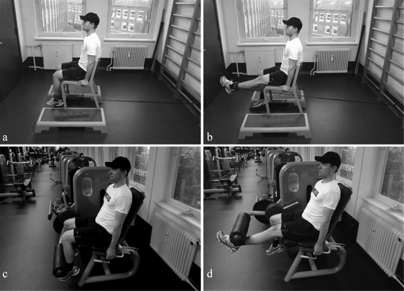
A week prior to testing, the participants performed a 10 repetition maximum (10 RM) test for the two exercises. During the elastic resistance exercise the 10 RM loading was found using one or a combination of several elastic tubes with resistances ranging from light to very heavy (red, green, blue, black, gray colors). All exercises were performed unilaterally using the dominant leg (preferred leg) as the exercising leg. A week later, on the day of EMG measurements, participants warmed up with submaximal loads, and then performed three consecutive repetitions for each exercise, using the predetermined 10 RM load, to avoid the influence of fatigue on the subsequent exercises. Exercises were performed in a controlled manner at a slow constant speed [participants attempted to perform each repetition in ∼3 sec (eccentric phase: ∼1.5 sec and concentric phase: ∼1.5 sec)]. The order of exercises was randomized for each subject, and the rest period between different exercises was approximately five minutes. The exercises are shown in Figure 1 and described in detail below:
Figure 1.
(a) Start position for knee extension against elastic resistance. (b) End position for knee extension against elastic resistance. (c) Start position for knee extension performed on the isotonic machine. (d) End position for knee extension performed on the isotonic machine.
Knee extension with elastic resistance (Fig. 1a & 1b).
The participant was sitting on a high chair, facing away, from a wooden bar (elastic fixation point located 10 cm above the floor and with a horizontal distance of ∼1.5 m from the chair to the bar) with both legs flexed at ∼90° knee joint angle and a 90° hip flexion. The elastic tubing was fixated to the participant's ankle on one end (1 finger above the medial malleoli), and the other end was attached to the bar. The elastic band was then stretched to ∼200% of the initial length. The participant started extending the knee from the flexed knee position (∼90° knee joint angle) (concentric phase) until full extension (∼0° knee joint angle), and then returned to the flexed knee position (eccentric phase).
Isolated knee extension in machine (Fig. 1c & 1d).
The participant was seated in a Technogym knee extension machine with the leg flexed at ∼90° knee joint angle and a 100° hip flexion. The machine's lever arm was fixated 1 finger above the medial malleoli. The participant started by extending the knee (concentric phase) until full extension was achieved (∼0° knee joint angle), and then flexed the knee (eccentric phase) returning to the ∼90° knee joint angle.
Inclinometer sampling and analysis
Knee joint angle was continuously measured using two electronic inclinometers (2D DTS inclination sensor, Noraxon, Arizona, USA) placed at the lateral side of the tibia and femur, respectively. The inclinometer data were synchronously sampled with the EMG data, using the 16‐channel 16‐bit PC‐interface receiver (TeleMyo DTS Telemetry, Noraxon, Arizona, USA). The dimension of the probes was 3.4 cm × 2.4 cm × 3.5 cm. During subsequent analysis, the inclinometer signals were digitally lowpass filtered using a 4th order zero‐lag Butterworth filter (3 Hz cutoff frequency).
The momentary knee joint angle was calculated as the difference in angular position, with respect to the gravitational line, between the tibia and femur inclinometers. Knee joint angles ranged from a 908 flexed position to a 0° full knee extension. The concentric and eccentric phases were defined as periods with negative or positive angular velocity, respectively, (going from 90°‐0° or 0°‐90°, respectively). Angle at peak EMG was calculated within the concentric and eccentric phase.
EMG signal sampling and analysis
EMG signals were recorded from 10 leg, abdominal, and lower back muscles, including: vastus medialis (VM), vastus lateralis (VL), rectus femoris (RF) (prime movers for knee extension) and biceps femoris, semitendinosus, adductors, gluteus medius, right erector spinae, right external oblique and the right rectus abdominis (non‐prime movers for knee extension). A bipolar surface EMG configuration (Blue Sensor N‐00‐S, Ambu A/S, Ballerup, Denmark) with an inter‐electrode distance of 2 cm were used.23,27 Before affixing the electrodes, the skin of the respective area was prepared with scrubbing gel (Acqua gel, Meditec, Parma, Italy) to effectively lower the impedance to less than 10 kΩ.21 Electrode placements for all muscles followed SENIAM recommendations (www.seniam.org).
The EMG electrodes were connected directly to wireless probes that pre‐amplified the signal (gain 400) and transmitted data in real‐time to a 16–channel 16‐bit PC‐interface receiver (TeleMyo DTS Telemetry, Noraxon, Arizona, USA). The dimension of the probes was 3.4 cm × 2.4 cm × 3.5 cm. Data was collected at a sampling rate of 1500 Hz. Common mode rejection ratio was higher than 100 dB.
During later off‐line analysis, all raw EMG signals obtained during MVCs as well as during the exercises were digitally filtered by a Butterworth 4th order high‐pass filter (10 Hz cutoff frequency). For each individual muscle, maximal moving root mean square (RMS) (500 ms constant) EMG was used to identify peak EMG within the concentric and eccentric phase whereas the RMS of the highpass filtered EMG signal was calculated within each 10° angle interval (0°‐10°, 10°‐20°,… 80°‐90°) of the concentric and eccentric phases and then normalized to the maximal moving RMS (500‐ms time constant) EMG obtained during MVC.21,27,28 Contraction time was calculated according to procedures previously described in.27
Maximal voluntary isometric contraction (MVC)
Prior to the dynamic exercises described above, isometric MVCs were performed, according to standardized procedures during 1) static knee extension and 2) flexion manoeuvres (positioned in a Biodex dynamometer: knee angle: 70° and hip angle: 110°), 3) hip adduction (lying flat on the back and pressing the knees against a solid ball), 4) hip abduction (lying flat on the back and pressing the knees outwards against a rigid band) and 5) hip extension (lying flat on the stomach with the knee flexed (90°) and pressing the foot upwards against the instructors hands), and 6) trunk extension and 7) trunk flexion (in standing posture and pelvis fixated the trunk was extended against a rigid band) to induce a maximal EMG response in the tested muscles.29 Two MVCs were performed for each muscle, and the trial with the highest RMS EMG value was subsequently used for normalization of the RMS EMG signals obtained in the resistance exercises. During the MVCs, subjects were instructed to gradually increase muscle contraction force towards maximum over a period of two seconds, sustain the MVC for three seconds, and then slowly release the force again. Strong and standardized verbal encouragement was given during all trials.
Perceived loading
Immediately after each set of exercise, the Borg CR10 scale30 (Appendix 1) was used to rate perceived loading during the resistance exercise. We have previously validated this scale in the evaluation of neck/shoulder resistance exercises with elastic resistance.21
Statistical analysis
A two‐way repeated measures analysis of variance (Proc Mixed, SAS version 9, SAS Institute, Cary, NC) was used to determine if differences existed between exercises and range of knee joint motion for each muscle and contraction mode (concentric or eccentric), vastus medialis to vastus lateralis activation ratio, perceived loading (BORG) and contraction time. Factors included in the model were Exercise (elastic resistance and machine) and knee joint angle (0‐90 degrees), as well as Exercise by knee joint angle interaction. The analysis was controlled for gender and age. Normalized EMG was the dependent variable. Values are reported as least square means (SE) unless otherwise stated. P‐values ≤0.05 were considered statistically significant.
A priori power analysis showed that 16 participants in this paired design were sufficient to obtain a statistical power of 80% at a minimal relevant difference of 10% and a type I error probability of 1%, assuming standard deviation of 10% based on previous research in the authors laboratory.31
RESULTS
Normalized EMG
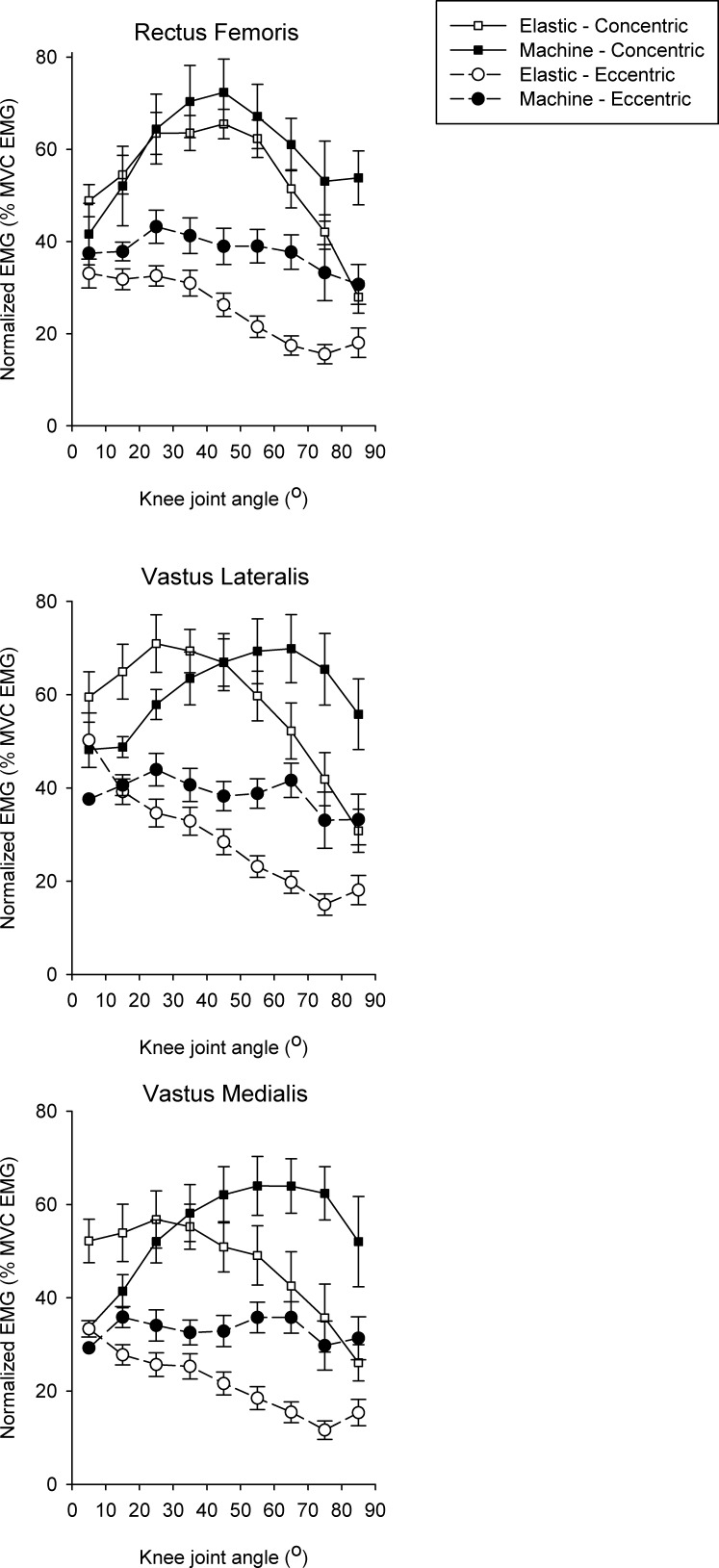
Figure 2 shows the normalized EMG‐angle relationship for the quadriceps muscles during knee extension exercises performed in machine or with elastic resistance during the 0‐90° knee joint range of motion. There was no significant difference in maximal EMG between machine and elastic resistance exercise for the prime movers for knee extension (rectus femoris, vastus lateralis, vastus medialis) during the concentric contraction phase (Table 2). However, during the eccentric phase, peak EMG was significantly higher (p<0.01) in RF and VM when performing knee extensions using the training machine compared with elastic resistance.
Figure 2.
EMG data for Quadriceps during the four different conditions, represented as means and standard deviations.
Table 2.
Maximal nEMG (% of max) and angle at maximal nEMG obtained during the concentric and eccentric phase of knee extensions performed with machine and elastic resistance.
| Muscle | nEMG(%of max) | Angle at peak EMG (°) | |||||||||
|---|---|---|---|---|---|---|---|---|---|---|---|
| Elastic | Machine | Elastic | Machine | ||||||||
| Mean | SE | Mean | SE | P | Mean | SE | Mean | SE | P | ||
| Rectus femoris | CONCENTRIC | 76.7 | 4.6 | 83.9 | 4.6 | .24 | 38.9 | 4.3 | 55.0 | 4.3 | <.01 |
| ECCENTRIC | 45.1 | 3.6 | 60.7 | 3.5 | <.01 | 15.5 | 4.9 | 52.4 | 4.9 | <.01 | |
| Vastus lateralis | CONCENTRIC | 83.7 | 4.6 | 84.7 | 4.6 | .87 | 33.8 | 4.3 | 60.2 | 4.3 | <.01 |
| ECCENTRIC | 53.1 | 3.6 | 61.6 | 3.5 | .08 | 12.7 | 4.9 | 60.5 | 4.9 | <.01 | |
| Vastus medialis | CONCENTRIC | 71.5 | 4.6 | 81.8 | 4.6 | .10 | 34.3 | 4.3 | 64.3 | 4.3 | <.01 |
| ECCENTRIC | 43.2 | 3.6 | 57.3 | 3.5 | <.01 | 15.6 | 4.9 | 59.6 | 4.9 | <.01 | |
| Adductors | CONCENTRIC | 17.2 | 4.5 | 20.2 | 4.6 | .61 | 31.0 | 4.3 | 58.3 | 4.3 | <.01 |
| ECCENTRIC | 11.4 | 3.4 | 15.2 | 3.5 | .43 | 18.1 | 4.8 | 49.9 | 4.9 | <.01 | |
| Gluteus medius | CONCENTRIC | 13.2 | 4.5 | 13.2 | 4.6 | .99 | 30.0 | 4.3 | 42.9 | 4.3 | <.01 |
| ECCENTRIC | 8.5 | 3.4 | 8.4 | 3.5 | .98 | 12.4 | 4.8 | 24.6 | 4.9 | .04 | |
| Erector spinae | CONCENTRIC | 13.3 | 4.5 | 21.2 | 4.6 | .19 | 31.9 | 4.3 | 62.4 | 4.3 | <.01 |
| ECCENTRIC | 12.0 | 3.4 | 17.2 | 3.5 | .28 | 39.7 | 4.8 | 48.8 | 4.9 | .12 | |
| Obliques externus | CONCENTRIC | 13.2 | 4.5 | 20.0 | 4.6 | .26 | 29.4 | 4.3 | 46.8 | 4.3 | <.01 |
| ECCENTRIC | 9.3 | 3.4 | 12.8 | 3.5 | .46 | 20.6 | 4.8 | 36.9 | 4.9 | <.01 | |
| Rectus abdominis | CONCENTRIC | 7.0 | 4.5 | 11.3 | 4.6 | .47 | 27.2 | 4.3 | 48.1 | 4.3 | <.01 |
| ECCENTRIC | 4.6 | 3.4 | 6.2 | 3.5 | .74 | 23.6 | 4.8 | 40.7 | 4.9 | <.01 | |
| Biceps femoris | CONCENTRIC | 13.6 | 4.5 | 17.3 | 4.6 | .53 | 43.2 | 4.3 | 68.5 | 4.3 | <.01 |
| ECCENTRIC | 8.6 | 3.4 | 13.2 | 3.5 | .34 | 24.8 | 4.8 | 64.8 | 4.9 | <.01 | |
| Semi tendinosus | CONCENTRIC | 7.9 | 4.5 | 7.3 | 4.6 | .93 | 36.1 | 4.3 | 67.0 | 4.3 | <.01 |
| ECCENTRIC | 5.3 | 3.4 | 5.4 | 3.5 | .97 | 16.4 | 4.8 | 62.8 | 4.9 | <.01 | |
Data presented as mean and SE (of the least square means and p‐values denotes differences between the elastic and machine exercise.
There was a significant exercise by knee joint angle interaction (P<0.01). The EMG‐knee joint angle relationships for the two investigated vasti were different between training modalities. For the machine, the concentric phase EMG‐amplitude peaked near maximal knee flexion (60.2°±4.3 and 60.5°±4.3 for the VL and VM, respectively) and decreased towards full knee extension, whereas the opposite pattern was seen for the elastic tubing (angle at peak EMG: 33.8°±4.3 and 34.3°±4.3 for the VL and VM, respectively). Irrespectively of training condition, angle and contraction mode, the VM to VL EMG ratio never exceeded 1.00 and was similar between the two exercise conditions.
All muscles besides the prime movers RF, VL and VM demonstrated low peak EMG values (<21% of nEMG). However, these followed a comparable EMG‐angle pattern as VL and VM of their respective training condition (i.e. using elastic resistance the EMG increased towards knee extension whereas EMG increased towards knee flexion during the machine exercise). Accordingly, significant differences (p<0.01) were observed in angle at peak EMG between the two exercise conditions.
External load and contraction time
The average load of the machine exercise was 28±2.31 kg, ranging from 15‐50 kg. The 10 RM elastic resistance ranged from a combination of 1xSilver, 1xBlue and 1xGreen to 3xSilver, 1xBlack, 1xBlue, 1xGreen and 1xRed (TheraBand elastic tubes).
Irrespectively of training condition there was no significant difference in contraction time (i.e. time under tension) during the knee extension exercise. Contraction times for machine and elastic resistance were 1824±111 ms and 1834±103 ms respectively, and for contraction modes concentric vs. eccentric were 1733±80 ms and 1572±75 ms, respectively.
Perceived loading and influence of age and gender
Perceived loading assessed with the Borg CR10 rating scale was similar (p=0.67) during knee extensions performed with elastic bands (5.72±0.57) compared with knee extensions performed in training machine (5.87±0.47). There were no significant effects of age and gender on muscle activity (p=0.71 and p=0.72, respectively).
DISCUSSION
The main finding of this study was that knee extensions performed with elastic tubing induces similar quadriceps EMG muscular activity as knee extensions using an isotonic training machine. However, different EMG‐angle patterns existed between exercise conditions.
Irrespectively of loading modality (machine or elastic), there was no significant difference in maximal quadriceps (rectus femoris (RF), vastus lateralis (VL), vastus medialis (VM)) EMG during the concentric contraction phase. The EMG‐angle pattern of the VL and VM, however, demonstrated quite a different pattern when comparing the two exercise conditions. When using elastic resistance, the EMG activity of the concentric phase increased from knee flexion position and peaked towards full knee extension whereas a reciprocal behavior was observed during the machine exercise, where the EMG activity increased from extension and peaked closer to knee flexion position. This difference may be explained by the elastic force generation (i.e. external loading) being greatest at the more extended knee angles whereas the intramuscular force induced by the external loading may be greater at the more flexed knee angles during the machine exercise.
A recent study by Aboodarda et al. compared the EMG angle‐relationship of knee extensions performed using elastic resistance and a Nautilus machine and showed that the average vastus ‐lateralis muscle activity was similar during the two exercise modalities.24 As previously indicated, Aboodarda et al. demonstrated that the applied force exerted during the elastic resistance exercise increases from knee flexion to knee extension, while the Nautilus machine provided a more constant load throughout ROM.24 Although, Aboodarda et al. observed significantly higher exerted forces in the first 66% of ROM (44°‐100°) using the Nautilus, the EMG‐angle pattern was quite similar (in intensity and shape) displaying an increase towards full knee extension during both exercise modalities. Accordingly, the present EMG‐angle pattern using elastic resistance seems comparable with the findings of Aboodarda et al., whereas a reciprocal EMG‐angle pattern seems to exist when comparing the Nautilus machine with the machine used in the present study.
Open‐chain exercises are generally better tolerated in early rehabilitation of postoperative patients when closed‐chain exercises such as a squat are not feasible. Despite conflicting guidelines regarding the impact of patellofemoral stress in extended knee positions32–34 studies have shown that open‐chain protocols are effective and safe near full knee extension.35,36 Accordingly, greater quadriceps muscle activity during extended knee positions, as observed using elastic resistance compared with machines, may be particularly beneficial for rehabilitation of knee pathologies such as ACL injury and following total knee arthroplasty where strength deficits have been observed to be present during the most extended knee angles.37,38 Thus, the observed reciprocal EMG‐angle pattern between the two training modalities may have clinical relevance when designing specific rehabilitation and strengthening programmes.
When comparing the two exercise modalities, the RF muscle demonstrated a somewhat similar curvilinear EMG‐angle relationship peaking in the mid region (39‐55°) of the concentric phase. This similarity in RF EMG‐angle pattern between the two types of exercise indicates less dependence on the specific type of regional external loading. This behavior may be explained by the bi‐articular function of the RF working as a knee extensor as well as a hip flexor consequently leading to enhanced activation in the middle part of range of motion irrespectively of exercise modality.
Knee extensions using the isotonic machine resulted in higher eccentric activation values compared to elastic resistance. During the machine exercise the eccentric EMG‐pattern was rather constant throughout ROM, whereas the elastic resistance showed an increase in EMG towards knee extension. This may indicate that the higher and more constant eccentric activation during the machine exercise makes this a more effective training modality for rehabilitation of muscular strain injuries.39,40 However, this assertion should be tested in a randomized controlled trial.
An imbalance in the synergetic stabilization ratio of the VM and VL may contribute to knee injuries such as patellofemoral pain syndrome.25,26 Atrophy of the VM is often the cause of an imbalance in the VM to VL ratio,41–43 consequently, making the VM muscle an important target for achieving a balanced knee joint. Thus, exercises with higher VM to VL ratios may be preferred during rehabilitation.23 Nevertheless, in line with previous findings of conventional knee extensions performed in machine23 the VM:VL‐ratio never exceeded 1:00 throughout ROM and was quite similar in the two training modalities. Although none of the exercises seems to preferentially activate VM over the VL, the VM:VL‐ratio never fell below 0:85 making both exercises optimal for maintaining patellar joint alignment.
The activation of the non‐prime movers (all muscles besides RF, VL and VM) was rather low for both exercise modalities, however, these followed a comparable EMG‐angle pattern as VL and VM during their respective training conditions. Accordingly, this indicates that increases in prime mover activity during knee extension are accompanied by a simultaneous increase in synergetic and antagonist muscle activation to preserve fixation and control of the knee and surrounding joints.
Perceived loading was similar between machine and elastic resistance exercises. This is in line with the comparable peak EMG and contraction time (time under tension) values observed during performance of the two exercises. Importantly, the exercises induce high muscle activity regardless of gender and age. Thus, these exercises can be used beneficially for both younger and elderly individuals, as well as men and women.
The knee extension exercise performed with elastic resistance seems to be a feasible and simple method, regardless of age and gender, for achieving high muscle activity potentially stimulating muscular hypertrophy and strength gains in the quadriceps muscles. Its portability makes it ideal for work site training, rehabilitation in hospitals, at home or in training fields where there may be few resources for large training equipment. A future randomized controlled study is needed to investigate the ability of elastic resistance exercise to increase knee‐extension strength over time.
Limitations
The fact that the chair has to be high (or raised), to prevent foot and ground contact when performing the knee extensions using elastic tubing, may limit the feasibility of the exercise. Alternatively, the exercise can be performed with the distal femur elevated e.g. with a pillow or triangular box beneath the thigh to ensure free movement of the lower leg. As this slightly changes the joint angles, it should be noted, that this may alter the muscle activity pattern. However, when considering this suggestion, the difference in hip angle between the elastic and machine exercise (90° vs. 100°) as well as the change in the elastic's angle of pull throughout the ROM should be considered when interpreting the EMG‐angle pattern of the two exercise conditions.
CONCLUSION
In untrained individuals, knee extensions performed with elastic tubing induces similar quadriceps EMG muscle activity during the concentric contraction phase, but slightly lower activity during the eccentric contraction phase, as knee extensions using an isotonic training machine. During the concentric contraction phase the two modalities displayed reciprocal EMG activity patterns during the range of motion. This reciprocal behaviour may have clinical relevance when designing specific rehabilitation and strengthening programmes.
APPENDIX 1
Borg CR10 scale
Use this scale to rate the load you experience (i.e. weight in the machine or resistance of the elastic band) in relation to your muscle strength.
0= Nothing at all
10= The maximal load that you can imagine if you use all your muscle strength
| Number | Load |
|---|---|
| 0 | Nothing at all |
| 0.3 | |
| 0.5 | Just noticeable |
| 0.7 | |
| 1 | Very light |
| 1.5 | |
| 2 | Light |
| 2.5 | |
| 3 | Moderate |
| 4 | |
| 5 | Heavy |
| 6 | |
| 7 | Very heavy |
| 8 | |
| 9 | |
| 10 | Maximal |
Borg CR 10 Scale
REFERENCES
- 1.Kaya D, Citaker S, Kerimoglu U, et al. Women with patellofemoral pain syndrome have quadriceps femoris volume and strength deficiency. Knee Surg Sports Traumatol Arthrosc. 2011;19(2):242–247 [DOI] [PubMed] [Google Scholar]
- 2.Holm B, Kristensen MT, Bencke J, Husted H, Kehlet H, Bandholm T. Loss of knee‐extension strength is related to knee swelling after total knee arthroplasty. Arch Phys Med Rehabil. 2010;91(11):1770–1776 [DOI] [PubMed] [Google Scholar]
- 3.Mizner RL. Emerging perspectives related to quadriceps central activation deficits in patients with total knee arthroplasty. Exerc Sport Sci Rev. 2012;40(2):61–62 [DOI] [PubMed] [Google Scholar]
- 4.Hart JM, Pietrosimone B, Hertel J, Ingersoll CD. Quadriceps activation following knee injuries: a systematic review. J Athl Train. 2010;45(1):87–97 [DOI] [PMC free article] [PubMed] [Google Scholar]
- 5.Baugher WH, Warren RF, Marshall JL, Joseph A. Quadriceps atrophy in the anterior cruciate insufficient knee. Am J Sports Med. 1984;12(3):192–195 [DOI] [PubMed] [Google Scholar]
- 6.Williams GN, Buchanan TS, Barrance PJ, Axe MJ, Snyder‐Mackler L. Quadriceps weakness, atrophy, and activation failure in predicted noncopers after anterior cruciate ligament injury. Am J Sports Med. 2005;33(3):402–407 [DOI] [PubMed] [Google Scholar]
- 7.Roos EM. Joint injury causes knee osteoarthritis in young adults. Curr Opin Rheumatol. 2005;17(2):195–200 [DOI] [PubMed] [Google Scholar]
- 8.Andersen LL, Mortensen OS, Hansen JV, Burr H. A prospective cohort study on severe pain as a risk factor for long‐term sickness absence in blue‐ and white‐collar workers. Occup Environ Med. 2011;68(8):590–592 [DOI] [PubMed] [Google Scholar]
- 9.Andersen LL, Clausen T, Mortensen OS, Burr H, Holtermann A. A prospective cohort study on musculoskeletal risk factors for long‐term sickness absence among healthcare workers in eldercare. International Archives of Occupational and Environmental Health. 2011. Available at: http://www.ncbi.nlm.nih.gov/pubmed/21986907 Accessed November 13, 2011. [DOI] [PubMed]
- 10.Aagaard P, Andersen JL, Dyhre‐Poulsen P, et al. A mechanism for increased contractile strength of human pennate muscle in response to strength training: changes in muscle architecture. J. Physiol. (Lond.). 2001;534(Pt. 2):613–623 [DOI] [PMC free article] [PubMed] [Google Scholar]
- 11.Narici MV, Roi GS, Landoni L, Minetti AE, Cerretelli P. Changes in force, cross‐sectional area and neural activation during strength training and detraining of the human quadriceps. Eur J Appl Physiol Occup Physiol. 1989;59(4):310–319 [DOI] [PubMed] [Google Scholar]
- 12.Andersen LL, Tufekovic G, Zebis MK, et al. The effect of resistance training combined with timed ingestion of protein on muscle fiber size and muscle strength. Metab. Clin. Exp. 2005;54(2):151–156 [DOI] [PubMed] [Google Scholar]
- 13.Andersen LL, Andersen JL, Magnusson SP, et al. Changes in the human muscle force‐velocity relationship in response to resistance training and subsequent detraining. J. Appl. Physiol. 2005;99(1):87–94 [DOI] [PubMed] [Google Scholar]
- 14.Kraemer WJ, Fleck SJ, Evans WJ. Strength and power training: physiological mechanisms of adaptation. Exerc Sport Sci Rev. 1996;24:363–397 [PubMed] [Google Scholar]
- 15.Aagaard P, Simonsen EB, Andersen JL, Magnusson SP, Halkjaer‐Kristensen J, Dyhre‐Poulsen P. Neural inhibition during maximal eccentric and concentric quadriceps contraction: effects of resistance training. J. Appl. Physiol. 2000;89(6):2249–2257 [DOI] [PubMed] [Google Scholar]
- 16.Andersen LL, Andersen JL, Magnusson SP, Aagaard P. Neuromuscular adaptations to detraining following resistance training in previously untrained subjects. Eur. J. Appl. Physiol. 2005;93(5‐6):511–518 [DOI] [PubMed] [Google Scholar]
- 17.Häkkinen K, Alén M, Komi PV. Changes in isometric force‐ and relaxation‐time, electromyographic and muscle fibre characteristics of human skeletal muscle during strength training and detraining. Acta Physiol. Scand. 1985;125(4):573–585 [DOI] [PubMed] [Google Scholar]
- 18.Andersen LL, Andersen CH, Zebis MK, Nielsen PK, Søgaard K, Sjøgaard G. Effect of physical training on function of chronically painful muscles: a randomized controlled trial. J. Appl. Physiol. 2008;105(6):1796–1801 [DOI] [PubMed] [Google Scholar]
- 19.Zebis MK, Andersen LL, Pedersen MT, et al. Implementation of neck/shoulder exercises for pain relief among industrial workers: a randomized controlled trial. BMC Musculoskelet Disord. 2011;12:205. [DOI] [PMC free article] [PubMed] [Google Scholar]
- 20.Thorborg K, Bandholm T, Petersen J, et al. Hip abduction strength training in the clinical setting: with or without external loading? Scand J Med Sci Sports. 2010;20 Suppl 2:70–77 [DOI] [PubMed] [Google Scholar]
- 21.Andersen LL, Andersen CH, Mortensen OS, Poulsen OM, Bjørnlund IBT, Zebis MK. Muscle activation and perceived loading during rehabilitation exercises: comparison of dumbbells and elastic resistance. Phys Ther. 2010;90(4):538–549 [DOI] [PubMed] [Google Scholar]
- 22.Andersen LL, Saervoll CA, Mortensen OS, Poulsen OM, Hannerz H, Zebis MK. Effectiveness of small daily amounts of progressive resistance training for frequent neck/shoulder pain: randomised controlled trial. Pain. 2011;152(2):440–446 [DOI] [PubMed] [Google Scholar]
- 23.Andersen LL, Magnusson SP, Nielsen M, Haleem J, Poulsen K, Aagaard P. Neuromuscular activation in conventional therapeutic exercises and heavy resistance exercises: implications for rehabilitation. Phys Ther. 2006;86(5):683–697 [PubMed] [Google Scholar]
- 24.Aboodarda SJ, Shariff MAH, Muhamed AMC, Ibrahim F, Yusof A. Electromyographic Activity and Applied Load During High Intensity Elastic Resistance and Nautilus Machine Exercises. Journal of Human Kinetics. 2011;30(‐1):5–12 [DOI] [PMC free article] [PubMed] [Google Scholar]
- 25.Powers CM. Patellar kinematics, part I: the influence of vastus muscle activity in subjects with and without patellofemoral pain. Phys Ther. 2000;80(10):956–964 [PubMed] [Google Scholar]
- 26.Souza DR, Gross MT. Comparison of vastus medialis obliquus: vastus lateralis muscle integrated electromyographic ratios between healthy subjects and patients with patellofemoral pain. Phys Ther. 1991;71(4):310–316; discussion 317–320 [DOI] [PubMed] [Google Scholar]
- 27.Jakobsen MD, Sundstrup E, Andersen CH, Zebis MK, Mortensen P, Andersen LL. Evaluation of muscle activity during a standardized shoulder resistance training bout in novice individuals. Journal of Strength and Conditioning Research / National Strength & Conditioning Association. 2011. Available at: http://www.ncbi.nlm.nih.gov/pubmed/22067242 Accessed November 11, 2011. [DOI] [PubMed]
- 28.Sundstrup E, Jakobsen MD, Andersen CH, Zebis MK, Mortensen OS, Andersen LL. Muscle activation strategies during strength training with heavy loading versus repetitions to failure. Journal of Strength and Conditioning Research / National Strength & Conditioning Association. 2011. Available at: http://www.ncbi.nlm.nih.gov/pubmed/21986694 Accessed November 7, 2011. [DOI] [PubMed]
- 29.Zebis MK, Bencke J, Andersen LL, et al. The effects of neuromuscular training on knee joint motor control during sidecutting in female elite soccer and handball players. Clin J Sport Med. 2008;18(4):329–337 [DOI] [PubMed] [Google Scholar]
- 30.Borg G. Perceived Exertion and Pain Scales. Human Kinetics. 1998;(Champaign, IL: ). [Google Scholar]
- 31.Andersen LL, Kjaer M, Andersen CH, et al. Muscle activation during selected strength exercises in women with chronic neck muscle pain. Phys Ther. 2008;88(6):703–711 [DOI] [PubMed] [Google Scholar]
- 32.Cohen ZA, Roglic H, Grelsamer RP, et al. Patellofemoral stresses during open and closed kinetic chain exercises. An analysis using computer simulation. Am J Sports Med. 2001;29(4):480–487 [DOI] [PubMed] [Google Scholar]
- 33.Hungerford DS, Barry M. Biomechanics of the patellofemoral joint. Clin. Orthop. Relat. Res. 1979;(144):9–15 [PubMed] [Google Scholar]
- 34.Steinkamp LA, Dillingham MF, Markel MD, Hill JA, Kaufman KR. Biomechanical considerations in patellofemoral joint rehabilitation. Am J Sports Med. 1993;21(3):438–444 [DOI] [PubMed] [Google Scholar]
- 35.Escamilla RF, Fleisig GS, Zheng N, Barrentine SW, Wilk KE, Andrews JR. Biomechanics of the knee during closed kinetic chain and open kinetic chain exercises. Med Sci Sports Exerc. 1998;30(4):556–569 [DOI] [PubMed] [Google Scholar]
- 36.Wild JJ, Jr, Franklin TD, Woods GW. Patellar pain and quadriceps rehabilitation. An EMG study. Am J Sports Med. 1982;10(1):12–15 [DOI] [PubMed] [Google Scholar]
- 37.Thomas AC, Stevens‐Lapsley JE. Importance of attenuating quadriceps activation deficits after total knee arthroplasty. Exerc Sport Sci Rev. 2012;40(2):95–101 [DOI] [PMC free article] [PubMed] [Google Scholar]
- 38.Eitzen I, Eitzen TJ, Holm I, Snyder‐Mackler L, Risberg MA. Anterior cruciate ligament‐deficient potential copers and noncopers reveal different isokinetic quadriceps strength profiles in the early stage after injury. Am J Sports Med. 2010;38(3):586–593 [DOI] [PMC free article] [PubMed] [Google Scholar]
- 39.Alfredson H, Pietilä T, Jonsson P, Lorentzon R. Heavy‐load eccentric calf muscle training for the treatment of chronic Achilles tendinosis. Am J Sports Med. 1998;26(3):360–366 [DOI] [PubMed] [Google Scholar]
- 40.Askling C, Karlsson J, Thorstensson A. Hamstring injury occurrence in elite soccer players after preseason strength training with eccentric overload. Scand J Med Sci Sports. 2003;13(4):244–250 [DOI] [PubMed] [Google Scholar]
- 41.Grana WA, Kriegshauser LA. Scientific basis of extensor mechanism disorders. Clin Sports Med. 1985;4(2):247–257 [PubMed] [Google Scholar]
- 42.Lieb FJ, Perry J. Quadriceps function. An electromyographic study under isometric conditions. J Bone Joint Surg Am. 1971;53(4):749–758 [PubMed] [Google Scholar]
- 43.Reynolds L, Levin TA, Medeiros JM, Adler NS, Hallum A. EMG activity of the vastus medialis oblique and the vastus lateralis in their role in patellar alignment. Am J Phys Med. 1983;62(2):61–70 [PubMed] [Google Scholar]


