Abstract
Vaccinia virus (VV) has been effectively utilized as a live vaccine against smallpox as well as a vector for vaccine development and immunotherapy. Increasingly there is a need for a new generation of highly attenuated and efficacious VV vaccines, especially in light of the AIDS pandemic and the threat of global bioterrorism. We therefore developed recombinant VV (rVV) vaccines that are significantly attenuated and yet elicit potent humoral and cell-mediated immune responses. B13R (SPI-2) and B22R (SPI-1) are two VV immunomodulating genes with sequence homology to serine protease inhibitors (serpins) that possess antiapoptotic and anti-inflammatory properties. We constructed and characterized rVVs that have the B13R or B22R gene insertionally inactivated (vΔB13R and vΔB22R) and coexpress the vesicular stomatitis virus glycoprotein (v50ΔB13R and v50ΔB22R). Virulence studies with immunocompromised BALB/cBy nude mice indicated that B13R or B22R gene deletion decreases viral replication and significantly extends time of survival. Viral pathogenesis studies in immunocompetent CB6F1 mice further demonstrated that B13R or B22R gene inactivation diminishes VV virulence, as measured by decreased levels of weight loss and limited viral spread. Finally, rVVs with B13R and B22R deleted elicited potent humoral, T-helper, and cytotoxic T-cell immune responses, revealing that the observed attenuation did not reduce immunogenicity. Therefore, inactivation of immunomodulating genes such as B13R or B22R represents a general method for enhancing the safety of rVV vaccines while maintaining a high level of immunogenicity. Such rVVs could serve as effective vectors for vaccine development and immunotherapy.
Vaccinia virus (VV), the prototypical member of the poxvirus family, served as an effective vaccine in the global eradication of smallpox and has been engineered as a vaccine vector against a myriad of infectious diseases and malignancies (15, 23, 42). Many recombinant VV (rVV) vectors have been shown to elicit potent and protective humoral and cell-mediated immune responses (6, 22, 34). Although VV has not been directly associated with any specific disease, serious complications such as dermatologic and central nervous system disorders have occurred, primarily in immunosuppressed populations (4, 5, 18, 44). Increased immunosuppression as a result of human immunodeficiency virus infection, cancer treatments, and organ transplantation, in addition to the possible vaccination of the general public due to the emerging threat of smallpox bioterrorism, underscores the need for the development of safer yet efficacious live VV vaccine and immunotherapeutic vectors.
Numerous approaches have been taken to enhance the safety of poxviruses. These include the replication-deficient modified VV Ankara (MVA) (35), nonreplicating defective VV (dVV) (41), host cell-restricted vectors such as avipoxviruses (ALVAC and fowlpox) (38, 45, 52), and poxvirus vectors with deletions in nonessential or host range genes, such as the NYVAC strain, which has deletions in 18 genes (13, 14, 51). Although these VV vectors have been shown to be relatively safe, they replicate to low levels or do not replicate at all, which may compromise vaccine efficacy.
Poxviruses are among many infectious agents that suppress host immunity by encoding proteins that interfere with the inflammatory response. These include soluble cytokine receptor homologs, such as the gamma interferon (IFN-γ) receptor homolog (1, 17, 39, 50), complement control protein homologs (25, 28), and serine protease inhibitor (serpin) homologs (B13R and B22R) (10, 27, 29, 48). Serpins play an important role in the regulation of immune and inflammatory responses as well as cell death by acting as pseudosubstrates for their target proteinases, binding irreversibly and inhibiting their activity. B13R (SPI-2) is a serpin homolog that interferes with host inflammatory responses by inhibiting the proteolytic activity of caspase-1, also known as interleukin-1β-converting enzyme, as well as granzyme B (29, 31). B22R (SPI-1) is nonessential for viral replication in vitro and plays a role in reducing the host's immune responses to the virus. The rabbitpox equivalent of the VV B22R gene has been shown to inhibit apoptosis in a caspase-independent manner and increase host range (12, 37).
In an effort to develop safer and more efficacious live vaccines, we constructed rVVs that have either the B13R or B22R viral immunomodulating gene inactivated and coexpress the glycoprotein of vesicular stomatitis virus (VSV-G) at the thymidine kinase (TK) site. VSV-G is an ideal model antigen, since both humoral and cell-mediated immune responses can be easily assessed (49, 56). We performed a series of virulence and immunogenicity assays with BALB/cBy immunodeficient mice as well as the CB6F1 strain of immunocompetent mice. With our model system, we were able to show that rVVs having an inactivated B13R or B22R serpin homolog gene were attenuated and yet quite immunogenic, eliciting strong humoral, T-helper, and cytotoxic T-cell immune responses.
MATERIALS AND METHODS
Cells and viruses.
African green monkey kidney cells (BS-C-1 and BS-C-40), murine L929 cells, hamster BHK-21 cells, as well as human A549 and HeLa S3 cells were grown in Dulbecco's modified Eagle's medium (DMEM) supplemented with 10% fetal bovine serum (FBS) at 37°C in 5% CO2. The Western Reserve strain of VV (WR) (11), obtained from B. Moss (National Institute of Allergy and Infectious Diseases, Bethesda, Md.), v50 (33), and their derivatives were propagated in HeLa S3 cells, and the titer was determined in BS-C-1 cells. The New Jersey serotype of VSV (56) was propagated and the titer was determined in BHK-21 cells.
Animals.
Athymic nude (nu/nu) BALB/cBy mice were purchased from the Jackson Laboratory (Bar Harbor, Maine), and female (BALB/c × C57BL/6) F1 normal hybrid mice (CB6F1) were purchased from Harlan Sprague Dawley (Indianapolis, Ind.). All animals were maintained according to National Institutes of Health guidelines. Animal care protocols were approved by the Animal Use and Care Administrative Advisory Committee at the University of California, Davis.
Construction of the VV transfer vectors.
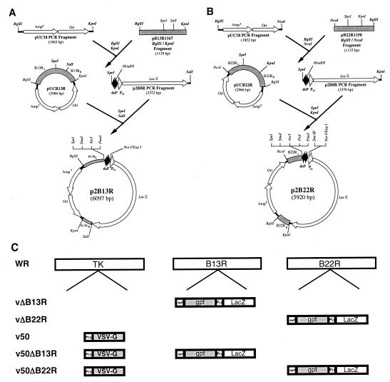
The strategies utilized to construct the p2B13R and p2B22R transfer vectors are shown in Fig. 1A and B. Standard PCR techniques were employed in vector construction with Vent DNA polymerase (New England Biolabs, Beverly, Mass.). The B13R and B22R genes were amplified with primers B13RF, B13RR, B22RF, and B22RR (Table 1), designed from published sequences (47). They were cloned into the SrfI site of pCR-Script (Stratagene, La Jolla, Calif.), producing plasmids pB13R1167 and pB22R1150. Briefly, p2B13R was developed as follows: a pUC18 PCR fragment containing the origin of replication (Ori) and the β-lactamase gene that confers ampicillin resistance (Ampr) was amplified from plasmid pUC18 (55) with primers pUC18F and pUC18/B13RR (Table 1). The PCR fragment and pB13R1167 were digested with BglII and KpnI and ligated, creating plasmid pUCB13R. A p2B8R PCR fragment was amplified from the p2B8R plasmid (53) with primers p2B8RF and p2B8R/B13RR (Table 1). The p2B8R PCR fragment and pUCB13R were digested with SpeI and SalI and ligated, producing the vector p2B13R. p2B22R was constructed as follows: a pUC18 PCR fragment was amplified from pUC18 with primers pUC18F and pUC18/B22RR (Table 1). This pUC18 PCR fragment and pB22R1150 were digested with BglII and NcoI and ligated, creating plasmid pUCB22R. Then a p2B8R PCR fragment was amplified from p2B8R with the primers p2B8RF and p2B8R/B22RR (Table 1). The p2B8R PCR fragment and pUCB22R were digested with SpeI and KpnI and ligated, producing p2B22R. Finally, p2B13Rgpt and p2B22Rgpt transfer vectors were constructed to further facilitate rVV selection by using the Escherichia coli xanthine-guanine phosphoribosyl transferase (gpt) gene under one of the two back-to-back synthetic VV promoters (dsP) (16). The gpt PCR fragment, flanked by SmaI, was amplified from plasmid pMSG (Pharmacia, Alameda, Calif.) with primers gptF and gptR (Table 1), digested with SmaI, and inserted into the SmaI site of p2B22R, producing p2B22Rgpt. The cloned gpt PCR fragment was sequenced with primers gptS1 and gptS2 (Table 1) and was subcloned into p2B13R from p2B22Rgpt by SmaI digestion.
FIG. 1.
Construction of rVVs with a deletion in the B13R or B22R gene. (A) The VV transfer vector p2B13R was generated by joining pUC18, pB13R1167, and p2B8R PCR fragments. (B) The VV transfer vector p2B22R was derived by joining pUC18, pB22R1150, and p2B8R PCR fragments. For both vectors, the two back-to-back synthetic VV promoters (dsP) are active in both early and late stages of infection. The multiple cloning sites adjacent to each side of the dsP facilitate cloning of heterologous genes (only unique sites are shown). (C) The WR strain of VV was used for the generation of recombinants with a deletion in the TK and B13R or B22R gene. The gpt gene was subcloned into the p2B13R and p2B22R transfer vectors, which were then used for the insertional inactivation and partial deletion of either the VV B13R or the B22R gene by homologous recombination. The gpt gene, utilized for rVV selection, is under one of the two dsPs, and the lacZ gene for β-galactosidase screening of rVVs is under the control of the VV P11 late promoter. v50, used as a parental virus, expresses VSV-G under the VV P7.5 promoter and is inserted into the TK site. The symbol Δ denotes insertional inactivation/deletion.
TABLE 1.
Oligonucleotide primers utilized in plasmid constructions in this study
| Name | Sequence (5′→3′)a |
|---|---|
| B13RF | TACACGACCAAGATCTATTACTATG |
| B13RR | TGAAAAGACGTGGGTACCAAAGTT |
| B22RF | GGTGGATCCCATGGATATCTTTAAA |
| B22RR | TATCAGATCTAGTTTATACTATTAC |
| pUC18F | TTCTTAGATCTCAGGTGGCACTT |
| pUC18/B13RR | AATACGGGTACCCACAGAATCAGG |
| pUC18/B22RR | ACAGAACCATGGGATAACGCAGG |
| p2B8RF | TAACAAACTAGTCCATAAGATCT CCC |
| p2B8R/B13RR | TTGTTTGTCGACTCTTATTATTTTTGACAC |
| p2B8R/B22RR | GTTTGCGGTACCTTATTATTTTTGACAC |
| gptF | ATAAACACCCGGGGACACTTCACAT |
| gptR | ACAACGCCCGGGAGTGCCAGG |
| gptS1 | AATATGAAAGTGGTGATTGTGA |
| gptS2 | ATGGGTTAAGACAATGACAAAT |
Engineered restriction endonuclease sites are underlined on the primer sequences.
Generation of rVVs.
rVVs were generated by standard homologous recombination with cationic liposome-mediated transfection of the transfer vectors p2B13Rgpt or p2B22Rgpt into BS-C-1 cell monolayers infected at 0.05 PFU per cell with VV 2 h earlier. Recombinant gpt-positive VVs were plaque purified on BS-C-40 cells from transfection supernatants, using gpt selection medium (25-μg/ml mycophenolic acid, 250-μg/ml xanthine, and 15-μg/ml hypoxanthine) (20). Blue plaques were visualized with X-Gal (5-bromo-4-chloro-3-indolyl-d-galactopyranoside) to detect the lacZ marker gene (19). The expression of the lacZ gene by the final plaque-purified rVVs was tested by cytochemical staining of infected cell monolayers as previously described (32). High-titer stocks were generated by infecting HeLa S3 cells with the rVVs at a multiplicity of infection (MOI) of 1. Infected cells were harvested 3 days postinfection by centrifugation at 200 ×g for 10 min. Cells were then lysed by freezing and thawing, sonicated, and trypsinized (19). Finally, cell lysates were clarified to remove contaminating cell debris by a second round of sonication and centrifugation at 200 × g for 5 min. The overall genomic structure of each rVV was determined by restriction analysis of rVV DNA, which was purified by a small-scale method employing micrococcal nuclease (30).
Viral growth kinetics.
Viral replication was determined by generating growth curves at low MOI. Briefly, triplicate monolayers of BS-C-1 or A549 cells were infected at a MOI of 0.01 for 1 h in 12-well plates, and virus replication was determined as previously described (53). At each time point, supernatants were collected, centrifuged (to pellet detached cells), and transferred to a new tube (the extracellular virus fraction), containing mostly extracellular enveloped virions (EEV). Cells in the well were resuspended in 1 ml of DMEM, scraped, and added to the pellet of detached cells (the intracellular fraction), containing mostly intracellular mature virions (IMV). To visualize plaques, monolayers of BS-C-1 and A549 cells were infected at 20 PFU per well for 1 h in six-well plates. Photographs were taken 36 h postinfection with a Kodak DC290 digital camera.
Caspase-1 assay.
HeLa S3 cells were infected with various rVVs at an MOI of 10 for 1 h in six-well plates in triplicates. The cells were washed, resuspended in 2 ml of DMEM-2.5% FBS, and incubated for 24 h at 37°C. Then, a caspase-1 colorimetric assay (Chemicon, Temecula, Calif.) was performed. Briefly, cells were lysed, and supernatants (cytosolic extract) were exposed to 4 mM YVAD-pNA substrate and incubated at 37°C for 2 h. Samples were read at 405 nm in a microtiter plate reader, and background readings from media and buffers were subtracted. HeLa S3 cells exposed to UV for 2 h and incubated at 37°C for 18 h were used as a positive control. Mock-infected HeLa S3 cells served as a negative control. Caspase-1 activity was expressed as optical density (OD).
Virulence and clearance studies in immunocompetent and immunodeficient mice.
Groups of CB6F1 normal immunocompetent mice (8- to 9-week-old females) were given 105 PFU of rVV intranasally in a final volume of 10 μl of sterile phosphate-buffered saline (PBS), under light anesthesia. Animals were examined and weighed daily. Survival was measured in groups of immunodeficient BALB/cBy nude mice (7- to 8-week-old males) inoculated intraperitoneally (i.p.) with 107 PFU of each rVV in a final volume of 250 μl of sterile PBS. Animals were examined twice daily. For clearance studies, groups of BALB/cBy nude mice and CB6F1 normal mice (7- to 8-week-old females) were inoculated i.p. with 107 PFU of rVV in a final volume of 250 μl of sterile PBS. Ovaries and spleens were removed, weighed, homogenized, and resuspended in DMEM at 10% (wt/vol). The tissues were then lysed by freeze-thawing and trypsinization. Viral titers were determined by plaque assay on BS-C-1 cell monolayers.
Humoral studies.
Groups of 11 CB6F1 mice (6- to 7-week-old females), under light anesthesia, were immunized intramuscularly (i.m.) with 105 PFU of rVV in a final volume of 50 μl of sterile PBS. Four weeks postvaccination, animals were boosted i.p. with 100 μl of an optimized dose (2 × 10⁵ PFU) of VSV stock recovered from VSV-inoculated mouse brains. Animals were monitored for 14 days postboost and euthanized at the end of the observation period. Mice were bled at 0, 2, 4, and 6 weeks postinfection. Serum samples were pooled for each group. Antibody titers to VSV and VV were determined by enzyme-linked immunosorbent assay (ELISA).
VSV and VV ELISAs.
To detect VSV- or VV-specific antibodies by ELISA, 96-well microtiter plates (Nunc, Roskilde, Denmark) were coated overnight with 100 μl of pelleted VSV virus per well (1/25,600 dilution) or cytosolic extracts from WR-infected HeLa S3 cells (1/500 dilution) suspended in PBS. These dilutions were determined to give the highest readings with positive control samples and the lowest background readings with naïve serum samples. The HeLa S3 cytosolic extracts (comprised mainly of IMV particles) were clarified by low-speed centrifugation to remove cell debris. Plates coated with mock-infected HeLa S3 cytosolic extracts were used as an additional control to rule out immune responses to HeLa S3 cell proteins in sera of vaccinated animals. After overnight incubation at 4°C, plates were washed twice with 50 mM Tris-0.2% Tween 20 and blocked for 1 h at room temperature with 5% nonfat dry milk in PBS. The plates were then washed twice and incubated for 1 h at room temperature with twofold serial dilutions of pooled sera from each rVV-inoculated group. Next, plates were washed twice, and antibody binding was detected with horseradish peroxidase-conjugated goat anti-mouse immunoglobulin G (heavy and light chains) antiserum (1/3,000 dilution; Bio-Rad, Hercules, Calif.). After 1 h, plates were washed three times and reacted with tetramethyl benzidine peroxidase substrate solution. After a 10-min incubation, the reaction was stopped by the addition of 2 N H2SO4, and A450 was read. Sample dilutions were considered positive if the OD recorded for that dilution was at least twofold higher than the OD recorded for a control serum sample (naïve CB6F1 mice) at the same dilution. Titers were expressed as the reciprocal of the highest dilution of samples scoring positive.
T-helper proliferation studies.
Groups of four to six CB6F1 (haplotype H-2b/d) mice (7-week-old females) were immunized i.p. with 107 PFU of each rVV in a final volume of 250 μl of sterile PBS. Splenocytes were harvested 10 days postvaccination. Triplicate cultures containing 105 splenocytes in complete RPMI 1640 medium supplemented with 50 μM 2-β-mercaptoethanol were seeded onto 96-microwell plates in concentrations of 0, 0.25, and 0.5 μg of baculovirus-expressed VSV-G/ml. The mitogen concanavalin A at 2 μg/ml served as a positive control. Splenocytes were incubated for 4 days at 37°C. On the 4th day, 0.5 μCi of [3H]thymidine per well was added, and cells were incubated for 18 h at 37°C. Cells were then harvested to determine incorporation of radioactivity, and data were expressed as cpm from triplicate samples.
Cytotoxic T-cell studies.
Splenocytes harvested for proliferation studies were also utilized for cytotoxic T-lymphocyte (CTL) studies. Effector splenocytes (5 × 10⁴ to 5 × 10⁵ cells) in complete RPMI 1640 medium supplemented with 50 μM 2-β-mercaptoethanol were stimulated for 4 h in U-bottom 96-well plates with 104 target splenocytes previously infected with UV-treated VSV. Cytotoxicity was measured with the Cytotox 96 nonradioactive cytotoxicity assay (Promega, Madison, Wis.). Infected L929 cells (haplotype H-2k) served as a negative target control. Percent specific cytolysis was calculated as (OD of experimental-effector spontaneous − target spontaneous)/(OD of target maximum − target spontaneous) × 100.
Data analysis.
Statistical analyses were performed with the statistical software program SAS, release 8.2 (SAS Institute, Cary, N.C.) or GraphPad Prism, version 4.0 (GraphPad Software Inc., San Diego, Calif.).
RESULTS
Generation and characterization of rVVs.
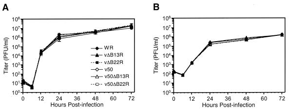
The p2B13R and p2B22R transfer vectors (Fig. 1A and B) direct homologous recombination with either the B13R or B22R region of the VV genome (Fig. 1C), causing deletions of 392 and 237 bp, respectively, which includes the putative active site of each gene (27). The rVVs vΔB13R and vΔB22R have only the B13R or the B22R gene inactivated, whereas v50ΔB13R and v50ΔB22R are derived from v50 and thus also have the TK locus insertionally inactivated by VSV-G (Fig. 1C). Extensive X-Gal cytochemical staining of infected cell monolayers indicated that all of the rVVs were stable and free of the parental strain (WR or v50) after four or more passages in tissue culture. HindIII restriction analysis of rVV-purified DNA samples confirmed the insertional inactivation of the B13R, B22R, and TK regions by the E. coli lacZ and gpt genes, as well as by the VSV-G gene (data not shown). Previously it had been demonstrated that VV serpin gene deletion does not affect viral replication kinetics in primate BS-C-1 cells (27). Our recombinants having either the B22R or B13R gene inactivated had virtually identical replication profiles in BS-C-1 cells for both intracellular and extracellular viral fractions, revealing no defects in IMV or EEV formation (Fig. 2). Finally, all B13R and B22R recombinants were able to replicate to levels identical to those of parental viruses in the murine L929 cells (data not shown).
FIG. 2.
B22R and B13R gene inactivation does not affect VV in vitro replication. Monolayers of BS-C-1 cells were infected at an MOI of 0.01, and at the indicated time points, intracellular (pelleted cells) (A) and extracellular (B) viral fractions (cell supernatants) were collected and their titers were determined. The data shown represent the mean values from triplicate samples assayed in duplicate; error bars indicate standard deviation.
B22R and B13R gene function is inactivated in rVVs with deleted serpin genes.
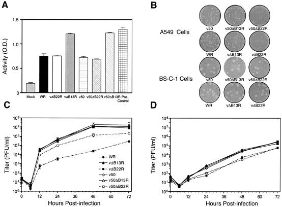
Since the B13R gene has been implicated in inhibiting the proteolytic activity of caspase-1 in vitro (26), functional inactivation of the gene was confirmed with a caspase-1 apoptosis assay. HeLa S3 cells were infected with the various rVVs, and caspase-1 activity was measured. VV infection induced a significant level of caspase-1 activity (Fig. 3A). Moreover, upon B13R gene inactivation (vΔB13R and v50ΔB13R), caspase-1 bioactivity increased to levels similar to those in a positive apoptotic sample (UV-treated HeLa S3 cells; Fig. 3A). On the other hand, parental and B22R-inactivated recombinants did not display such elevated levels of caspase-1 activity.
FIG. 3.
B13R and B22R gene function is inactivated in rVVs with deleted serpin genes. (A) B13R gene inactivation restores caspase-1 activity. To assess caspase-1 activity, HeLa S3 cells were infected with the various rVVs to induce apoptosis. UV-exposed HeLa S3 cells served as a positive control, and mock-infected HeLa S3 cells were used as a negative control. (B) B22R gene inactivation reduces plaque numbers by approximately fourfold in A549 cells. Plaques of rVV-infected BS-C-1 and A549 cell monolayers (20 PFU per well) were photographed at 36 h postinfection. (C and D) B22R gene inactivation reduces viral replication in A549 cells. Monolayers of A549 cells were infected at an MOI of 0.01, and at the indicated time points, intracellular (pelleted cells) (C) and extracellular (cell supernatants) (D) viral fractions were collected and their titers were determined. The data shown represent the mean values from triplicate samples assayed in duplicate; error bars indicate standard deviation.
Inactivation of the B22R gene results in greatly reduced viral yields compared to wild-type WR virus in human A549 cells (46). To demonstrate the functional inactivation of the VV B22R gene, we conducted growth kinetics studies with A549 cells. As expected, viral yields for both recombinants with the B22R gene deleted were diminished in human A549 cells, with vΔB22R showing greatly reduced levels and v50ΔB22R representing a less marked but nonetheless significant decrease in replication (Fig. 3C and D). Moreover, an approximately fourfold decrease in plaque numbers was observed in A549 cells for both vΔB22R and v50ΔB22R, while plaque numbers remained unaffected in BS-C-1 cells (Fig. 3B). Plaque phenotype and viral replication of B13R-inactivated rVVs were unaltered in A549 cells (Fig. 3B, C, and D).
Inactivation of the B13R or B22R gene attenuates VV virulence in nude mice.
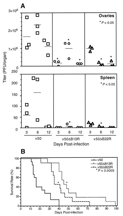
Female immunodeficient BALB/cBy mice were inoculated i.p. with 107 PFU of v50, v50ΔB13R, or v50ΔB22R, and levels of viral replication were measured in ovaries and spleen. v50 viral titers were high, with the ovaries being the initial and major site of viral replication followed by the spleen (Fig. 4A and Table 2). On the other hand, viral replication of serpin-inactivated rVVs (v50ΔB13R and v50ΔB22R) was reduced in both the ovaries and spleen. Significant reduction in viral replication was observed in the ovaries 3, 6, and 12 days postinfection for v50ΔB13R-inoculated mice and 6 and 12 days postinfection for v50ΔB22R-inoculated mice (Mann-Whitney test, P < 0.05). Moreover, viral replication was significantly reduced in the spleen 6 days postinfection for both v50ΔB13R- and v50ΔB22R-inoculated mice (P < 0.05).
FIG. 4.
Serpin-inactivated rVVs are attenuated in immunodeficient mice. (A) Serpin gene deletion decreases viral replication in VV-infected immunodeficient mice. Female BALB/cBy athymic mice were inoculated i.p. with 107 PFU of each rVV. Ovaries and spleen were removed at 3, 6, and 12 days postinfection. Significant reduction in viral replication was observed in the ovaries on 3, 6, and 12 days postinfection for v50ΔB13R-inoculated mice and on 6 and 12 days postinfection for v50ΔB22R-inoculated mice (Mann-Whitney test, P < 0.05). Viral replication was significantly reduced in the spleen 6 days postinfection for both v50ΔB13R- and v50ΔB22R-inoculated mice (P < 0.05). (B) Serpin gene deletion increases survival of VV-infected nude mice. Male athymic mice inoculated i.p. with 107 PFU of v50 (n = 15), v50ΔB13R (n = 11), or v50ΔB22R (n = 11) had median survival rates of 16, 45, and 35 days, respectively. Statistical analysis of survival time (log-rank test) yielded P < 0.0005 for v50ΔB13R or v50ΔB22R versus v50.
TABLE 2.
VV mean titers in ovaries 3 days postinoculation
| VVa | BALB/cBy athymic mice (n = 3)
|
CB6F1 normal mice (n = 5)
|
||
|---|---|---|---|---|
| PFU/organ | P value vs parental VVb | PFU/organ | P value vs parental VVb | |
| WR | NDc | 5.36 × 10⁶ | ||
| vΔB13R | ND | 4.23 × 10⁵ | 0.004 vs WR | |
| vΔB22R | ND | 1.06 × 10⁶ | 0.004 vs WR | |
| v50 | 1.67 × 10⁶ | 1.09 × 10⁶ | ||
| v50ΔB13R | 1.70 × 10⁵ | 0.050 vs v50 | 5.34 × 10⁵ | 0.028 vs v50 |
| v50ΔB22R | 9.92 × 10⁵ | 0.100 vs v50 | 8.90 × 10⁵ | 0.274 vs v50 |
Mice were inoculated i.p. with 107 PFU of each VV.
Mann-Whitney test.
ND, not determined.
Attenuation was further assessed by survival analysis. Male BALB/cBy immunodeficient mice, inoculated i.p. with 107 PFU of v50ΔB13R and v50ΔB22R, survived significantly longer than mice inoculated with v50 (log-rank test, P < 0.0005). Median survival rates for v50ΔB13R, v50ΔB22R, and v50 were 45, 35, and 16 days, respectively (Fig. 4B). The difference in survival rates between the v50ΔB13R- and v50ΔB22R-inoculated groups was not statistically significant (P = 0.1). Mice inoculated with sterile PBS (n = 4) remained healthy throughout the observation period of 120 days (data not shown).
Inactivation of the B13R or B22R gene decreases VV virulence in normal mice.
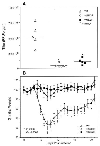
In order to show the relative differences in pathogenicity between parental and rVVs with deleted serpin genes, virulence studies were conducted with CB6F1 immunocompetent mice. Previously we have shown that rVVs with the TK gene inactivated are very attenuated in these mice (53). Even with very high doses, we did not observe any differences in virulence among the recombinants. Therefore, female CB6F1 mice were inoculated i.p. with 107 PFU of WR, vΔB13R, and vΔB22R, all having an intact TK gene. Three days postinfection, the mice were sacrificed, and ovaries were removed for virus titration (Table 2). Significantly lower levels of virus were detected in the ovaries of mice inoculated with vΔB13R or vΔB22R (Mann-Whitney test, P < 0.004) compared to the WR parental virus (Fig. 5A).
FIG. 5.
Inactivation of B13R or B22R decreases rVV virulence in immunocompetent mice. (A) Female CB6F1 mice were inoculated i.p. with 107 PFU of WR, vΔB13R, or vΔB22R. Three days postinfection, the mice were sacrificed, ovaries were removed, and viral titers (PFU per organ) were determined. A bar indicates the mean titer per group. A significant reduction in viral titers was found in the ovaries of mice inoculated with vΔB13R or vΔB22R (Mann-Whitney test, P < 0.004) compared to those inoculated with WR parental virus. (B) Normal mice (CB6F1, n = 11) were inoculated intranasally with 105 PFU of WR, vΔB13R, or vΔB22R. Each animal was weighed and monitored daily for signs of disease. The mean group weight (± standard error) was expressed as the percentage of the mean weight of that group of animals immediately after infection. vΔB13R- or vΔB22R-inoculated mice did not display significant weight loss, whereas mice infected with wild-type WR virus exhibited marked weight loss. This difference was statistically significant (analysis of variance followed by Tukey's studentized range test) from days 5 through 21 postinfection (P < 0.05) and was highly significant at days 6 through 10 (P < 0.0005).
VV virulence in immunocompetent CB6F1 mice was also assessed by weight loss. Mice were inoculated intranasally with 105 PFU of either vΔB13R or vΔB22R. As expected, no mortality was observed with this dose of the viruses (53). Mice receiving the recombinants vΔB13R and vΔB22R did not display significant weight loss (Fig. 5B), whereas mice infected with the same dose of the wild-type WR virus had marked weight loss. This difference was statistically significant (analysis of variance followed by Tukey's studentized range test) from days 5 through 21 postinfection (P < 0.05) and was highly significant at days 6 through 10 (P < 0.0005).
B13R- and B22R-inactivated rVVs induce strong humoral immune responses.
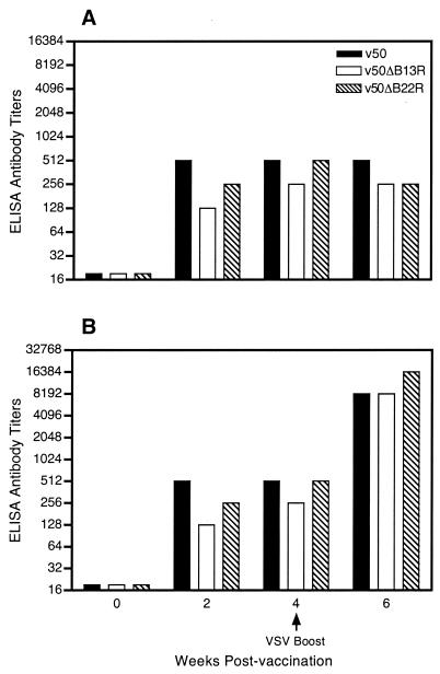
Historically, i.m. inoculation has been utilized to study VV humoral responses in mice (58). Groups of normal mice were inoculated i.m. with 105 PFU of v50, v50ΔB13R, or v50ΔB22R. Serum was collected at 0, 2, 4, and 6 weeks postinfection from each animal, and titers of antibodies to VV and VSV were determined by ELISA. VV plaque reduction and VSV serum neutralization assays were also performed as previously described (53), providing similar results (data not shown). Although there was a significant increase in attenuation, no differences were observed in the humoral immune responses to either homologous (VV) or heterologous (VSV-G) antigens in mice inoculated with the respective rVVs (Fig. 6). Animals were boosted with VSV i.p. 4 weeks postvaccination to monitor anamnestic responses. All animals survived this nonlethal VSV boost. Strong anamnestic responses to VSV were observed, and titers at termination reached a similar level for all vaccinated groups.
FIG. 6.
Serpin gene inactivation does not alter humoral immune responses to rVV. Groups of normal mice (11 animals per group) were inoculated i.m. with 105 PFU of the rVVs v50, v50ΔB13R, and v50ΔB22R. Animals were boosted with VSV i.p. at 4 weeks postvaccination. Antibody titers to VV (A) and VSV (B) were determined by ELISA from pooled samples assayed in duplicate.
Serpin-inactivated rVVs induce strong cell-mediated immune responses.
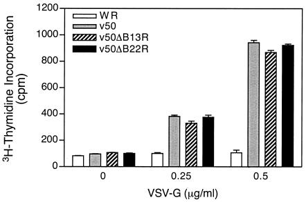
Since the i.p. route is routinely utilized to measure VV-mediated cellular immune responses (3), groups of normal CB6F1 (H-2b/d) mice were inoculated i.p. with 107 PFU of v50, v50ΔB13R, or v50ΔB22R. Relative T-helper-cell proliferative responses were measured against a concentration of 0, 0.25, and 0.5 μg of soluble VSV-G protein per ml by the incorporation of [3H]thymidine. Proliferation was determined to be dose dependent, with 0.5 μg/ml providing an optimal response (Fig. 7). The measured T-helper proliferative responses to the wild type, v50, and recombinants v50ΔB13R and v50ΔB22R with deleted serpin genes did not differ statistically. T-cell proliferative responses of the three groups were, however, 8 to 9 times higher than that of a WR-infected control at 0.5 μg of VSV-G per ml.
FIG. 7.
VVs having an inactivated B13R or B22R gene elicit strong T-helper responses. Female CB6F1 mice were immunized i.p. with 107 PFU of each rVV. Splenocytes were harvested 10 days postvaccination and stimulated with 0, 0.25, and 0.5 μg of VSV-G per ml for 4 days and pulsed with [3H]thymidine. Data analysis was based on cpm in triplicates, and error bars represent the standard error of the mean.
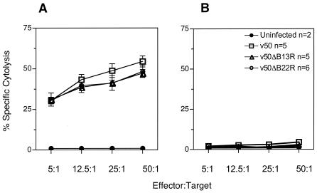
Finally, VSV-G-specific CTL responses were determined. CTL responses of parental v50 and the B13R- and B22R-inactivated recombinants were similar, reaching as high as 54.5% (Fig. 8A). Infected L929 cells (H-2k) served as negative target controls. Lysis values for negative control targets were <5% (Fig. 8B).
FIG. 8.
rVVs with an inactivated serpin gene induce potent cytotoxic T-cell responses. CB6F1 mice were inoculated i.p. with 107 PFU of v50, v50ΔB13R, or v50ΔB22R. CTL immune responses were measured 10 days postvaccination. (A) Effector splenocytes were stimulated with haplotype-matched (H-2b/d) target splenocytes that were infected with UV-treated VSV. (B) Infected L929 cells (H-2k) served as negative target controls. The lysis values for negative control targets were <5%. Specific cytolysis values at the indicated effector/target cell ratios represent the means of triplicate experiments assayed in duplicate. Error bars represent standard deviation.
DISCUSSION
Increasingly, a need has arisen for attenuated yet highly immunogenic VV vaccine vectors. In an attempt to further improve rVV safety and efficacy, we deleted either the VV B13R or B22R immunomodulating gene and showed that it does not affect in vitro viral replication in BS-C-1 cells (Fig. 2). Characterization studies substantiated the functional inactivation of the B13R and B22R genes (Fig. 3). It has been well established that the cowpox virus cytokine response modifier A (crmA) protein and the VV crmA equivalent, B13R (SPI-2), mediate potent anti-inflammatory properties by inhibiting the proteolytic activity of caspase-1 (26, 29, 31). Our recombinants vΔB13R and v50ΔB13R had similar properties, because inactivation of the B13R gene restored caspase-1 activity. Furthermore, we demonstrated the functional inactivation of the B22R gene by measuring viral replication levels in human A549 cells, since previously it has been shown that B22R inactivation results in greatly diminished viral yields in human A549 cells compared to wild-type WR virus (46).
The increased safety and efficacy of rVVs with deleted serpin genes were tested in vivo. We were able to show that B13R or B22R gene deletion reduces rVV virulence and pathogenicity in both immunocompetent and immunodeficient mice. Intraperitoneal inoculation of immunodeficient nude mice is an established model for studying disseminated VV infection (21, 24, 43). To study attenuation in immunodeficient mice, we used TK-negative rVVs (v50, v50ΔB13R, and v50ΔB22R), which have a lower starting virulence and therefore are more suitable for use in nude mice (8). We demonstrated that nude mice inoculated with the TK-negative virus v50ΔB13R or v50ΔB22R survived significantly longer (4 and 3 weeks, respectively) and were able to more effectively control viral replication than those inoculated with the control virus, v50 (Fig. 4). The abrogation of either B13R- or B22R-mediated anti-inflammatory responses may have bolstered innate immune responses in the immunodeficient mice, allowing for better control of viral replication and spread and ultimately resulting in increased time of survival. Although the difference in survival rate between the v50ΔB13R- and v50ΔB22R-inoculated groups was not statistically significant, these TK-negative recombinants displayed somewhat different viral replication kinetics (Fig. 4A and Table 2). B13R gene inactivation significantly reduced viral replication in the ovaries as early as 3 days postinfection, whereas the decrease in viral replication mediated by B22R gene inactivation was delayed, commencing at 6 days postinfection. These differences could be explained by the fact that the B13R gene product has been shown to have immediate and direct antiapoptotic properties by inhibiting caspase-1 activity, while the B22R gene product has been shown to have a role in the efficient expression of viral intermediate and late mRNAs, viral late protein synthesis, and virion morphogenesis (46). Therefore, in an attenuated (TK deleted) background, these differences in serpin gene function may be reflected in the various viral replication kinetics in immunodeficient mice, with v50ΔB13R being slightly more attenuated than v50ΔB22R. Similarly, viral titers in ovaries of immunocompetent mice (CB6F1) inoculated i.p. with the rVVs with deleted serpin genes (vΔB13R and vΔB22R) were significantly lower than those of the WR parental virus (Fig. 5A and Table 2), whereas in the TK gene deletion background, the difference was significant only for v50ΔB13R compared to the v50 parental virus (Table 2).
The murine intranasal model of VV infection has previously been reported to be a sensitive route for the study of pathogenesis and virulence (54). Using the CB6F1 strain of immunocompetent mice, we were able to show the contribution of the B13R and B22R genes to VV virulence in vivo. In our study, CB6F1 mice inoculated with the WR strain of VV displayed significant weight loss and were slower to clear the virus, compared to mice inoculated with recombinants having a deletion in the B13R or B22R serpin homolog gene (Fig. 5A and B). These results, however, differ from previous findings, where virulence was indistinguishable in BALB/c mice inoculated intranasally with wild-type WR and B13R or B22R (27) deletion mutants. This discrepancy may be due to differences in the degree of susceptibility of each mouse strain to VV. The CB6F1 mice used in this study, inoculated intranasally with 105 PFU of WR, suffered only weight loss with no mortality (53), while the same dose results in 100% mortality in BALB/c mice (27, 50). Studies with the related ectromelia virus may explain these observations (13). Ectromelia virus infection of BALB/c and DBA/2 mice is rapid and severe. In contrast, C57BL/6 and AKR mice are barely susceptible to infection, show no lesions, and survive. The CB6F1 mice used in these studies are a cross between BALB/c and C57BL/6 mice and therefore seem to manifest an intermediate level of susceptibility that allowed detection of differences in virulence between WR and B13R or B22R deletion mutants.
Previously, in an in vitro model, Blake et al. (9) showed that rVVs with large deletions in the B region of the VV genome, containing many host immune evasion genes including the B13R and B22R serpin homolog genes, the B8R IFN-γ receptor homolog gene, and the B15R IL-1β receptor homolog, do not impede major histocompatibility complex (MHC) class I antigen presentation. Using influenza virus CTL clones, this group was able to delineate that such VV immunomodulating genes do not interfere with viral peptide processing and subsequent cytotoxic T-cell responses. Moreover, Mullbacher et al. have shown that serpins encoded by ectromelia virus and cowpox virus do not limit MHC-restricted CTL responses (40). Here, in our in vivo murine model, we demonstrated that the specific inactivation of either the B13R or B22R gene affects neither humoral nor cell-mediated immune responses in vivo. In fact, such rVVs with deleted serpin genes elicited strong cell-mediated and humoral immune responses that did not differ from those induced by the parental virus (Fig. 6, 7, and 8). Formerly, it had been reported that VVs lacking the B13R or B22R gene induce enhanced humoral immune responses, whereas no enhancement was seen with viruses lacking the TK gene (7, 57). For our studies, we constructed rVVs that have the TK gene inactivated, as well as the B13R or B22R serpin homolog gene (v50ΔB13R and v50ΔB22R). Since recombinants with the TK gene inactivated are attenuated, a subsequent serpin gene deletion should enhance rVV attenuation even further. In fact, we were able to demonstrate that both the VV B13R and B22R genes act as virulence factors in CB6F1 mice and that their deletion attenuates VV. Therefore, any serpin deletion-mediated immune enhancements could in turn be masked by the observed attenuation. These competing actions may simultaneously cancel each other out and account for our observed maintenance of strong humoral and cell-mediated immune responses. Moreover, the choice of mouse strain could also play a factor. Since CB6F1 mice are less susceptible to VV infection than the BALB/c mice utilized in the previous studies, reduced viral replication could account for the observed lack of enhanced humoral responses.
Recently, multiple groups have shown that heterologous prime-boost vaccination regimens, such as DNA priming followed by a recombinant poxvirus (e.g., MVA) boost, induce strong and long-lasting immune responses in both nonhuman primate and human models (2, 36). An exciting prospect for rVV vectors with a deletion in serpin homolog genes is that they may be utilized in a similar DNA prime and VV boost vaccination regimen. The added advantage of rVVs with deleted serpin genes over MVA is that while they are significantly attenuated, they retain the ability to replicate, allowing for increased heterologous antigen expression and enhanced immunogenicity.
In conclusion, our studies indicate that B13R or B22R serpin homolog gene inactivation attenuates VV by significantly increasing the safety of the virus without compromising the strength of the elicited immune responses. Therefore, the inactivation of VV immunomodulating genes, such as B13R or B22R, can greatly improve upon the usefulness of VV as vectors for vaccine development and immunotherapy.
Acknowledgments
This work was supported by an NIH grant awarded to T.D.Y. (AI37182) and a grant awarded to L.A.J. by The Harold Wetterberg Foundation. Other support included NIH grants AI47025, AI53811, and AI54951 to T.D.Y. F.A.L. was supported by the Floyd and Mary Schwall Fellowship, the Jastro Shields Scholarship, the Peter J. Shields Block Grant, and the UC Davis Biotechnology Program Fellowship.
We are grateful to the members of the International Laboratory of Molecular Biology for Tropical Disease Agents, especially Fan-Ching Lin, Julia A. Collins, Ian Crossley, Lael Brown, and Troy Crowder for their assistance; Shabbir Ahmad for providing the recombinant baculovirus expressing VSV-G; and Sally Owens for helpful discussions.
REFERENCES
- 1.Alcamí, A., and G. L. Smith. 1995. Vaccinia, cowpox, and camelpox viruses encode soluble gamma interferon receptors with novel broad species specificity. J. Virol. 69:4633-4639. [DOI] [PMC free article] [PubMed] [Google Scholar]
- 2.Amara, R. R., F. Villinger, S. I. Staprans, J. D. Altman, D. C. Montefiori, N. L. Kozyr, Y. Xu, L. S. Wyatt, P. L. Earl, J. G. Herndon, H. M. McClure, B. Moss, and H. L. Robinson. 2002. Different patterns of immune responses but similar control of a simian-human immunodeficiency virus 89.6P mucosal challenge by modified vaccinia virus Ankara (MVA) and DNA/MVA vaccines. J. Virol. 76:7625-7631. [DOI] [PMC free article] [PubMed] [Google Scholar]
- 3.Andrew, M. E., B. E. Coupar, and D. B. Boyle. 1989. Humoral and cell-mediated immune responses to recombinant vaccinia viruses in mice. Immunol. Cell Biol. 67:331-337. [DOI] [PubMed] [Google Scholar]
- 4.Arita, I., and F. Fenner. 1985. Complications of smallpox vaccination, p. 49-60. In G. V. Quinnan, Jr. (ed.), Vaccinia viruses as vectors for vaccine antigens: proceedings of the Workshop on Vaccinia Viruses as Vectors for Vaccine Antigens. Elsevier, New York, N.Y.
- 5.Baxby, D. 1991. Safety of recombinant vaccinia vaccines. Lancet 337:913. [DOI] [PubMed] [Google Scholar]
- 6.Belyakov, I. M., B. Moss, W. Strober, and J. A. Berzofsky. 1999. Mucosal vaccination overcomes the barrier to recombinant vaccinia immunization caused by preexisting poxvirus immunity. Proc. Natl. Acad. Sci. USA 96:4512-4517. [DOI] [PMC free article] [PubMed] [Google Scholar]
- 7.Bennett, A. M., T. Lescott, R. J. Phillpotts, M. Mackett, and R. W. Titball. 1999. Recombinant vaccinia viruses protect against Clostridium perfringens alpha-toxin. Viral Immunol. 12:97-105. [DOI] [PubMed] [Google Scholar]
- 8.Binns, M. M., and G. L. Smith (ed.). 1992. Recombinant poxviruses, p. 81-162. CRC Press, Boca Raton, Fla.
- 9.Blake, N. W., S. Kettle, K. M. Law, K. Gould, J. Bastin, A. R. Townsend, and G. L. Smith. 1995. Vaccinia virus serpins B13R and B22R do not inhibit antigen presentation to class I-restricted cytotoxic T lymphocytes. J. Gen. Virol. 76:2393-2398. [DOI] [PubMed] [Google Scholar]
- 10.Boursnell, M. E., I. J. Foulds, J. I. Campbell, and M. M. Binns. 1988. Non-essential genes in the vaccinia virus HindIII K fragment: a gene related to serine protease inhibitors and a gene related to the 37K vaccinia virus major envelope antigen. J. Gen. Virol. 69:2995-3003. [DOI] [PubMed] [Google Scholar]
- 11.Bronson, L. H., and R. F. Parker. 1941. The neutralization of vaccine virus by immune serum: titration by the intracerebral inoculation of mice. J. Bacteriol. 41:56-57. [Google Scholar]
- 12.Brooks, M. A., A. N. Ali, P. C. Turner, and R. W. Moyer. 1995. A rabbitpox virus serpin gene controls host range by inhibiting apoptosis in restrictive cells. J. Virol. 69:7688-7698. [DOI] [PMC free article] [PubMed] [Google Scholar]
- 13.Buller, R. M., and G. J. Palumbo. 1991. Poxvirus pathogenesis. Microbiol. Rev. 55:80-122. [DOI] [PMC free article] [PubMed] [Google Scholar]
- 14.Buller, R. M., G. L. Smith, K. Cremer, A. L. Notkins, and B. Moss. 1985. Decreased virulence of recombinant vaccinia virus expression vectors is associated with a thymidine kinase-negative phenotype. Nature 317:813-815. [DOI] [PubMed] [Google Scholar]
- 15.Carroll, M. W., W. W. Overwijk, D. R. Surman, K. Tsung, B. Moss, and N. P. Restifo. 1998. Construction and characterization of a triple-recombinant vaccinia virus encoding B7-1, interleukin 12, and a model tumor antigen. J. Natl. Cancer Inst. 90:1881-1887. [DOI] [PMC free article] [PubMed] [Google Scholar]
- 16.Chakrabarti, S., J. R. Sisler, and B. Moss. 1997. Compact, synthetic, vaccinia virus early/late promoter for protein expression. BioTechniques 23:1094-1097. [DOI] [PubMed] [Google Scholar]
- 17.Colamonici, O. R., P. Domanski, S. M. Sweitzer, A. Larner, and R. M. Buller. 1995. Vaccinia virus B18R gene encodes a type I interferon-binding protein that blocks interferon alpha transmembrane signaling. J. Biol. Chem. 270:15974-15978. [DOI] [PubMed] [Google Scholar]
- 18.Cooney, E. L., A. C. Collier, P. D. Greenberg, R. W. Coombs, J. Zarling, D. E. Arditti, M. C. Hoffman, S. L. Hu, and L. Corey. 1991. Safety of and immunological response to a recombinant vaccinia virus vaccine expressing HIV envelope glycoprotein. Lancet 337:567-572. [DOI] [PubMed] [Google Scholar]
- 19.Earl, P. L., and B. Moss. 1994. Generation of recombinant vaccinia viruses, p. 16.17.1-16.17.16. In F. M. Ausubel, R. Brent, R. E. Kingston, D. D. Moore, J. G. Seidman, J. A. Smith, and K. Struhl (ed.), Current protocols in molecular biology, vol. 2. John Wiley & Sons, New York, N.Y. [Google Scholar]
- 20.Falkner, F. G., and B. Moss. 1988. Escherichia coli gpt gene provides dominant selection for vaccinia virus open reading frame expression vectors. J. Virol. 62:1849-1854. [DOI] [PMC free article] [PubMed] [Google Scholar]
- 21.Flexner, C., A. Hugin, and B. Moss. 1987. Prevention of vaccinia virus infection in immunodeficient mice by vector-directed IL-2 expression. Nature 330:259-262. [DOI] [PubMed] [Google Scholar]
- 22.Gherardi, M. M., J. C. Ramirez, D. Rodriguez, J. R. Rodriguez, G. Sano, F. Zavala, and M. Esteban. 1999. IL-12 delivery from recombinant vaccinia virus attenuates the vector and enhances the cellular immune response against HIV-1 Env in a dose-dependent manner. J. Immunol. 162:6724-6733. [PubMed] [Google Scholar]
- 23.Giavedoni, L., L. Jones, C. Mebus, and T. Yilma. 1991. A vaccinia virus double recombinant expressing the F and H genes of rinderpest virus protects cattle against rinderpest and causes no pock lesions. Proc. Natl. Acad. Sci. USA 88:8011-8015. [DOI] [PMC free article] [PubMed] [Google Scholar]
- 24.Giavedoni, L. D., L. Jones, M. B. Gardner, H. L. Gibson, C. T. Ng, P. J. Barr, and T. Yilma. 1992. Vaccinia virus recombinants expressing chimeric proteins of human immunodeficiency virus and gamma interferon are attenuated for nude mice. Proc. Natl. Acad. Sci. USA 89:3409-3413. [DOI] [PMC free article] [PubMed] [Google Scholar]
- 25.Isaacs, S. N., G. J. Kotwal, and B. Moss. 1992. Vaccinia virus complement-control protein prevents antibody-dependent complement-enhanced neutralization of infectivity and contributes to virulence. Proc. Natl. Acad. Sci. USA 89:628-632. [DOI] [PMC free article] [PubMed] [Google Scholar]
- 26.Kettle, S., A. Alcamí, A. Khanna, R. Ehret, C. Jassoy, and G. L. Smith. 1997. Vaccinia virus serpin B13R (SPI-2) inhibits interleukin-1b-converting enzyme and protects virus-infected cells from TNF- and Fas-mediated apoptosis, but does not prevent IL-1b-induced fever. J. Gen. Virol. 78:677-685. [DOI] [PubMed] [Google Scholar]
- 27.Kettle, S., N. W. Blake, K. M. Law, and G. L. Smith. 1995. Vaccinia virus serpins B13R (SPI-2) and B22R (SPI-1) encode Mr 38.5 and 40K, intracellular polypeptides that do not affect virus virulence in a murine intranasal model. Virology 206:136-147. [DOI] [PubMed] [Google Scholar]
- 28.Kotwal, G. J., and B. Moss. 1988. Vaccinia virus encodes a secretory polypeptide structurally related to complement control proteins. Nature 335:176-178. [DOI] [PubMed] [Google Scholar]
- 29.Kotwal, G. J., and B. Moss. 1989. Vaccinia virus encodes two proteins that are structurally related to members of the plasma serine protease inhibitor superfamily. J. Virol. 63:600-606. (Erratum, 64:966, 1990.) [DOI] [PMC free article] [PubMed] [Google Scholar]
- 30.Lai, A. C., and Y. Chu. 1991. A rapid method for screening vaccinia virus recombinants. BioTechniques 10:564-565. [PubMed] [Google Scholar]
- 31.Macen, J. L., R. S. Garner, P. Y. Musy, M. A. Brooks, P. C. Turner, R. W. Moyer, G. McFadden, and R. C. Bleackley. 1996. Differential inhibition of the Fas- and granule-mediated cytolysis pathways by the orthopoxvirus cytokine response modifier A/SPI-2 and SPI-1 protein. Proc. Natl. Acad. Sci. USA 93:9108-9113. [DOI] [PMC free article] [PubMed] [Google Scholar]
- 32.MacGregor, G. R., G. P. Nolan, S. Fiering, M. Roederer, and L. A. Herzenberg. 1991. Use of E. coli lacZ (β-galactosidase) as a reporter gene, p. 217-235. In E. J. Murray (ed.), Gene transfer and expression protocols, vol. 7. Humana Press, Clifton, N.J. [DOI] [PubMed] [Google Scholar]
- 33.Mackett, M., T. Yilma, J. K. Rose, and B. Moss. 1985. Vaccinia virus recombinants: expression of VSV genes and protective immunization of mice and cattle. Science 227:433-435. [DOI] [PubMed] [Google Scholar]
- 34.Marshall, J. L., R. J. Hoyer, M. A. Toomey, K. Faraguna, P. Chang, E. Richmond, J. E. Pedicano, E. Gehan, R. A. Peck, P. Arlen, K. Y. Tsang, and J. Schlom. 2000. Phase I study in advanced cancer patients of a diversified prime-and-boost vaccination protocol using recombinant vaccinia virus and recombinant nonreplicating avipox virus to elicit anti-carcinoembryonic antigen immune responses. J. Clin. Oncol. 18:3964-3973. [DOI] [PubMed] [Google Scholar]
- 35.Mayr, A., H. Stickl, H. K. Muller, K. Danner, and H. Singer. 1978. The smallpox vaccination strain MVA: marker, genetic structure, experience gained with the parenteral vaccination and behavior in organisms with a debilitated defence mechanism. Zentbl. Bakteriol. B 167:375-390. (Author's translation.) [PubMed]
- 36.McConkey, S. J., W. H. Reece, V. S. Moorthy, D. Webster, S. Dunachie, G. Butcher, J. M. Vuola, T. J. Blanchard, P. Gothard, K. Watkins, C. M. Hannan, S. Everaere, K. Brown, K. E. Kester, J. Cummings, J. Williams, D. G. Heppner, A. Pathan, K. Flanagan, N. Arulanantham, M. T. Roberts, M. Roy, G. L. Smith, J. Schneider, T. Peto, R. E. Sinden, S. C. Gilbert, and A. V. Hill. 2003. Enhanced T-cell immunogenicity of plasmid DNA vaccines boosted by recombinant modified vaccinia virus Ankara in humans. Nat. Med. 9:729-735. [DOI] [PubMed] [Google Scholar]
- 37.Moon, K. B., P. C. Turner, and R. W. Moyer. 1999. SPI-1-dependent host range of rabbitpox virus and complex formation with cathepsin G is associated with serpin motifs. J. Virol. 73:8999-9010. [DOI] [PMC free article] [PubMed] [Google Scholar]
- 38.Moss, B. 1996. Genetically engineered poxviruses for recombinant gene expression, vaccination, and safety. Proc. Natl. Acad. Sci. USA 93:11341-11348. [DOI] [PMC free article] [PubMed] [Google Scholar]
- 39.Mossman, K., C. Upton, R. M. Buller, and G. McFadden. 1995. Species specificity of ectromelia virus and vaccinia virus interferon-gamma binding proteins. Virology 208:762-769. [DOI] [PubMed] [Google Scholar]
- 40.Mullbacher, A., R. Wallich, R. W. Moyer, and M. M. Simon. 1999. Poxvirus-encoded serpins do not prevent cytolytic T cell-mediated recovery from primary infections. J. Immunol. 162:7315-7321. [PubMed] [Google Scholar]
- 41.Ober, B. T., P. Brühl, M. Schmidt, V. Wieser, W. Gritschenberger, S. Coulibaly, H. Savidis-Dacho, M. Gerencer, and F. G. Falkner. 2002. Immunogenicity and safety of defective vaccinia virus Lister: comparison with modified vaccinia virus Ankara. J. Virol. 76:7713-7723. [DOI] [PMC free article] [PubMed] [Google Scholar]
- 42.Pastoret, P. P., and B. Brochier. 1996. The development and use of a vaccinia-rabies recombinant oral vaccine for the control of wildlife rabies; a link between Jenner and Pasteur. Epidemiol. Infect. 116:235-240. [DOI] [PMC free article] [PubMed] [Google Scholar]
- 43.Ramshaw, I. A., M. E. Andrew, S. M. Phillips, D. B. Boyle, and B. E. Coupar. 1987. Recovery of immunodeficient mice from a vaccinia virus/IL-2 recombinant infection. Nature 329:545-546. [DOI] [PubMed] [Google Scholar]
- 44.Redfield, R. R., D. C. Wright, W. D. James, T. S. Jones, C. Brown, and D. S. Burke. 1987. Disseminated vaccinia in a military recruit with human immunodeficiency virus (HIV) disease. N. Engl. J. Med. 316:673-676. [DOI] [PubMed] [Google Scholar]
- 45.Rodriguez, D., J. R. Rodriguez, J. F. Rodriguez, D. Trauber, and M. Esteban. 1989. Highly attenuated vaccinia virus mutants for the generation of safe recombinant viruses. Proc. Natl. Acad. Sci. USA 86:1287-1291. [DOI] [PMC free article] [PubMed] [Google Scholar]
- 46.Shisler, J. L., S. N. Isaacs, and B. Moss. 1999. Vaccinia virus serpin-1 deletion mutant exhibits a host range defect characterized by low levels of intermediate and late mRNAs. Virology 262:298-311. [DOI] [PubMed] [Google Scholar]
- 47.Smith, G. L., Y. S. Chan, and S. T. Howard. 1991. Nucleotide sequence of 42 kbp of vaccinia virus strain WR from near the right inverted terminal repeat. J. Gen. Virol. 72:1349-1376. [DOI] [PubMed] [Google Scholar]
- 48.Smith, G. L., S. T. Howard, and Y. S. Chan. 1989. Vaccinia virus encodes a family of genes with homology to serine proteinase inhibitors. J. Gen. Virol. 70:2333-2343. [DOI] [PubMed] [Google Scholar]
- 49.Sun, C. S., P. R. Wyde, S. Z. Wilson, and V. Knight. 1984. Efficacy of aerosolized recombinant interferons against vesicular stomatitis virus-induced lung infection in cotton rats. J. Interferon Res. 4:449-459. [DOI] [PubMed] [Google Scholar]
- 50.Symons, J. A., A. Alcami, and G. L. Smith. 1995. Vaccinia virus encodes a soluble type I interferon receptor of novel structure and broad species specificity. Cell 81:551-560. [DOI] [PubMed] [Google Scholar]
- 51.Tartaglia, J., M. E. Perkus, J. Taylor, E. K. Norton, J. C. Audonnet, W. I. Cox, S. W. Davis, J. van der Hoeven, B. Meignier, M. Riviere, B. Languet, and E. Paoletti. 1992. NYVAC: a highly attenuated strain of vaccinia virus. Virology 188:217-232. [DOI] [PubMed] [Google Scholar]
- 52.Taylor, J., R. Weinberg, J. Tartaglia, C. Richardson, G. Alkhatib, D. Briedis, M. Appel, E. Norton, and E. Paoletti. 1992. Nonreplicating viral vectors as potential vaccines: recombinant canarypox virus expressing measles virus fusion (F) and hemagglutinin (HA) glycoproteins. Virology 187:321-328. [DOI] [PubMed] [Google Scholar]
- 53.Verardi, P. H., L. A. Jones, F. H. Aziz, S. Ahmad, and T. D. Yilma. 2001. Vaccinia virus vectors with an inactivated gamma interferon receptor homolog gene (B8R) are attenuated in vivo without a concomitant reduction in immunogenicity. J. Virol. 75:11-18. [DOI] [PMC free article] [PubMed] [Google Scholar]
- 54.Williamson, J. D., R. W. Reith, L. J. Jeffrey, J. R. Arrand, and M. Mackett. 1990. Biological characterization of recombinant vaccinia viruses in mice infected by the respiratory route. J. Gen. Virol. 71:2761-2767. (Erratum, 72:474, 1991.) [DOI] [PubMed] [Google Scholar]
- 55.Yanisch-Perron, C., J. Vieira, and J. Messing. 1985. Improved M13 phage cloning vectors and host strains: nucleotide sequences of the M13mp18 and pUC19 vectors. Gene 33:103-119. [DOI] [PubMed] [Google Scholar]
- 56.Yilma, T., R. G. Breeze, S. Ristow, J. R. Gorham, and S. R. Leib. 1985. Immune responses of cattle and mice to the G glycoprotein of vesicular stomatitis virus. Adv. Exp. Med. Biol. 185:101-115. [DOI] [PubMed] [Google Scholar]
- 57.Zhou, J., L. Crawford, L. McLean, X. Y. Sun, M. Stanley, N. Almond, and G. L. Smith. 1990. Increased antibody responses to human papillomavirus type 16 L1 protein expressed by recombinant vaccinia virus lacking serine protease inhibitor genes. J. Gen. Virol. 71:2185-2190. [DOI] [PubMed] [Google Scholar]
- 58.Zinkernagel, R. M., and H. Hengartner. 1997. Antiviral immunity. Immunol. Today 18:258-260. [DOI] [PubMed] [Google Scholar]


