Abstract
Primary cilia are present on most cell types including chondrocytes. Dysfunction of primary cilia results in pleiotropic symptoms including skeletal dysplasia. Previously, we showed that deletion of Ift88 and subsequent depletion of primary cilia from chondrocytes resulted in disorganized columnar structure and early loss of growth plate. To understand underlying mechanisms whereby Ift88 regulates growth plate function, we compared gene expression profiles in normal and Ift88 deleted growth plates. Pathway analysis indicated that Hedgehog (Hh) signaling was the most affected pathway in mutant growth plate. Expression of the Wnt antagonist, Sfrp5, was also down-regulated. In addition, Sfrp5 was up-regulated by Shh in rib chondrocytes and regulation of Sfrp5 by Shh was attenuated in mutant cells. This result suggests Sfrp5 is a downstream target of Hh and that Ift88 regulates its expression. Sfrp5 is an extracellular antagonist of Wnt signaling. We observed an increase in Wnt/β-catenin signaling specifically in flat columnar cells of the growth plate in Ift88 mutant mice as measured by increased expression of Axin2 and Lef1 as well as increased nuclear localization of β-catenin. We propose that Ift88 and primary cilia regulate expression of Sfrp5 and Wnt signaling pathways in growth plate via regulation of Ihh signaling.
Keywords: primary cilia, Ift88, Hedgehog, Sfrp5, Wnt
Introduction
Primary cilia are nonmotile microtubule-based organelles present on almost every type of cell (1). The ciliary body is comprised of microtubule bundles elongating from one of the centrioles (basal body), covered by a specialized plasma membrane. Primary cilia are built up and maintained by a process called intraflagellar transport (IFT), in which IFT proteins associate with kinesin motors or dyneins to carry cargos into or out of primary cilia, respectively. Deletion of IFTs or motor proteins (e.g. Ift88 and Kif3a) results in depletion of primary cilia (2). Extensive studies in the past ten years have demonstrated that primary cilia act as the hub for several signaling pathways, including Hedgehog (Hh), Wnt, Platelet Derived Growth Factor, and fluid flow (3). Dysfunction of primary cilia is associated with a group of genetic diseases called ciliopathies, characterized by pleiotropic symptoms including skeletal dysplasias (4).
The role of primary cilia in skeletal development has been shown in studies using mouse models (5). In our previous study, we generated Col2aCre;Kif3afl/fl and Col2aCre;Ift88fl/fl mice in which primary cilia were specifically deleted in chondrocytes. The mutant mice showed postnatal cartilage phenotypes including disorganization of columnar structure and accelerated hypertrophic differentiation resulting in complete loss of growth plate around two-weeks of age (6). These mice also developed symptoms of early osteoarthritis (7).
Longitudinal bone growth results from the complex events of chondrocyte differentiation in the growth plate. Chondrocytes in the resting zone are stem-like, round-shaped cells that serve as a reservoir to generate proliferating chondrocytes. In the proliferative zone, cells are flat and arranged into a columnar structure. It has been suggested that the columnar structure is a result of a process called chondrocyte rotation. During this process, chondrocytes divide perpendicular to the long axis of the bone then migrate one on top of the other to form columns of cells (8). In addition to primary cilia (6), integrins (9), Hh (10), and Wnt/ PCP signaling (11) have been implicated in regulation of chondrocyte rotation. When the proliferating cells in the growth plate stop dividing they undergo hypertrophic differentiation. The Indian hedgehog (Ihh)/ Parathyroid Hormone-related Protein (PTHrP) feedback loop is a key regulator of hypertrophic differentiation (12). Ihh, a secreted ligand produced by prehypertrophic chondrocytes, stimulates perichondrial cells and periarticular chondrocytes to synthesize PTHrP, which acts on receptor expressing cells in the growth plate to delay hypertrophic differentiation. Wnt/ β-catenin signaling has also been shown to regulate hypertrophic differentiation and mice with activated β-catenin demonstrate premature closure of the growth plate in post-natal mice (13; 14).
In this study, we used microdissection and microarray technology to characterize global changes in gene expression in control and Ift88 depleted growth plates. We focused specifically on the columnar cells in the growth plate at postnatal day 7, when alterations in the Ift88 mutant growth plate were just beginning to be seen. We identified a list of genes whose expression was altered in mutant versus control growth plates. Altered expression was verified by real time RT- PCR for a subset of genes. Pathway analysis indicated that down-regulation of Hh signaling was the most significant alteration. Down-regulation of Sfrp5 was also observed in mutant cartilage. Sfrp5 is an extracelluar antagonist of Wnt signaling pathways (15; 16). We then demonstrated that Shh directly regulates Sfrp5 expression and that this regulation is dependent on Ift88. An increase in Wnt/β-catenin signaling was observed specifically in the flat columnar cells in mutant growth plates. We propose that Sfrp5, acting down-stream of Ift88 and Ihh signaling, at least partially regulates growth plate phenotype through regulation of Wnt signaling pathways.
Materials and Methods
Animals
All animal procedures were approved by the Institutional Animal Care and Use Committee (IACUC) of the University of Alabama at Birmingham. Ift88fl/fl mice were a gift from Dr. Bradley Yoder, University of Alabama at Birmingham, (17) Col2aCre mice were obtained from Jackson labs (stock No. 003554), (18). No haploinsufficiency was observed at the level of Ift88 protein expression or phenotype (data not shown); therefore, Col2aCre;Ift88fl/wt or Ift88fl/fl littermates were used as controls to compare with Col2aCre;Ift88fl/fl mutants.
Tissue preparation and RNA extraction
Cryosections with thickness of 40 mm were used. Slides were dehydrated in an ethanol series followed by xylene. Slides were air-dried before micro-dissection. Proliferating zone chondrocytes were cut out from proximal tibia and distal femur using an 18 gauge needle. RNA was isolated using the RNAqueous®-Micro Kit (Ambion, AM1931). Thirty-five sections from a combination of tibia and femur growth plates from each mouse were used for the RNA isolation.
Affymetrix microarrays
The Gene Expression Shared Facility, Heflin Center for Genomic Sciences at the University of Alabama, Birmingham performed the microarrays. Affymetrix Mouse 430 2.0 GeneChip Array was used. The quality of each RNA sample was determined using a 2100 Agilent Bioanalyzer prior to RNA labeling. Detailed procedures are presented in the manufacturer’s GeneChip Expression Technical Manual (Affymetrix). Gene expression levels were extracted using AGCC (Affymetrix GeneChip Command Console).
Microarray analysis
Statistical analysis and gene lists for the array experiments were generated using the software package GeneSprings (Agilent, Santa Clara, CA). Gene lists were generated from the raw GeneChip files (.cel) using the default settings. The wild type group was used as a baseline to calculate the intensity ratio/fold changes of the mutant versus the wild type groups. The p-values were obtained by unpaired T-test assuming unequal variance. Pathway analysis was done using Ingenuity Pathway Analysis (Redwood City, CA). Microarray data was deposited into the Gene Expression Omnibus (GEO; accession number GSE35003).
Costal chondrocyte culture
Chondrocytes were isolated from rib cages of mice that were less than 3 days old and cultured according to standard protocols (19). Cells were plated at a density of 2*105 cells/cm2. Culture medium contained DMEM/F12, 1 mM sodium pyruvate, 2 mM L-glutamine, 50 ug/mL L-ascorbate-2-phosphate (Fluka), 50 U penicillin, and 50 mg streptomycin.
Real-time RT-PCR
RNA was isolated from microdissected growth plate using the Ambion kit. For costal chondrocytes, RNA was extracted using Trizol reagent, (Invitrogen). RNA samples were DNaseI treated and real time RT-PCR was performed using the QuantiFast® SYBR Green RT-PCR kit (Qiagen) and LightCycler® 480 (Roche). Data was analyzed with REST 2009 software (www.qiagen.com/Products/REST2009Software.aspx?r=8042); (20). Reference genes were selected that were not regulated by treatment and expressed at a similar level to the test genes (www.qiagen.com/products/pcr/quantitect/housekeepinggenes.aspx). Beta-2-microglobulin (B2m) was used when assaying tissue and Hypoxanthine guanine phosphoribosyl transferase (Hprt) was used for cell culture experiments.
Immunostaining
Ten micron cryosections were fixed in ice-cold methanol and washed with PBS. Mouse anti-γ-tubulin antibody (Sigma, T6557), rabbit anti-Arl13b (a gift from Dr. Tamara Caspary, Emory University (21)), biotinylated anti-mouse IgG, biotinylated anti-rabbit IgG, Alexa488 conjugated or Cy3-conjugated streptavidin were used to stain for cilia. M.O.M blocking reagent (Vector Laboratories) was used. Polyclonal anti-β-catenin antibody (Cell signaling) was used to localize β-cateinin in tissue sections. For staining of primary cilia on cultured chondrocytes, mouse anti-acetylated tubulin (Sigma T6793) and rabbit anti-γ-tubulin (Sigma T3559) antibodies were used. Avidin/Biotin blocking kit (Vector Labs) was used before staining of the second antibody.
Results
Gene expression is altered in primary cilia depleted columnar growth plate chondrocytes
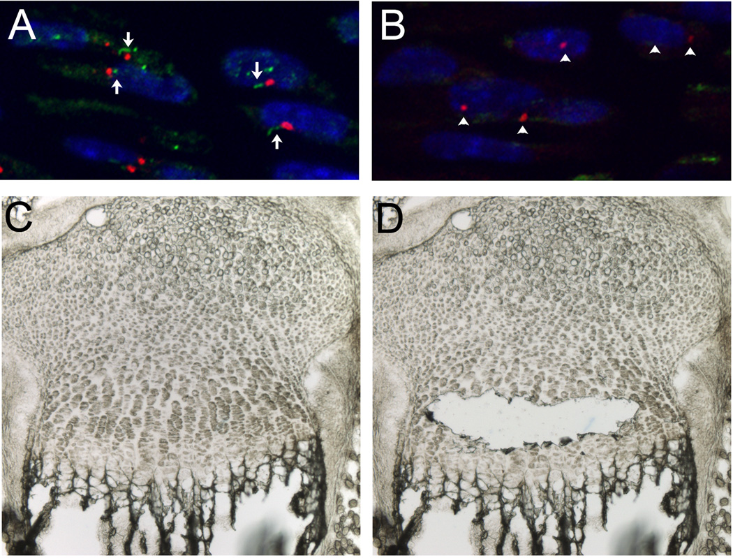
To begin to understand how Ift88 and depletion of primary cilia in the growth plate affects growth plate function, we compared gene expression in control and Col2aCre;Ift88fl/fl chondrocytes using Affymetrix gene arrays. First, depletion of primary cilia in the growth plate was verified by immunostaining to Arl13b, a cilia specific protein (21); (Fig. 1A and B). Next, the flat columnar cells of the of the growth plate was microdissected from thick sections of post-natal day 7 (P7) proximal tibiae and distal femur from control and mutant mice. This is the time the abnormalities in the growth plate first start and the columnar structure of the growth plate can be easily seen under a dissecting microscope (Fig. 1C and D). RNA was isolated from the dissected tissue and quality control tested. The high quality RNA from two controls and two mutants was amplified, labeled, and then hybridized each to separate Affymetrix Mouse 430 2.0 GeneChip Arrays. Microarray results were analyzed using GeneSpring software. A total of 48 genes showed at least a two-fold change of expression (up or down) in mutant columnar growth plate chondrocytes compared to controls (T-test, p<0.05), with 28 genes down-regulated (Table 1) and 20 genes up-regulated (Table 2). The change in expression of selected genes was verified by real time RT-PCR (Table 3) using separate RNA samples isolated from 3 control and 3 mutant mice. Combined PCR data from the three biological replicates was analyzed using REST software, which normalizes results and calculates the relative fold difference in gene expression, standard error (65% confidence interval), and statistical significance across experiments (20). The change in expression for all of the selected genes was consistent with the microarray result (Table 3). Pathway analysis using Ingenuity Pathway Analysis software indicated that Hh signaling was significantly down-regulated in the mutant cells relative to controls (p-value= 0.002). This was evident by down-regulation of and Gli1 and Ptch1, targets of Hh signaling. Cell death and cell growth pathways were also regulated (p-values range = 9.38 X 10⁻⁵ – 3.17 X 10⁻² and 9.52 X 10⁻⁵ – 2.77 X 10⁻²). We had previously reported that loss of cilia in the growth plate resulted in reduced chondrocyte proliferation and accelerated hypertrophic differentiation. Alterations in apoptosis as measured by TUNEL staining, which measures late stages of apoptosis, were not previously detected in Kif3a or Ift88 mutants (6). Down-regulation of Prelp, a glycosaminoglycan and collagen-binding anchor protein that is highly expressed in columnar growth plate chondrocytes (22), in mutant chondrocytes suggests that the extracellular matrix environment at P7 was already changing even though only minor differences in growth plate histology were observed. The results indicate that deletion of Ift88 in proliferating zone chondrocytes results in changes in gene expression that may provide clues about the mechanism of Ift88 and cilia function in growth plate chondrocytes.
Figure 1. Micro-dissection of postnatal day 7 growth plate.
Depletion of primary cilia was confirmed by immunostaining in growth plates from P7 control (A) and Col2aCre;Ift88fl/fl (B) mice. Ciliary bodies were stained with anti-Arl13b antibody (green), and the basal bodies were stained with anti-γ-tubulin antibody (red). The nuclei were stained with DAPI (blue). Images showing the cartilage tissue section on microscope slide before (C) and after (D) micro-dissection.
Table 1.
Genes down-regulated (T-test p value < 0.05, >2-fold change) in proliferative zone of mutant growth plate.
| Probe Set ID | Fold change down | Gene Symbol | Gene Title |
|---|---|---|---|
| 1436838_x_at | 2.010 | Cotl1 | coactosin-like 1 (Dictyostelium) |
| 1416142_at | 2.055 | Rps6 | ribosomal protein S6 |
| 1424308_at | 2.079 | Slc24a3 | solute carrier family 24 (sodium/potassium/calcium exchanger), member 3 |
| 1417464_at | 2.110 | Tnnc2 | troponin C2, fast |
| 1427356_at | 2.119 | Fam89a | family with sequence similarity 89, member A |
| 1420911_a_at | 2.135 | Mfge8 | milk fat globule-EGF factor 8 protein |
| 1438968_x_at | 2.198 | Spint2 | serine protease inhibitor, Kunitz type 2 |
| 1417069_a_at | 2.223 | Gmfb | glia maturation factor, beta |
| 1457111_at | 2.229 | AA415038 | expressed sequence AA415038 |
| 1426565_at | 2.248 | Igf1r | insulin-like growth factor I receptor |
| 1435338_at | 2.371 | Cdk6 | cyclin-dependent kinase 6 |
| 1438936_s_at | 2.627 | Ang | angiogenin, ribonuclease, RNase A family, 5 |
| 1417868_a_at | 2.992 | Ctsz | cathepsin Z |
| 1443837_x_at | 3.033 | Bcl2 | B-cell leukemia/lymphoma 2 |
| 1427247_at | 3.122 | D3Bwg0562e | DNA segment, Chr 3, Brigham & Women's Genetics 0562 expressed |
| 1419379_x_at | 3.219 | Fxyd2 | FXYD domain-containing ion transport regulator 2 |
| 1434677_at | 3.253 | Hps5 | Hermansky-Pudlak syndrome 5 homolog (human) |
| 1416321_s_at | 3.289 | Prelp | proline arginine-rich end leucine-rich repeat |
| 1416022_at | 3.773 | Fabp5 | fatty acid binding protein 5, epidermal |
| 1424196_at | 4.028 | Yipf1 | Yip1 domain family, member 1 |
| 1435554_at | 4.418 | Tmcc3 | transmembrane and coiled coil domains 3 |
| 1428853_at | 4.425 | Ptch1 | patched homolog 1 |
| 1451814_a_at | 5.275 | Htatip2 | HIV-1 tat interactive protein 2, homolog (human) |
| 1436075_at | 5.850 | Sfrp5 | secreted frizzled-related sequence protein 5 |
| 1443686_at | 6.104 | H2-DMb2 | histocompatibility 2, class II, locus Mb2 |
| 1449058_at | 6.155 | Gli1 | GLI-Kruppel family member GLI1 |
| 1436964_at | 6.239 | D7Ertd715e | DNA segment, Chr 7, ERATO Doi 715, expressed |
| 1416889_at | 9.012 | Tnni2 | troponin I, skeletal, fast 2 |
Table 2.
Genes up-regulated (T-test p value < 0.05, >2-fold change) in proliferative zone of mutant growth plate.
| Probe Set ID | Fold change up | Gene Symbol | Gene Title |
|---|---|---|---|
| 1442489_at | 2.016 | D1Ertd564e | DNA segment, Chr 1, ERATO Doi 564, expressed |
| 1439255_s_at | 2.125 | Gpr137b | G protein-coupled receptor 137B |
| 1417814_at | 2.130 | Pla2g5 | phospholipase A2, group V |
| 1452382_at | 2.168 | Dnm3os | dynamin 3, opposite strand |
| 1460453_at | 2.243 | Tagap1 | similar to T-cell activation Rho GTPase-activating protein |
| 1434667_at | 2.271 | Col8a2 | collagen, type VIII, alpha 2 |
| 1437506_at | 2.274 | Adamts6 | a disintegrin-like and metallopeptidase (reprolysin type) with thrombospondin type 1 motif, 6 |
| 1437128_a_at | 2.293 | A630033E08Rik | RIKEN cDNA A630033E08 gene |
| 1421861_at | 2.329 | Clstn1 | calsyntenin 1 |
| 1438441_at | 2.330 | Id4 | Inhibitor of DNA binding 4 (Id4), mRNA |
| 1426818_at | 2.346 | Arrdc4 | arrestin domain containing 4 |
| 1426774_at | 2.379 | Parp12 | poly (ADP-ribose) polymerase family, member 12 |
| 1448201_at | 2.544 | Sfrp2 | secreted frizzled-related protein 2 |
| 1458667_at | 2.643 | 4930519N13Rik | RIKEN cDNA 4930519N13 gene |
| 1426518_at | 2.672 | Tubgcp5 | tubulin, gamma complex associated protein 5 |
| 1459860_x_at | 2.693 | Trim2 | tripartite motif-containing 2 |
| 1438878_at | 3.220 | 6430537K16Rik | RIKEN cDNA 6430537K16 gene |
| 1453152_at | 3.504 | Mamdc2 | MAM domain containing 2 |
| 1435290_x_at | 6.859 | H2-Aa | histocompatibility 2, class II antigen A, alpha |
| 1447360_at | 14.370 | Tsc22d1 | TSC22-related inducible leucine zipper 1b (Tilz1b) |
Table 3.
Selected genes verified by real time RT-PCR.
| Gene | Expression mutant/control | Standard Error | P value | Result |
|---|---|---|---|---|
| Gli1 | 0.271 | 0.197 – 0.374 | 0.000 | DOWN |
| Ptch1 | 0.274 | 0.213 – 0.358 | 0.000 | DOWN |
| Sfrp5 | 0.253 | 0.160 – 0.401 | 0.000 | DOWN |
| Prelp | 0.380 | 0.255 – 0.555 | 0.000 | DOWN |
| Igf1r | 0.714 | 0.531 – 0.995 | 0.005 | DOWN |
| Bcl2 | 0.518 | 0.298 – 0.900 | 0.001 | DOWN |
| Sfrp2 | 1.730 | 1.206 – 2.502 | 0.002 | UP |
Sfrp5 is down-regulated in cilia depleted growth plate and canonical signaling is up-regulated
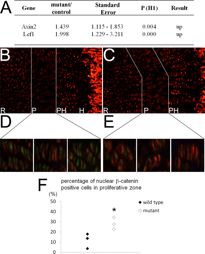
Two proteins that modulate Wnt signaling, Secreted Frizzled-related Proteins −5 and −2 (Sfrp5 and Sfrp2), were down-regulated and up-regulated, respectively, in mutant chondrocytes compared to controls (Tables 1–3) (15; 16). Sfrp5 is expressed in proliferating chondrocytes in the growth plate while Sfrp2 is more highly expressed in the developing joint and perichondrium (23). Since inappropriate activation of Wnt/β-catenin signaling promotes hypertrophic differentiation and induces premature closure of the growth plate (13; 14) similar to what is seen in the Ift88-deleted mice, we tested whether low levels of Sfrp5 in Ift88 mutants correlated with increased canonical Wnt signaling. Wnt/β-catenin signaling was measured as nuclear localization of β-catenin, and expression of Axin2 and Lef1, down-stream targets of β-catenin, in mutant and control growth plates (Fig. 2). Real time RT-PCR using RNA isolated from microdissected columnar growth plate chondrocytes indicated a small but significant up-regulation of both Axin2 and Lef1 (Figure 2A). In sections from P7 control limbs, β-catenin was strongly detected by immunoflourescence in the nucleus of resting and maturing prehypertrophic cells immediately adjacent to the hypertrophic zone. Few flat, columnar cells in the growth plate demonstrated nuclear β-catenin staining, similar to what has been previously reported (Fig. 2 B, D) (24). A similar staining pattern was observed in the resting and prehypertrophic cells in the Col2aCre;Ift88fl/fl limbs (Fig. 2C); however, there was an increase in nuclear β-catenin staining specifically in the flat columnar cells of the growth plate (Figure 2C, E) and the increase was statistically significant (Fig. 2F). We conclude that Wnt/β-catenin signaling is increased in the columnar cells of Ift88-deleted growth plate when Sfrp5 is reduced.
Figure 2. Canonical Wnt signaling in Col2aCre;Ift88fl/fl mutant cartilage.
(A) Expression levels of Axin2 and Lef1 were measured by quantitative real time RT-PCR using RNA from micro-dissected columnar growth plate chondrocytes. Three separate control and mutant mice were used. Data was analyzed by REST software. B2m was used for normalization. (B-E) Immunostaining for β-catenin (red) in sections from the distal femur in control (B, D) and mutant (C, E) mice (10X magnification). High magnification images (D, E; 40X) demonstrating nuclear versus cytoplasmic staining in flat columnar chondrocytes for three random fields in sections from control (D) and mutant (E) mice. Nuclei are stained green with YoPro. (F) Three separate control and mutant samples were used to count nuclear β-catenin staining. Ten to fifteen columns in columnar chondrocytes of the growth plate were counted for each sample (100–200 cells) and the percentage of cells with nuclear β-catenin staining was calculated as nuclear staining over total nuclei stained with YoPro in the field. Each point represents data from an individual mouse. The percentage of nuclear β-catenin was increased in mutant growth plate (χ-squared test, *p<0.05).
Sfrp5 is a downstream target of Hh signaling
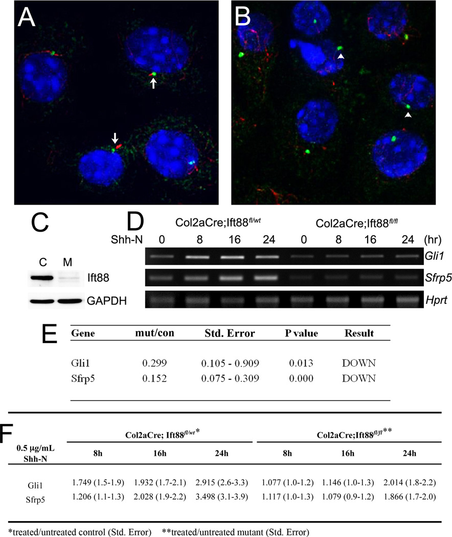
Since Hh was the most significantly affected signaling pathway in Ift88-deleted chondrocytes and it has been shown that loss of Ihh signaling in post-natal growth plate has similar phenotype to the Ift88-deleted mice (10; 25), we tested the hypothesis that Hh signaling regulates Sfrp5. Costal chondrocytes from newborn control and Col2aCre;Ift88fl/fl mice were isolated, placed in culture and treated with 0.5µg recombinant Shh-N protein/ ml for varying times (Fig. 3). RNA was isolated and used in real time RT-PCR to determine the relative expression levels of Gli1, a known target of Hh signaling, and Sfrp5. First, we confirmed using immunofluorescent staining that cilia were depleted in Ift88-deleted cells grown in culture (Fig. 3A, B) and by western blot that Ift88 protein was down-regulated (Fig. 3C). We then showed that, as expected, Gli1 and Sfrp5 were down-regulated in untreated mutant cells relative to controls (Fig. 3D, E). When control cells were treated with Shh-N protein, Gli1 was significantly up-regulated by 8 hours with highest up-regulation by 24 hours (Fig. 3D, F). Sfrp5 was significantly up regulated by 16 hours with the highest expression at 24 hours after treatment (Fig. 3D, F). In contrast, in mutant cells, up-regulation of Gli1 and Sfrp5 by Shh treatment was delayed and attenuated (Fig. 3D, F). Significant changes in gene expression were not detected in mutant cells until 24 hours and the level of induction was reduced relative to control cells. The response to Shh in mutant cells at 24 hours could result from a few cells that may retain cilia in the cultures. The results suggest that Sfrp5 is a downstream target of Hh signaling and that down-regulation of Sfrp5 in Ift88-deleted growth plate is likely due to regulation of Hh signaling by primary cilia. Two putative Gli binding sites (26) were identified in the Sfrp5 promoter region supporting this hypothesis (supplemental Fig. 1). This is the first report showing that Sfrp5 is regulated by Hh.
Figure 3. Gli1 and Sfrp5 expression in Shh-N treated rib chondrocytes.
Deletion of primary cilia was confirmed by immunostaining using anti-acetylated tubulin (red) and anti-γ-tubulin antibodies (green) in control (A) and mutant (B) cells. (C) Ift88 protein levels in control (c) and mutant (m) cells as measured by western blot. (D) Semi-quantitative RT-PCR measuring relative levels of Gli1 and Sfrp5 in control (Col2aCre;Ift88fl/wt) and mutant (Col2aCre;Ift88fl/fl) cells treated with 0.5µg/ml Shh-N for varying times (hr). Hprt was used as a loading control. (E) Real time RT-PCR comparing relative expression of Gli1 and Sfrp5 in untreated control and mutant cells. (F) Real time RT-PCR comparing untreated control cells to control cells treated with Shh-N for varying times and untreated mutant cells to mutant cells treated with Shh-N for varying times. RT-PCR data was analyzed using REST software, expression levels of treated/ untreated samples with the standard error are shown. A representative of two separate experiments is shown. Each sample was analyzed in triplicate with Hprt used for normalization.
Discussion
To understand the mechanisms of cilia action in the growth plate, we compared gene expression patterns in control and Col2aCre;Ift88fl/fl growth plates. The RNA samples used were prepared from micro-dissected columnar growth plate chondrocytes at postnatal day 7 when alterations in histology were just beginning. Analysis using micro-dissected tissue sections avoids contamination from surrounding cells, which could mask changes in gene expression in a specific cell type. Previously, we reported that Hh signaling was not altered at P10 in growth plate from Col2aCre;Kif3afl/fl mice because we did not detect changes in Ptch1 expression by semi-quantitative RT-PCR using samples isolated from total metaphysis and epiphysis (6). While this may indicate some molecular differences in how Kif3a and Ift88 function, it is more likely due to how the experiments were done. In this study, with samples isolated specifically from columnar growth plate chondrocytes, we found that Hh signaling was significantly affected. This result fits well with the current model that primary cilia are required for ligand-dependent Hh signaling (3).
The importance of primary cilia in Hh signaling has been demonstrated by several studies. Primary cilia are required for ligand-dependent activation of Hh signaling and ligand-independent proteolytic processing of full length Gli3 activator to repressor (5). Without primary cilia, both ligand-mediated activation and Gli3-mediated repression of Hh signaling are disrupted. Depending on whether Gli activator or repressor functions are dominant, loss of primary cilia has varying effects on different tissues. Here we show that ligand-dependent Hh signaling is disrupted in postnatal Col2aCre;Ift88fl/fl growth plate as evidenced by reduced expression of Ptc1 and Gli1 mRNA. In addition, the response to Shh in cilia-depleted costal chondrocytes in culture is attenuated. Mice with a conditional deletion of Ihh in the postnatal growth plate, Col2aCreER;Ihhfl/fl, have a similar phenotype to Col2aCre;Ift88fl/fl mice including accelerated hypertrophic differentiation and disorganization of the columnar structure of the growth plate (25). These results together suggest that ligand-dependent Gli activator function is dominant in the post-natal growth plate and Ift88 is required for activator functions. In contrast, we recently showed that deletion of Ift88 in articular cartilage results in up-regulation of Hh signaling, reduced Gli3 repressor to activator ratio, and symptoms of early osteoarthritis (7). Up-regulation of Hh signaling has been associated with osteoarthritis in human patients and mouse models of osteoarthritis (27). In embryonic growth plate, Hh inhibits hypertrophic differentiation though regulation of PTHrP expression. In contrast, in articular cartilage, a PTHrP-independent pathway becomes dominant and Hh signaling promotes chondrocyte hypertrophy (28). Therefore, low levels of Hh signaling are required in articular cartilage where Gli3 repressor function is dominant. The results fit well with what is known about the molecular differences between articular and growth plate cartilage and point to a role for Ift88 in this regulation.
In this study, we identified Sfrp5 as a downstream target of cilia and Hh signaling in the growth plate. The Sfrp gene family contains five members that are classified into Sfrp1 and Frzb subfamilies. Sfrp1/2/5 belong to the Sfrp1 subfamily (29). In embryonic cartilage development, Sfrp2 is expressed in synovial joints and Sfrp5 is expressed in proliferating chondrocytes (23). In our study, Sfrp2 was up-regulated and Sfrp5 was down-regulated in Ift88 deficient growth plate. Sfrp5 was up-regulated by Hh in chondrocytes in culture while Sfrp2 expression was not altered in Ift88-deleted costal chondrocytes nor was Sfrp2 regulated by Shh (not shown). Mice with activated β-catenin demonstrate accelerated hypertrophic differentiation and premature closure of the growth plate (13; 14). Deletion of Sfrp1, the Sfrp with the highest homology to Sfrp5, in mice also resulted in accelerated hypertrophic differentiation and activation of Wnt/β-catenin signaling in the growth plate similar to what we see in the Ift88-deleted mice (30). We hypothesize that Ihh regulates Sfrp5 expression through Ift88/ primary cilia and that Sfrp5 regulates Wnt/β-catenin activity, hypertrophic differentiation, and premature closure of the growth plate. The idea that Wnt signaling is regulated indirectly by Ift88/ cilia through Hh signals could explain some of the conflicting results regarding whether or not β-catenin signaling is regulated by primary cilia (3).
Deletion of Ift88 and disruption of Hh signaling in growth plate both also result in disorganized columnar structure in the growth plate (6; 25) but the mechanism of how these signals regulate chondrocyte orientation is not clear. The method to generate columnar structure in the growth plate was first proposed by Dodds in 1930 (8) based on microscopic observations of tissue sections. Proliferating chondrocytes divide perpendicularly to the axis of long bone, then migrate one on the top of another. This process, called chondrocytes rotation, is similar to the convergent extension movements that take place during embryonic gastrulation (31). Wnt/PCP signaling is a key regulator for this process (32) and it was recently shown that chondrocyte rotation is regulated by Wnt/PCP signaling (11). Sfrps genetically interact with the core PCP component Vangl2 (16), and function redundantly in regulation of canonical Wnt/β-catenin and noncanonical Wnt/PCP signaling (16). It has been suggested that primary cilia restrain canonical Wnt/β-catenin signaling and act as a molecular switch between canonical Wnt and noncanonical Wnt/PCP signaling ((33). Since the Wnt/ PCP pathway and Ihh regulate organization of the growth plate (11), we speculate that in addition to regulating Wnt/βcatenin signaling and hypertrophic differentiation, Sfrp5 acts downstream of Ihh to regulate organization of the growth plate through PCP. Sfrp5 may be a molecular switch between Wnt signaling in the context of growth plate. At this time, there are no good down-stream markers of PCP signaling in the growth plate to test this hypothesis directly.
Supplementary Material
(A) The consensus Gli binding motif is GACCACCCA. (B) The partial gene sequence of Sfrp5. Red sequence is a partial sequence of exon 1. The site of start codon is marked with ( ). Two putative Gli binding sites are underlined
Acknowledgements
We would like to thank Drs. Michael Crowley and David Crossman at the Heflin Center for Genetics, University of Alabama at Birmingham, for help with the microarray and data analysis. We also thank Dr. Bradley Yoder, Department of Cell Biology, University of Alabama at Birmingham, for providing the Ift88fl/fl mice. Funding for this study was through NIH grants AR055110 and AR053860 to RS.
Footnotes
None of the authors have any conflicts to declare.
References
- 1.Ishikawa H, Marshall WF. Ciliogenesis: Building the cell's antenna. Nat Rev Mol Cell Biol. 2011;12:222–234. doi: 10.1038/nrm3085. [DOI] [PubMed] [Google Scholar]
- 2.Marshall WF. Basal bodies platforms for building cilia. Curr Top Dev Biol. 2008;85:1–22. doi: 10.1016/S0070-2153(08)00801-6. [DOI] [PubMed] [Google Scholar]
- 3.Goetz SC, Anderson KV. The primary cilium: A signalling centre during vertebrate development. Nat Rev Genet. 2010;11:331–344. doi: 10.1038/nrg2774. [DOI] [PMC free article] [PubMed] [Google Scholar]
- 4.Sharma N, Berbari NF, Yoder BK. Ciliary dysfunction in developmental abnormalities and diseases. Curr Top Dev Biol. 2008;85:371–427. doi: 10.1016/S0070-2153(08)00813-2. [DOI] [PubMed] [Google Scholar]
- 5.Haycraft CJ, Serra R. Cilia involvement in patterning and maintenance of the skeleton. Curr Top Dev Biol. 2008;85:303–332. doi: 10.1016/S0070-2153(08)00811-9. [DOI] [PMC free article] [PubMed] [Google Scholar]
- 6.Song B, Haycraft CJ, Seo HS, et al. Development of the post-natal growth plate requires intraflagellar transport proteins. Dev Biol. 2007;305:202–216. doi: 10.1016/j.ydbio.2007.02.003. [DOI] [PMC free article] [PubMed] [Google Scholar]
- 7.Chang CF, Ramaswamy G, Serra R. Depletion of primary cilia in articular chondrocytes results in reduced gli3 repressor to activator ratio, increased hedgehog signaling, and symptoms of early osteoarthritis. Bone. 2012 doi: 10.1016/j.joca.2011.11.009. [DOI] [PMC free article] [PubMed] [Google Scholar]
- 8.Dodds GS. Row formation and other types of arrangement of cartilage cells in endochondral ossifcation. Anat Rec. 1930;46:385–399. [Google Scholar]
- 9.Aszodi A, Hunziker EB, Brakebusch C, Fassler R. Beta1 integrins regulate chondrocyte rotation, g1 progression, and cytokinesis. Genes Dev. 2003;17:2465–2479. doi: 10.1101/gad.277003. [DOI] [PMC free article] [PubMed] [Google Scholar]
- 10.St-Jacques B, Hammerschmidt M, McMahon AP. Indian hedgehog signaling regulates proliferation and differentiation of chondrocytes and is essential for bone formation. Genes Dev. 1999;13:2072–2086. doi: 10.1101/gad.13.16.2072. [DOI] [PMC free article] [PubMed] [Google Scholar]
- 11.Li Y, Dudley AT. Noncanonical frizzled signaling regulates cell polarity of growth plate chondrocytes. Development. 2009;136:1083–1092. doi: 10.1242/dev.023820. [DOI] [PMC free article] [PubMed] [Google Scholar]
- 12.Kronenberg HM. Developmental regulation of the growth plate. Nature. 2003;423:332–336. doi: 10.1038/nature01657. [DOI] [PubMed] [Google Scholar]
- 13.Yuasa T, Kondo N, Yasuhara R, et al. Transient activation of wnt/{beta}-catenin signaling induces abnormal growth plate closure and articular cartilage thickening in postnatal mice. Am J Pathol. 2009;175:1993–2003. doi: 10.2353/ajpath.2009.081173. [DOI] [PMC free article] [PubMed] [Google Scholar]
- 14.Tamamura Y, Otani T, Kanatani N, et al. Developmental regulation of wnt/beta-catenin signals is required for growth plate assembly, cartilage integrity, and endochondral ossification. J Biol Chem. 2005;280:19185–19195. doi: 10.1074/jbc.M414275200. [DOI] [PubMed] [Google Scholar]
- 15.Li Y, Rankin SA, Sinner D, et al. Sfrp5 coordinates foregut specification and morphogenesis by antagonizing both canonical and noncanonical wnt11 signaling. Genes Dev. 2008;22:3050–3063. doi: 10.1101/gad.1687308. [DOI] [PMC free article] [PubMed] [Google Scholar]
- 16.Satoh W, Matsuyama M, Takemura H, et al. Sfrp1, sfrp2, and sfrp5 regulate the wnt/beta-catenin and the planar cell polarity pathways during early trunk formation in mouse. Genesis. 2008;46:92–103. doi: 10.1002/dvg.20369. [DOI] [PubMed] [Google Scholar]
- 17.Haycraft CJ, Zhang Q, Song B, et al. Intraflagellar transport is essential for endochondral bone formation. Development. 2007;134:307–316. doi: 10.1242/dev.02732. [DOI] [PubMed] [Google Scholar]
- 18.Ovchinnikov DA, Deng JM, Ogunrinu G, Behringer RR. Col2a1-directed expression of cre recombinase in differentiating chondrocytes in transgenic mice. Genesis. 2000;26:145–146. [PubMed] [Google Scholar]
- 19.Thirion S, Berenbaum F. Culture and phenotyping of chondrocytes in primary culture. Methods Mol Med. 2004;100:1–14. doi: 10.1385/1-59259-810-2:001. [DOI] [PubMed] [Google Scholar]
- 20.Pfaffl MW, Horgan GW, Dempfle L. Relative expression software tool (rest) for group-wise comparison and statistical analysis of relative expression results in real-time pcr. Nucleic Acids Res. 2002;30:e36. doi: 10.1093/nar/30.9.e36. [DOI] [PMC free article] [PubMed] [Google Scholar]
- 21.Caspary T, Larkins CE, Anderson KV. The graded response to sonic hedgehog depends on cilia architecture. Dev Cell. 2007;12:767–778. doi: 10.1016/j.devcel.2007.03.004. [DOI] [PubMed] [Google Scholar]
- 22.Grover J, Roughley PJ. Characterization and expression of murine prelp. Matrix Biol. 2001;20:555–564. doi: 10.1016/s0945-053x(01)00165-2. [DOI] [PubMed] [Google Scholar]
- 23.Witte F, Dokas J, Neuendorf F, et al. Comprehensive expression analysis of all wnt genes and their major secreted antagonists during mouse limb development and cartilage differentiation. Gene Expr Patterns. 2009;9:215–223. doi: 10.1016/j.gep.2008.12.009. [DOI] [PubMed] [Google Scholar]
- 24.Hens JR, Wilson KM, Dann P, et al. Topgal mice show that the canonical wnt signaling pathway is active during bone development and growth and is activated by mechanical loading in vitro. J Bone Miner Res. 2005;20:1103–1113. doi: 10.1359/JBMR.050210. [DOI] [PubMed] [Google Scholar]
- 25.Maeda Y, Nakamura E, Nguyen MT, et al. Indian hedgehog produced by postnatal chondrocytes is essential for maintaining a growth plate and trabecular bone. Proc Natl Acad Sci U S A. 2007;104:6382–6387. doi: 10.1073/pnas.0608449104. [DOI] [PMC free article] [PubMed] [Google Scholar]
- 26.Sasaki H, Hui C, Nakafuku M, Kondoh H. A binding site for gli proteins is essential for hnf-3beta floor plate enhancer activity in transgenics and can respond to shh in vitro. Development. 1997;124:1313–1322. doi: 10.1242/dev.124.7.1313. [DOI] [PubMed] [Google Scholar]
- 27.Lin AC, Seeto BL, Bartoszko JM, et al. Modulating hedgehog signaling can attenuate the severity of osteoarthritis. Nat Med. 2009;15:1421–1425. doi: 10.1038/nm.2055. [DOI] [PubMed] [Google Scholar]
- 28.Mak KK, Kronenberg HM, Chuang PT, et al. Indian hedgehog signals independently of pthrp to promote chondrocyte hypertrophy. Development. 2008;135:1947–1956. doi: 10.1242/dev.018044. [DOI] [PMC free article] [PubMed] [Google Scholar]
- 29.Bovolenta P, Esteve P, Ruiz JM, et al. Beyond wnt inhibition: New functions of secreted frizzled-related proteins in development and disease. J Cell Sci. 2008;121:737–746. doi: 10.1242/jcs.026096. [DOI] [PubMed] [Google Scholar]
- 30.Gaur T, Rich L, Lengner CJ, et al. Secreted frizzled related protein 1 regulates wnt signaling for bmp2 induced chondrocyte differentiation. J Cell Physiol. 2006;208:87–96. doi: 10.1002/jcp.20637. [DOI] [PubMed] [Google Scholar]
- 31.Wallingford JB, Fraser SE, Harland RM. Convergent extension: The molecular control of polarized cell movement during embryonic development. Dev Cell. 2002;2:695–706. doi: 10.1016/s1534-5807(02)00197-1. [DOI] [PubMed] [Google Scholar]
- 32.Kiefer JC. Planar cell polarity: Heading in the right direction. Dev Dyn. 2005;233:695–700. doi: 10.1002/dvdy.20359. [DOI] [PubMed] [Google Scholar]
- 33.He X. Cilia put a brake on wnt signalling. Nat Cell Biol. 2008;10:11–13. doi: 10.1038/ncb0108-11. [DOI] [PubMed] [Google Scholar]
Associated Data
This section collects any data citations, data availability statements, or supplementary materials included in this article.
Supplementary Materials
(A) The consensus Gli binding motif is GACCACCCA. (B) The partial gene sequence of Sfrp5. Red sequence is a partial sequence of exon 1. The site of start codon is marked with ( ). Two putative Gli binding sites are underlined


