Abstract
Objective
To present the accuracy and safety of cervical pedicle screw insertion using the technique with direct exposure of the pedicle by laminoforaminotomy.
Methods
We retrospectively reviewed 12 consecutive patients. A total of 104 subaxial cervical pedicle screws in 12 patients had been inserted. We also assessed the clinical and radiological outcomes and analyzed the direction and grade of pedicle perforation (grade 0: no perforation, 1: <25%, 2: 20% to 50%, 3: >50% of screw diameter) on the postoperative vascular-enhanced computed tomography scans. Grade 2 and 3 were considered as incorrect position.
Results
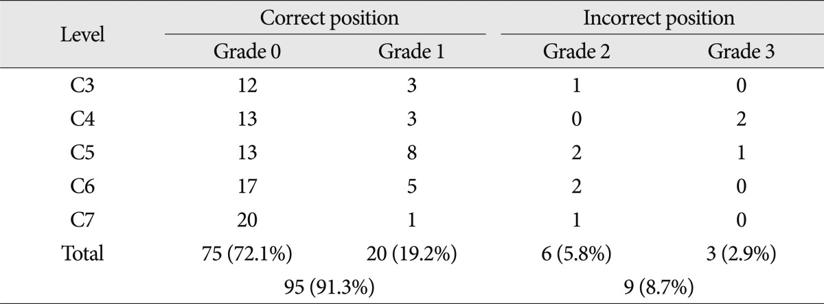
The correct position was found in 95 screws (91.3%); grade 0-75 screws, grade 1-20 screws and the incorrect position in 9 screws (8.7%); grade 2-6 screws, grade 3-3 screws. There was no neurovascular complication related with cervical pedicle screw insertion.
Conclusion
This technique (technique with direct exposure of the pedicle by laminoforaminotomy) could be considered relatively safe and easy method to insert cervical pedicle screw.
Keywords: Cervical pedicle screw, Laminoforaminotomy, Pedicle perforation
INTRODUCTION
Because a cervical pedicle screw has more rigid fixation than other posterior fixation techniques, including posterior wiring, lateral mass screw, or facet screw fixation. Cervical pedicle screw not only allow for shorter instrumentation with sagittal correction11-13,19,31,34), but also are valuable for simultaneous posterior decompression and reconstruction1-3). Then, the cervical pedicle screw insertion has become popular in a wide range of cervical spine related disorder (traumatic, degenerative, inflammatory, and neoplastic conditions). However, cervical pedicle screw insertion is technically demanding because of the anatomical variations in cervical pedicle size, lack of anatomical landmarks, small pedicle diameter, and the large convergence angle of cervical pedicles14,22). The potential risk of neurovascular injury remain the issue preventing wide acceptance17,24). Therefore, accurate and safe cervical pedicle screw insertion techniques are necessary.
Following the first description of cervical pedicle screw insertion by Abumi et al.1), different surgical techniques have been developed and evaluated, including the techniques relying on anatomical landmarks for cervical pedicle screw insertion , techniques with direct exposure of the pedicle by laminoforaminotomy to palpate the medial and superior pedicle walls, by the funnel technique, and the computer-assisted navigation system5-8,15,16,18,21,23,25,33,37).
The accuracy of cervical pedicle screw insertion varied significantly in literature, ranging from 16.8 to 97%16,21,23,29,32). The navigation system has been shown to improve screw accuracy significantly, but has limited application due to its high cost and lengthy registration procedure. Furthermore, surgical skills and experience are still needed and the surgeon should never solely rely on the navigation system32,33).
Although a number of studies have evaluated the morphometry of cervical pedicles to support accurate placement of pedicle screws, the results are inconclusive14,27). Therefore, several techniques and computer-assisted navigation systems for cervical pedicle screw insertion have been advocated.
The objective of this study was to present the cervical pedicle screw insertion technique with direct exposure of the pedicle by laminoforaminotomy, and evaluate the accuracy of pedicle screw placement and validity of pedicle screw fixation in patients.
But, the authors did not evaluate the accuracy and safety of pedicle screw placement in cadavers before applying this technique to patients. This is limitation of our study.
MATERIALS AND METHODS
A total of 12 consecutive cases in which 104 cervical pedicle screws had been inserted from C3 to C7 were reviewed for this study. The ratio of men to women was 3 : 1 and the mean age was 54 years (range, 31-75 y). Preoperative diagnosis was myelopathy due to cervical spondylosis in 6 cases, ossification of the posterior longitudinal ligament in 3 cases, traumatic lesion in 3 cases. Our institutional ethics committee has approved the human protocol for this investigation that all investigations were conducted in conformity with ethical principles of research, and that informed consent was obtained.
Preoperative examination
To evaluate the anomaly and dominant side of vertebral artery, we checked vascular-enhanced computed tomography (CT) scans in all patients. On preoperative CT scans, the convergence angle and the minimal diameter of the pedicle on the axial plane were measured for each cervical vertebra. The length of the screw was decided to reach the anterior one-third of the vertebral body on the preoperative CT images. Those with a pedicle diameter of less than 3.5 mm or a pedicle convergence angle of over 60 degrees were excluded from cervical pedicle screw insertion.
Surgical technique
The authors considered every pedicle to be built as a funnel with a wide posterior base, which narrows towards the isthmus. A distinct characteristic of the cervical pedicles is that the lateral pedicle wall is always the thinnest cortex14,27). Therefore, the medial pedicle cortex was used as a safe guide into the pedicle isthmus, then through it and then into the vertebral body. The arch of the medial pedicle wall, under which advancement into the pedicle proceeded, was always identified.
First step : finding of entry point
Patients were placed in the prone position with the head fixed using a Mayfield clamp. A standard midline incision was made and paravertebral muscles were dissected and retracted laterally to expose facet joints.
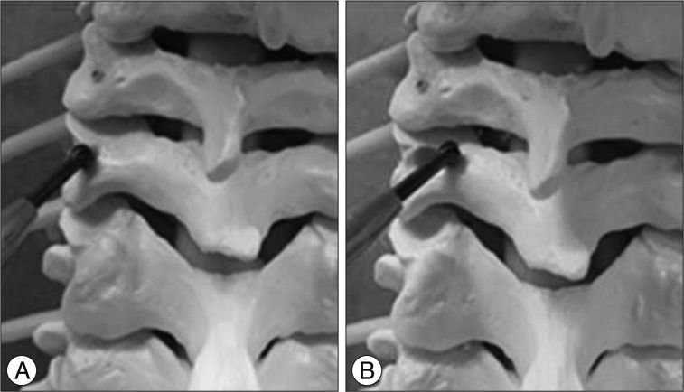
Under the lateral fluoroscopic guide and the basis of the lateral mass depth, and the convergence angle of the pedicle obtained from a preoperative computed tomography (CT) scans, 3 mm cutting burr was used to remove the outer cortex of the lateral mass over the pedicle entrance in the slightly lateral to the center of the facet, close to posterior margin of superior articular surface, the appropriate convergence angle to visualize the entrance of the pedicle (Fig. 1A). The pedicle screw insertion point is indicated by a cancellous bone at the pedicle entrance, which is commonly observed to be reddish because of a bloody cancellous bone.
Fig. 1.
A : 3 mm cutting burr is used to remove the outer cortex of the lateral mass over the pedicle entrance. B : the inferior aspect of the superior lamina and the superior aspect of the inferior lamina for the laminoforaminotomy.
Second step : identification of the pedicle medial and superior walls
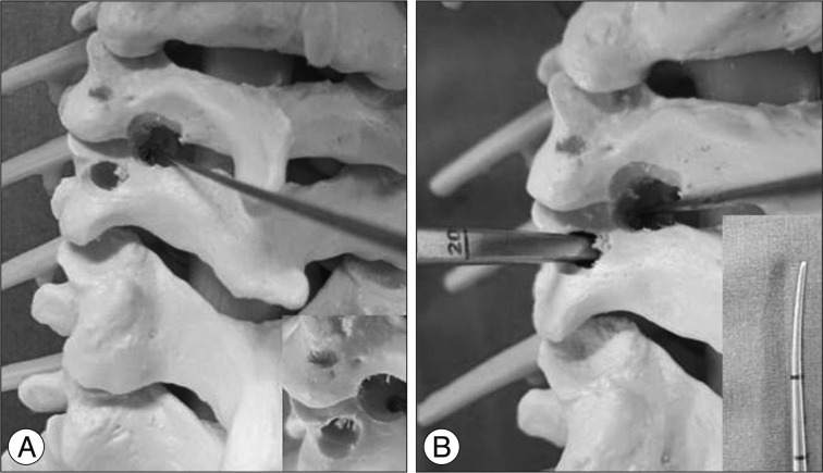
A laminoforaminotomy was performed to provide visual and tactile cues regarding the orientation of the pedicle medial and superior walls. The ligamentum flavum at each level was gently dissected free from the inferior aspect of the superior laminar arch and from the superior aspect of the inferior laminar arch using a small curved curette. Thereafter, the inferior aspect of the superior lamina and the superior aspect of the inferior lamina were removed in varying amounts using a 3 mm cutting burr followed by 1 mm and 2 mm Kerrison punches (Fig. 1B). The ball-tip hook then could be used to identify the medial and superior walls of the pedicle before the insertion of each screw (Fig. 2A).
Fig. 2.
A : A laminoforaminotomy provides visual identification of the medial and superior walls of the pedicle. B : The pedicle is probed as close to the medial wall as possible by gentle manual pressure using a 15-degree, 2 mm-diameter curved gear shift probe.
Third step : screw insertion
The direction in the sagittal plane of the pedicle probe and screw was intraoperatively determined using lateral fluoroscopic imaging. The pedicle was probed as close to the medial wall as possible by gentle manual pressure using a 15-degree, 2 mm-diameter curved gear shift probe (Fig. 2B). After probing about 2 cm depth, lack of pedicle perforation was confirmed using a ball-tip probe.
If perforation was detected within the pedicle, the trajectory was changed or the segment was skipped. Then sequential drilling, tapping, and screwing were performed. For preventing iatrogenic neural tissue injury during rod connection procedure, Rods were connected to the screws by appropriately sized slotted connectors before posterior decompression. Laminectomy was conducted for posterior decompression. Local bone harvested from the decompression site was grafted in the lateral mass bone defect.
Radiographic analysis
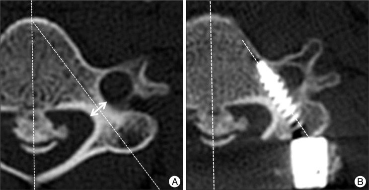
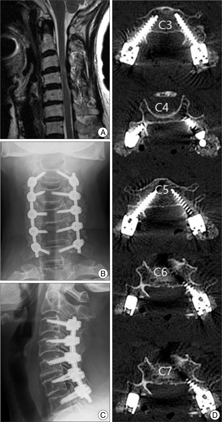
Using preoperative and postoperative vascular enhanced CT axial images, the diameter and convergence angle of the each pedicle and the screws were measured and the difference between pedicles and screws in diameter and convergence angle were analyzed (Fig. 3). The degree of perforation was classified as grade 0 if the screw was located within the pedicle; grade 1, if perforation was made by the screw by less than 25% of the screw diameter; grade 2, if perforation was made by 25% to 50% of the screw diameter; and grade 3, if perforation was made by over 50% of the screw diameter (Fig. 4). Grade 0 and 1 were classified as the correct position and the others, as the incorrect position (Fig. 5). The direction of pedicle perforation was assessed; medial, lateral, cranial, and caudal.
Fig. 3.
Preoperative and postoperative axial CT images on same cervical spine showing the diameter (arrow) and the convergence angle of the pedicle (A), and the convergence angle of the screw (B).
Fig. 4.
Grading system of pedicle perforation. Grade 0 : the screw is located within the pedicle. Grade 1 : perforation less than 25% of the screw diameter. Grade 2 : 25% to 50% of the screw diameter. Grade 3 : over 50%.
Fig. 5.
Preoperative sagittal MRI image (A) of a 71-year-old female myelopathic patient showing severe cervical cord compression. Posterior decompression with pedicle screw fixation from C3 to C7 was performed (B and C). Postoperative axial CT images show all grade 0 screw position without pedicle perforation (D).
Interobserver and intraobserver error analysis
The intraobserver and interobserver agreement rate and k values were obtained to check errors between 2 observers in grading of the pedicle perforation.
Clinical analysis
Operative time, intraoperative blood loss, and pedicle screw-related complications such as vertebral artery injury, nerve root injury, or irritation sign were analyzed.
RESULTS
The interobserver agreement rate was 87% for the grade of pedicle perforation (mean k=0.65), and intraobserver agreement rate was 93% (mean k=0.77).
The intraobserver and interobserver error analyses showed good agreement. The mean number of subaxial fusion segments was 3.33±0.6. In all cases cervical screw insertion was acperformed before total laminectomy. The number of screws inserted was 16 at C3, 18 at C4, 24 at C5, 24 at C6, and 22 at C7 (Table 1). Laminectomy with fusion was performed in 8 cases, anterior cervical discectomy and fusion in 1, cervicothoracic fusion in 3. The mean axial diameter of cervical pedicles was 5.0±1.0 mm and the mean convergence angle was 41.2±5.1 degrees. All the screws had a diameter of 4 mm and the mean convergence angle of the screws inserted was 36.6±4.2 degrees. The mean difference between the preoperative convergence angle of the pedicles and the convergence angle of the inserted screws was 4.57±4.3 degrees.
Table 1.
Total number of cervical pedicle screw and pedicle perforation
Perforation of cervical pedicles occurred in 29 screws (27.9%) : grade 1 in 20 (19.2%), grade 2 in 6 (5.8%), and grade 3 in 3 (2.9%) (Table 2). Overall, the correct position (perforation of grade 0, 1) was found in 95 screws (91.3%) and the incorrect position (perforation of grade 2, 3) in 9 screws (8.7%).
Table 2.
Grade of pedicle perforation
The mean difference between the convergence angle of pedicles and that of screws was 3.67±2.7 degrees in the correct position group and 10.04±3.4 degrees in the incorrect position group. Incorrect position occurred in 1 screw (6.2%) at C3, 2 (11.1%) at C4, 3 (12.5 %) at C5, 2 (8.3%) at C6, and 1 (4.5%) at C7 (Table 1). The direction of perforation was lateral in 21 (72.4%), followed by medial in 6 (20.6%), and caudal in 2(6.8%) screw. No perforation was directed toward cranial side (Table 3).
Table 3.
Direction of perforation
The mean operative time was 185±80 minutes. The mean intraoperative blood loss was 285±87 mL. Blood transfusion was performed in 6 cases. Vertebral artery injury or nerve root irritation symptom was not observed in any case. Postoperative vascular-enhanced CT scan confirmed that the blood flow of the vertebral artery maintained in all cases.
DISCUSSION
Cervical pedicle screw insertion is advantageous for certain pathologies of the cervical spine. Biomechanical studies have reported that cervical pedicle screws provide greater stability than other posterior cervical fixation procedures12,13,30). However, cervical pedicle screw insertion is more technically demanding than in thoracic or lumbar vertebrae because of the smaller pedicle dimension, the individual variations in pedicle anatomy, and the potential risk of injury to neurovascular structures4,14,24,26,28,35).
The cervical pedicle screw insertion technique was first described by Abumi et al.1). Since Abumi technique of cervical pedicle screw insertion, several reports have discussed various methods to insert cervical pedicle screw in easier and safer techniques. However, the methods for precisely and reproducibly determining the ideal entry points and trajectories are yet to be defined.
Recently, an anatomical study of subaxial cervical pedicles and lateral masses using CT scans of adult volunteers provided entry points and trajectories for subaxial cervical pedicle screw insertion. Zheng et al.38) reported a high success rate using subaxial cervical pedicle screw insertion, with an overall accuracy of 83.3%, including a non-critical perforation of 13.3% and a critical perforation of 3.3%. This success can be achieved using the recently proposed guidelines and the oblique view obtained through fluoroscopy. However, landmarks to the cervical pedicle entrance alone are insufficient for achieving accurate cervical pedicle screw insertion because of the variation in pedicle anatomy15,22). Then, several insertion techniques using direct pedicle exposure have been advocated in clinical or cadaver studies14,16,22,23).
Abumi et al.1) described a technique in which the cortex at the insertion point is penetrated using a high speed burr, resulting in direct observation of the pedicle entrance. They also reported a pedicle-perforation rate of 6.7% in another clinical report4). Karaikovic et al.16) used the funnel technique, in which the entrance into the pedicle and vertebral body was identified by removing the outer cortex. They used the medial cervical pedicle cortex as a guide into the vertebral body through the pedicle isthmus. This study reported a screw perforation rate of 16.8%, including non-critical perforations of 9.7% and critical perforations of 7.1%, using the funnel technique. Miller et al.23) used partial laminectomy and a tapping technique in which the entrance point for screw insertion and angulations for screw placement were guided by direct determination of the superior, medial, and inferior borders of the pedicle through the laminar window opening. Ludwig et al.21) inserted a cervical pedicle screw insertion after a laminoforaminotomy, which provides visual and tactile cues regarding orientation of the medial, and superior pedicle walls. Yoshimoto et al.36) observed incomplete perforation in 7.3% and complete perforation in 3.7%. In 100 cases of cervical pedicle screw insertion using an oblique view in the year 2006, Yukawa et al.37) reported incomplete perforation in 10.3% and complete perforation in 4%. In 2005, Neo et al.24) mainly applied the Abumi technique to 18 cases and found pedicle perforation in 25%, a rate higher than previous reports.
On the other hand, several authors have reported cervical pedicle screw insertion techniques using computer-assisted navigation systems and computer-assisted navigation systems have lower pedicle screw perforation than free-hand techniques (direct pedicle exposure techniques)9,18,32). Kotani et al.18) reported that the screw misplacement rate was significantly lower in a computer-assisted group (1.2%) than in a free-hand group (6.7%). Increasing use of three-dimensional fluoroscopy-based computer-assisted navigation systems has recently been reported9,10). Three-dimensional fluoroscopy is superior to conventional CT-based image guidance because anatomical registration is not required and real-time updates of the spine position can be obtained intraoperatively. Ito et al.10) reported that the rate of pedicle perforations was 2.8% in the absence of clinically significant perforation when a three-dimensional fluoroscopy-based navigation system was used. Ishikawa et al.9) reported that the prevalence of pedicle perforation was 18.7% in a three-dimensional fluoroscopy-based navigation group and 27% in a conventional free-hand group. Although computer-assisted navigation systems can improve the accuracy of cervical pedicle screw insertion, there are also some disadvantages, such as the requirement of very expensive system costs.
These results cannot be compared directly because the criteria for assessing development and degree of pedicle perforation vary. The absolute rate of pedicle perforation was 27.9% in our study. We classified perforation of less than 1 mm as the correct position and perforation of over 1 mm, as the incorrect position. Incorrect position was 8.7%. This result seems higher perforation rate than some of previous studies, because we included pedicle perforation of less than 1 mm at classification. Pedicle perforation of less than 1 mm during insertion of screws was usually insignificant. The perforation rate could vary according to each grading system, which has not standardized yet.
The method used in this study is basically a modification to the Abumi technique. The difference lies in the laminoforaminotomy performed to provide visual and tactile cues regarding the orientation of the pedicle medial and superior walls. In addition, the use of a 15-degree, 2 mm-diameter curved gear shift probe is expected to reduce the risk of lateral perforation by guiding the trajectory along the strong medial cortex of the pedicle, which is about two times thicker than lateral cortex. The method used in this study requires removal of the bone in the lateral mass, similar to the Abumi and funnel techniques. Therefore expected outcome includes a significant reduction of screw fixation strength. However, Kowalski et al.20) reported no significant difference in the biomechanical pullout strength of cervical pedicle screw when the lateral mass cancellous bone is removed.
The cervical pedicle screw insertion technique with direct exposure of the pedicle by laminoforaminotomy did not require special preoperative preparation or an intraoperative environment and showed that good screw position placement and strong screw fixation stability.
CONCLUSION
We performed cervical pedicle screw insertion using the technique with direct exposure of the pedicle by laminoforaminotomy and with 91.3% correct position without clinical complications. This technique could be considered relatively safe and easy method to insert cervical pedicle screw.
References
- 1.Abumi K, Itoh H, Taneichi H, Kaneda K. Transpedicular screw fixation for traumatic lesions of the middle and lower cervical spine : description of the techniques and preliminary report. J Spinal Disord. 1994;7:19–28. doi: 10.1097/00002517-199407010-00003. [DOI] [PubMed] [Google Scholar]
- 2.Abumi K, Kaneda K. Pedicle screw fixation for nontraumatic lesions of the cervical spine. Spine (Phila Pa 1976) 1997;22:1853–1863. doi: 10.1097/00007632-199708150-00010. [DOI] [PubMed] [Google Scholar]
- 3.Abumi K, Kaneda K, Shono Y, Fujiya M. One-stage posterior decompression and reconstruction of the cervical spine by using pedicle screw fixation systems. J Neurosurg. 1999;90:19–26. doi: 10.3171/spi.1999.90.1.0019. [DOI] [PubMed] [Google Scholar]
- 4.Abumi K, Shono Y, Ito M, Taneichi H, Kotani Y, Kaneda K. Complications of pedicle screw fixation in reconstructive surgery of the cervical spine. Spine (Phila Pa 1976) 2000;25:962–969. doi: 10.1097/00007632-200004150-00011. [DOI] [PubMed] [Google Scholar]
- 5.Albert TJ, Klein GR, Joffe D, Vaccaro AR. Use of cervicothoracic junction pedicle screws for reconstruction of complex cervical spine pathology. Spine (Phila Pa 1976) 1998;23:1596–1599. doi: 10.1097/00007632-199807150-00017. [DOI] [PubMed] [Google Scholar]
- 6.Barrey C, Cotton F, Jund J, Mertens P, Perrin G. Transpedicular screwing of the seventh cervical vertebra : anatomical considerations and surgical technique. Surg Radiol Anat. 2003;25:354–360. doi: 10.1007/s00276-003-0163-5. [DOI] [PubMed] [Google Scholar]
- 7.Ebraheim NA, Xu R, Knight T, Yeasting RA. Morphometric evaluation of lower cervical pedicle and its projection. Spine (Phila Pa 1976) 1997;22:1–6. doi: 10.1097/00007632-199701010-00001. [DOI] [PubMed] [Google Scholar]
- 8.Hardy RW., Jr The posterior surgical approach to the cervical spine. Neuroimaging Clin N Am. 1995;5:481–490. [PubMed] [Google Scholar]
- 9.Ishikawa Y, Kanemura T, Yoshida G, Ito Z, Muramoto A, Ohno S. Clinical accuracy of three-dimensional fluoroscopy-based computer-assisted cervical pedicle screw placement : a retrospective comparative study of conventional versus computer-assisted cervical pedicle screw placement. J Neurosurg Spine. 2010;13:606–611. doi: 10.3171/2010.5.SPINE09993. [DOI] [PubMed] [Google Scholar]
- 10.Ito Y, Sugimoto Y, Tomioka M, Hasegawa Y, Nakago K, Yagata Y. Clinical accuracy of 3D fluoroscopy-assisted cervical pedicle screw insertion. J Neurosurg Spine. 2008;9:450–453. doi: 10.3171/SPI.2008.9.11.450. [DOI] [PubMed] [Google Scholar]
- 11.Jang WY, Kim IS, Lee HJ, Sung JH, Lee SW, Hong JT. A computed tomography-based anatomic comparison of three different types of c7 posterior fixation techniques : pedicle, intralaminar, and lateral mass screws. J Korean Neurosurg Soc. 2011;50:166–172. doi: 10.3340/jkns.2011.50.3.166. [DOI] [PMC free article] [PubMed] [Google Scholar]
- 12.Johnston TL, Karaikovic EE, Lautenschlager EP, Marcu D. Cervical pedicle screws vs. lateral mass screws : uniplanar fatigue analysis and residual pullout strengths. Spine J. 2006;6:667–672. doi: 10.1016/j.spinee.2006.03.019. [DOI] [PubMed] [Google Scholar]
- 13.Jones EL, Heller JG, Silcox DH, Hutton WC. Cervical pedicle screws versus lateral mass screws. Anatomic feasibility and biomechanical comparison. Spine (Phila Pa 1976) 1997;22:977–982. doi: 10.1097/00007632-199705010-00009. [DOI] [PubMed] [Google Scholar]
- 14.Karaikovic EE, Daubs MD, Madsen RW, Gaines RW., Jr Morphologic characteristics of human cervical pedicles. Spine (Phila Pa 1976) 1997;22:493–500. doi: 10.1097/00007632-199703010-00005. [DOI] [PubMed] [Google Scholar]
- 15.Karaikovic EE, Kunakornsawat S, Daubs MD, Madsen TW, Gaines RW., Jr Surgical anatomy of the cervical pedicles : landmarks for posterior cervical pedicle entrance localization. J Spinal Disord. 2000;13:63–72. doi: 10.1097/00002517-200002000-00013. [DOI] [PubMed] [Google Scholar]
- 16.Karaikovic EE, Yingsakmongkol W, Gaines RW., Jr Accuracy of cervical pedicle screw placement using the funnel technique. Spine (Phila Pa 1976) 2001;26:2456–2462. doi: 10.1097/00007632-200111150-00012. [DOI] [PubMed] [Google Scholar]
- 17.Kast E, Mohr K, Richter HP, Börm W. Complications of transpedicular screw fixation in the cervical spine. Eur Spine J. 2006;15:327–334. doi: 10.1007/s00586-004-0861-7. [DOI] [PMC free article] [PubMed] [Google Scholar]
- 18.Kotani Y, Abumi K, Ito M, Minami A. Improved accuracy of computer-assisted cervical pedicle screw insertion. J Neurosurg. 2003;99:257–263. doi: 10.3171/spi.2003.99.3.0257. [DOI] [PubMed] [Google Scholar]
- 19.Kothe R, Rüther W, Schneider E, Linke B. Biomechanical analysis of transpedicular screw fixation in the subaxial cervical spine. Spine (Phila Pa 1976) 2004;29:1869–1875. doi: 10.1097/01.brs.0000137287.67388.0b. [DOI] [PubMed] [Google Scholar]
- 20.Kowalski JM, Ludwig SC, Hutton WC, Heller JG. Cervical spine pedicle screws : a biomechanical comparison of two insertion techniques. Spine (Phila Pa 1976) 2000;25:2865–2867. doi: 10.1097/00007632-200011150-00005. [DOI] [PubMed] [Google Scholar]
- 21.Ludwig SC, Kramer DL, Balderston RA, Vaccaro AR, Foley KF, Albert TJ. Placement of pedicle screws in the human cadaveric cervical spine : comparative accuracy of three techniques. Spine (Phila Pa 1976) 2000;25:1655–1667. doi: 10.1097/00007632-200007010-00009. [DOI] [PubMed] [Google Scholar]
- 22.Ludwig SC, Kramer DL, Vaccaro AR, Albert TJ. Transpedicle screw fixation of the cervical spine. Clin Orthop Relat Res. 1999:77–88. doi: 10.1097/00003086-199902000-00009. [DOI] [PubMed] [Google Scholar]
- 23.Miller RM, Ebraheim NA, Xu R, Yeasting RA. Anatomic consideration of transpedicular screw placement in the cervical spine. An analysis of two approaches. Spine (Phila Pa 1976) 1996;21:2317–2322. doi: 10.1097/00007632-199610150-00003. [DOI] [PubMed] [Google Scholar]
- 24.Neo M, Sakamoto T, Fujibayashi S, Nakamura T. The clinical risk of vertebral artery injury from cervical pedicle screws inserted in degenerative vertebrae. Spine (Phila Pa 1976) 2005;30:2800–2805. doi: 10.1097/01.brs.0000192297.07709.5d. [DOI] [PubMed] [Google Scholar]
- 25.Onibokun A, Khoo LT, Bistazzoni S, Chen NF, Sassi M. Anatomical considerations for cervical pedicle screw insertion : the use of multiplanar computerized tomography measurements in 122 consecutive clinical cases. Spine J. 2009;9:729–734. doi: 10.1016/j.spinee.2009.04.021. [DOI] [PubMed] [Google Scholar]
- 26.Panjabi MM, Duranceau J, Goel V, Oxland T, Takata K. Cervical human vertebrae. Quantitative three-dimensional anatomy of the middle and lower regions. Spine (Phila Pa 1976) 1991;16:861–869. doi: 10.1097/00007632-199108000-00001. [DOI] [PubMed] [Google Scholar]
- 27.Panjabi MM, Shin EK, Chen NC, Wang JL. Internal morphology of human cervical pedicles. Spine (Phila Pa 1976) 2000;25:1197–1205. doi: 10.1097/00007632-200005150-00002. [DOI] [PubMed] [Google Scholar]
- 28.Reinhold M, Bach C, Audigé L, Bale R, Attal R, Blauth M, et al. Comparison of two novel fluoroscopy-based stereotactic methods for cervical pedicle screw placement and review of the literature. Eur Spine J. 2008;17:564–575. doi: 10.1007/s00586-008-0584-2. [DOI] [PMC free article] [PubMed] [Google Scholar]
- 29.Reinhold M, Magerl F, Rieger M, Blauth M. Cervical pedicle screw placement : feasibility and accuracy of two new insertion techniques based on morphometric data. Eur Spine J. 2007;16:47–56. doi: 10.1007/s00586-006-0104-1. [DOI] [PMC free article] [PubMed] [Google Scholar]
- 30.Rhee JM, Kraiwattanapong C, Hutton WC. A comparison of pedicle and lateral mass screw construct stiffnesses at the cervicothoracic junction : a biomechanical study. Spine (Phila Pa 1976) 2005;30:E636–E640. doi: 10.1097/01.brs.0000184750.80067.a1. [DOI] [PubMed] [Google Scholar]
- 31.Richter M, Amiot LP, Neller S, Kluger P, Puhl W. Computer-assisted surgery in posterior instrumentation of the cervical spine : an in-vitro feasibility study. Eur Spine J. 2000;9(Suppl 1):S65–S70. doi: 10.1007/PL00010024. [DOI] [PMC free article] [PubMed] [Google Scholar]
- 32.Richter M, Cakir B, Schmidt R. Cervical pedicle screws : conventional versus computer-assisted placement of cannulated screws. Spine (Phila Pa 1976) 2005;30:2280–2287. doi: 10.1097/01.brs.0000182275.31425.cd. [DOI] [PubMed] [Google Scholar]
- 33.Richter M, Mattes T, Cakir B. Computer-assisted posterior instrumentation of the cervical and cervico-thoracic spine. Eur Spine J. 2004;13:50–59. doi: 10.1007/s00586-003-0604-1. [DOI] [PMC free article] [PubMed] [Google Scholar]
- 34.Schmidt R, Wilke HJ, Claes L, Puhl W, Richter M. Pedicle screws enhance primary stability in multilevel cervical corpectomies : biomechanical in vitro comparison of different implants including constrained and nonconstrained posterior instumentations. Spine (Phila Pa 1976) 2003;28:1821–1828. doi: 10.1097/01.BRS.0000083287.23521.48. [DOI] [PubMed] [Google Scholar]
- 35.Shin EK, Panjabi MM, Chen NC, Wang JL. The anatomic variability of human cervical pedicles : considerations for transpedicular screw fixation in the middle and lower cervical spine. Eur Spine J. 2000;9:61–66. doi: 10.1007/s005860050011. [DOI] [PMC free article] [PubMed] [Google Scholar]
- 36.Yoshimoto H, Sato S, Hyakumachi T, Yanagibashi Y, Masuda T. Spinal reconstruction using a cervical pedicle screw system. Clin Orthop Relat Res. 2005;431:111–119. doi: 10.1097/01.blo.0000150321.81088.ab. [DOI] [PubMed] [Google Scholar]
- 37.Yukawa Y, Kato F, Ito K, Horie Y, Hida T, Nakashima H, et al. Placement and complications of cervical pedicle screws in 144 cervical trauma patients using pedicle axis view techniques by fluoroscope. Eur Spine J. 2009;18:1293–1299. doi: 10.1007/s00586-009-1032-7. [DOI] [PMC free article] [PubMed] [Google Scholar]
- 38.Zheng X, Chaudhari R, Wu C, Mehbod AA, Transfeldt EE. Subaxial cervical pedicle screw insertion with newly defined entry point and trajectory : accuracy evaluation in cadavers. Eur Spine J. 2010;19:105–112. doi: 10.1007/s00586-009-1213-4. [DOI] [PMC free article] [PubMed] [Google Scholar]


