Summary
Background
Epiploic appendagitis is an ischemic infarction of an epiploic appendage caused by torsion or spontaneous thrombosis of the central draining vein. Epiploic appendagitis is self-limited without surgery, and it is imperative for clinicians to be familiar with this entity.
Case Report
A healthy 27-year-old man was admitted due to acute right lower quadrant abdominal pain. Physical examination showed focal abdominal tenderness with slight rebound tenderness. Laboratory tests showed leukocytosis and an increased serum C-reactive protein level. Computed tomography (CT) showed a fatty ovoid pericolonic mass measuring 12 mm in diameter, with a circumferential hyperdense ring that abutted on the ascending colon and was surrounded by ill-defined fat stranding with a hyperdense ring. These findings were diagnostic of primary epiploic appendagitis. The patient was given high-dose antibiotics due to the secondary inflammation involving the parietal peritoneum.
Conclusions
Epiploic appendagitis presents with an abrupt onset of focal abdominal pain and tenderness without significant guarding or rigidity; it is an uncommon and difficult diagnosis. With awareness of this condition, however, evaluation by CT can provide an accurate diagnosis of epiploic appendagitis, distinguishing it from conditions with clinically overlapping manifestations.
Keywords: epiploic appendagitis, computed tomography, epiploic appendage
Background
Epiploic appendagitis is an ischemic infarction of an epiploic appendage caused by torsion or spontaneous thrombosis of the central draining vein [1–3]. Epiploic appendagitis presents with an abrupt onset of focal abdominal pain and tenderness without significant guarding or rigidity, and it is difficult to diagnose because of the lack of pathognomonic clinical features [1]. Right-sided epiploic appendagitis is often confused with acute appendicitis or diverticulitis, whereas left-sided epiploic appendagitis can mimic sigmoid diverticulitis [1,4]. In the past, diagnosis of epiploic appendagitis was often the result of an unexpected finding during an exploratory laparotomy [4]. However, epiploic appendagitis is self-limited and needs only conservative management without excessive intervention [1,2]. Thus, it is imperative for clinicians to be familiar with this entity.
Case Report
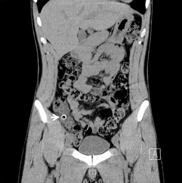
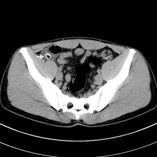
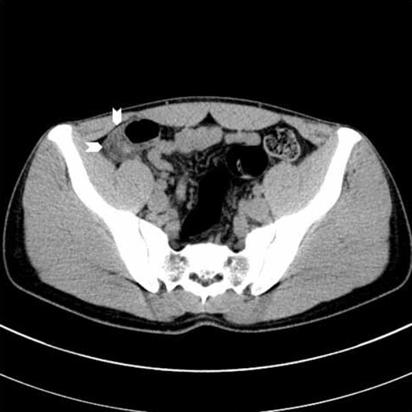
A healthy 27-year-old man was admitted to our hospital due to acute right lower quadrant abdominal pain. He denied nausea, vomiting, or diarrhea. On physical examination, the blood pressure was 111/64 mmHg, the heart rate was 75 beats per minute and regular, the respiratory rate was 16 breaths per minute, and the temperature was 37.9°C. Abdominal examination showed focal abdominal tenderness with slight rebound tenderness. Bowel sounds were normal, and no tumor was palpable. Laboratory tests showed a white blood cell count of 9000/mm3 (3900/mm3 to 8900/mm3) and a CRP of 8.7 mg/dL (<0.17 mg/dL). Otherwise, the laboratory data were within normal limits. The patient was treated with cefmetazole sodium (2 g/day) for 2 days, but the symptoms became worse. The antimicrobial dose was increased to 4 g/day for the subsequent 3 days. An abdominal series and ultrasound of the upper abdomen were performed and interpreted as normal. On the coronal section of computed tomography (CT), a fatty oval lesion measuring 12 mm in diameter with a circumferential hyperdense ring (arrow, Figure 1) was seen in the right lower abdomen. The transverse CT image showed that the ovoid, pericolonic mass abutted on the ascending colon and was surrounded by ill-defined fat stranding (arrow, Figure 2). Thickening of the parietal peritoneum was seen (arrow heads, Figure 3). There was neither free air nor ascites, and the appendix was normal. These findings were diagnostic of primary epiploic appendagitis. Antibiotics were discontinued. Oral loxoprofen sodium (60 mg) was prescribed twice before his symptoms and signs resolved with normalization of the laboratory results. The patient was doing well at the last outpatient follow-up visit.
Figure 1.

Computed tomography (CT), coronal section, shows a fatty oval lesion measuring 12 mm in diameter with a circumferential hyperdense ring in the right lower abdomen.
Figure 2.

The transverse CT image shows that the ovoid pericolonic mass abuts on the ascending colon and is surrounded by ill-defined fat stranding.
Figure 3.

Thickening of the parietal peritoneum is seen.
Discussion
In the present case, CT images showed a fatty, ovoid, pericolonic mass with a circumferential hyperdense ring, surrounded by ill-defined fat stranding. The thin hyperdense rim is called the hyperattenuating ring sign, which represents the inflamed peritoneal covering of the epiploic appendages [1]. The typical findings were diagnostic of primary epiploic appendagitis [1,2], which is caused by epiploic appendage torsion or spontaneous thrombosis of the draining vein resulting in vascular occlusion and focal inflammation [1]. Epiploic appendages are small, multiple, fat-filled, serosa-covered sacs, arranged in the tenia coli over the external surface. They number about 100, and their average size is about 3 cm [3]. Their function is unknown but considered to be as follows: buffering the blood flow of the colon, a defense mechanism like the epiploon, absorption of body fluid, fat storage, and a protective cushion for the colon. Their limited blood supply, together with their pedunculated shape, makes epiploic appendages prone to ischemic infarction [1–3].
Epiploic appendagitis presents with an abrupt onset of focal abdominal pain and tenderness without significant guarding or rigidity, and it is considered an uncommon and difficult diagnosis [1]. With increased awareness of this condition, however, evaluation by CT can provide an accurate diagnosis of epiploic appendagitis, distinguishing it from conditions with clinically overlapping manifestations such as diverticulitis, acute appendicitis, and acute cholecystitis [1,2]. Ultrasound coupled with awareness of this condition and/or performed by expertise can reveal epiploic appendagitis as an ovoid, non-compressible fatty mass with normal aspect of the adjacent colon which is adherent to the peritoneum during respiration [5]. However, in this case the expertise was not available and ultrasound was performed without awareness of epiploic appendagitis, thus ultrasound could not give the diagnosis in our patient.
Acute appendicitis occurs due to bacterial infections inside the vermiform appendix and can lead to complications like sepsis and perforation [6]. Acute appendicitis causes fever, abdominal tenderness, and rebound tenderness, just like epiploic appendagitis [1–3,6]. Some cases can be treated conservatively with drug therapy alone, but in an emergency, appendectomy is the standard treatment. CT scans show swelling of the appendix and an increase in surrounding fat density [6]. Acute cholecystitis is most often caused by gall stones, and the main symptoms are right upper abdominal tenderness, fever, nausea, vomiting, and jaundice. The serum transaminase, alkaline phosphatase, and bilirubin levels are sometimes elevated [7]. CT scan or ultrasonography shows gallbladder wall thickness, and Murphy’s sign is the typical clinical finding [7,8]. It is said that diverticulitis is caused by stagnation of stool in the diverticulum, and it may rarely lead to abscess and fistula formation [9]. When images are not typical, it is difficult to differentiate epiploic appendagitis from diverticulitis, and some cases of diverticulitis develop epiploic appendagitis when inflammation spreads to the epiploic appendages [1]. Less commonly, mesenteric panniculitis, primary tumors, and metastases to the peritoneum may be considered in the differential diagnosis [2,10].
Epiploic appendagitis is self-limited and needs only conservative management without excessive intervention [1–3]. Misdiagnosis may lead to unwarranted surgery, medical treatment, and hospitalization. Thus, it is imperative for clinicians to be familiar with this entity.
Conclusions
Epiploic appendagitis presents with an abrupt onset of focal abdominal pain and tenderness without significant guarding or rigidity, and it is considered an uncommon and difficult diagnosis. With awareness of this condition, however, evaluation by CT can provide an accurate diagnosis of epiploic appendagitis, distinguishing it from conditions with clinically overlapping manifestations.
Footnotes
Conflict of interest
None declared.
Written informed consent to present laboratory, clinical, and radiological data was obtained from the patients.
Source of support: Departmental sources
References
- 1.Almeida AT, Melão L, Viamonte B, et al. Epiploic appendagitis: an entity frequently unknown to clinicians – diagnostic imaging, pitfalls, and look-alikes. AJR Am J Roentgenol. 2009;193:1243–51. doi: 10.2214/AJR.08.2071. [DOI] [PubMed] [Google Scholar]
- 2.Singh AK, Gervais DA, Hahn PF, et al. Acute epiploic appendagitis and its mimics. Radiographics. 2005;25:1521–34. doi: 10.1148/rg.256055030. [DOI] [PubMed] [Google Scholar]
- 3.Blinder E, Ledbetter S, Rybicki F. Primary epiploic appendagitis. Emerg Radiol. 2002;9:231–33. doi: 10.1007/s10140-002-0235-6. [DOI] [PubMed] [Google Scholar]
- 4.Sand M, Gelos M, Bechara FG, et al. Epiploic appendagitis--clinical characteristics of an uncommon surgical diagnosis. BMC Surg. 2007;7:11. doi: 10.1186/1471-2482-7-11. [DOI] [PMC free article] [PubMed] [Google Scholar]
- 5.Schnedl WJ, Krause R, Tafeit E, et al. Insights into epiploic appendagitis. Nat Rev Gastroenterol Hepatol. 2011;8:45–49. doi: 10.1038/nrgastro.2010.189. [DOI] [PubMed] [Google Scholar]
- 6.Pinto Leite N, Pereira JM, Cunha R, et al. CT evaluation of appendicitis and its complications: imaging techniques and key diagnostic findings. AJR Am J Roentgenol. 2005;185:406–17. doi: 10.2214/ajr.185.2.01850406. [DOI] [PubMed] [Google Scholar]
- 7.Barie PS, Eachempati SR. Acute acalculous cholecystitis. Gastroenterol Clin North Am. 2010;39:343–57. doi: 10.1016/j.gtc.2010.02.012. [DOI] [PubMed] [Google Scholar]
- 8.Shakespear JS, Shaaban AM, Rezvani M. CT findings of acute cholecystitis and its complications. AJR Am J Roentgenol. 2010;194:1523–29. doi: 10.2214/AJR.09.3640. [DOI] [PubMed] [Google Scholar]
- 9.Heise CP. Epidemiology and pathogenesis of diverticular disease. J Gastrointest Surg. 2008;12:1309–11. doi: 10.1007/s11605-008-0492-0. [DOI] [PubMed] [Google Scholar]
- 10.Sabaté JM, Torrubia S, Maideu J, et al. Sclerosing mesenteritis: imaging findings in 17 patients. AJR Am J Roentgenol. 1999;172:625–29. doi: 10.2214/ajr.172.3.10063848. [DOI] [PubMed] [Google Scholar]


