Summary
Background
The visuospatial deficit is recognized as typical for dyslexia only in some definitions. However problems with visuospatial orientation may manifest themselves as difficulties with letter identification or the memorizing and recalling of sign sequences, something frequently experienced by dyslexics.
Material/Methods
The experimental group consisted of 62 children with developmental dyslexia. The control group consisted of 67 pupils with no diagnosed deficits, matched to the clinical group in terms of age. We used the Clock Drawing Test (CDT), the Spatial Span subtest from the Wechsler Memory Scale – third edition (WMS – III), the Rey-Osterrieth Complex Figure Test in order to analyze visuospatial functioning.
Results
The results show that dyslexics experienced problems with visuospatial functioning, however only while performing difficult tasks. Significant group differences were found for the Clock Drawing Test, Spatial Span – Backward and the precision of figure coping in the Rey-Osterrieth Test. In addition, the results of dyslexic boys were lower than those obtained by all other groups.
Conclusions
Our findings provide support for the hypothesis concerning visual deficit as characteristic for dyslexia.
Keywords: specific learning difficulties, visuospatial deficit
Background
Sight is the sense which plays a fundamental role in visuospatial orientation. The emplacement and sense of one’s body and other objects in space requires the establishing of the shapes constancy, sense of depth, distance and movement. The sense of perception includes the process of creating the recognized image of a person, place, object, situation or event in one’s mind [1]. The majority of processes occurring during the act of perception are subliminal, which means that we are not aware of their existence. The thing we perceive – the object, is the final product of a series of transformations occurring in our brain [2]. Although the status of the object of perception does not determine its reality, in most cases in healthy persons the normal process of visual perception lasts from the early phases of stimuli transformation to that if the object’s recognition [1].
Visuospatial competency is often mentioned by researchers dealing with issues connected with reading and writing skills. The constancy of size and shape, sense of depth and space should appear already in early childhood [3]. Unfortunately the disturbances in this sphere may result in later problems with letters identification, memorizing their sequences in words etc.. In professional literature difficulties of this type are described as: visuospatial deficit or visuospatial difficulties [4]. It is stressed that they are common in children with dyslexia [5,6], or in children with both reading difficulties and difficulties with learning mathematics [7].
An examination of the genetic background of visuospatial deficit is mainly based on neurodevelopmental disorders with selective cognitive impairment, such as William’s syndrome which provides a unique model for relating single genes to visual-spatial cognition. In this case one gene is suggested (GTF21RDI) to contribute to visual-spatial performance [8]. However, in the case of other neurodevelopmental disorders with global cognitive impairment (e.g. Down’s syndrome) or disorders which are suggested to be a ‘complex trait’ (e.g. dyslexia) genetic studies are particularly difficult.
It is the right hemisphere which is most of all responsible for proper spatial functioning. It “specializes” in such functions as: spatial localization of stimuli, describing the angle of depression and the estimation of so called spatial frequencies i.e. the number of elements constituting the particular part of a picture [1,9]. However, working with patients with aphasia suggests that for navigation skills the damage to the left hemisphere is also important.[1] It has been claimed that there exist quality differences between orientation disturbances connected with the damage of the right and left hemisphere [10].
An infant possesses neither structure nor constancy of space [11,12]. The first space it experiences is of a practical character – cognition is connected with acting. An infant cannot differentiate itself from its surroundings. Its sensations are not located in particular space. An infant does not feel its body as its own one nor does it treat the mother’s breast as something separate from its mouth [13].
The essence of space structure development is a child’s physical activity and visual perception [12]. Due to eyeball movement we drive our eye to a certain point, manipulate the picture, place the eye in a certain position and thus ensure a constancy of perception.
Step by step our body becomes something separate from the world and space turns into something that is within the child’s reach. Dissociation of perception and action appears when a child begins to notice objects which are not within its reach. A moving baby recognizes different elements of space as relatively constant points of reference. Space becomes independent of its activities. The basis for space orientation is the knowledge of one’s body scheme [1].
The research [14,15] shows the following sequence in space orientation development: knowledge of one’s body scheme, setting directions in space off one’s body, transferring one’s body scheme to another person and setting directions in space off this person’s body and space orientation skill on a sheet of paper. At the same time the results point out that only 6–7-year-old children have no difficulty with the first two skills. Space orientation on a sheet of paper is difficult and in fact not manageable for even 7-year-old children.
The ability to transfer one’s own body scheme to another person appears nearly immediately after learning one’s own body scheme and setting directions in space off one’s own body’s axle. Therefore the disorders called autopatognosia (from Greek auto “own” + topos “place, localization + agnosia” lack of knowledge) never appear in isolation but in connection with difficulties in recognizing the body parts of other people [16]. The disturbance on this level is undoubtedly equivalent to difficulties connected with the following steps of spatial functioning.
In the relevant literature the majority of data is concerned with the orientation disturbance in adults following brain damage and most of these data refer to patients with aphasia [17]. Spatial disturbances have also been reported in patients with schizophrenia [18].
One of the neurodevelopmental disturbances, ones usually typical of visuospatial deficit, are specific difficulties in learning as the most commonly diagnosed developmental disorder among school children [19]. European statistics on its frequency of occurrence point out that it afflicts about 10–15% of the population [20].
Modern definitions of specific difficulties in learning underline that this type of learning problem is caused by central nervous system dysfunction and as a result specific functioning during school skills acquisition. The term specific emphasizes the narrow range of difficulties when compared to generalized problems concerning all spheres i.e. the unspecific. As a result of the cooperation of European authors in creating the International Statistical Classification of Diseases and Related Health Problems, the ICD-10 approved by the World Health Organization [20], and the authors from the United States responsible for the the classification of American Psychiatric Association, the DSM-IV-TR [21] diagnostic criteria of specific learning difficulties are similar. The thing that differs in the classifications is the terminology: DSM-IV-TR stands for ‘learning disturbances’, whereas ISD-10 means ‘specific developmental learning skills disturbances’. Both classifications stress that these difficulties do not constitute a homogeneous symptom but they concern the group of disturbances connected with various important problems of speech, writing and arithmetic.
The biggest coverage, both in the form of scientific elaborations and handbooks for parents and teachers, has been given over to specific difficulties in reading and writing. This descriptive term is most often substituted by the notion of developmental dyslexia [22].
At the present moment there does not exist one single concept explaining the pathomechanism of dyslexia. Many pieces of research [23,24] point at the occurrence of dyslexia in a family. The most recent research estimates the hereditary factor to be 58% [25].
Owing to the application of the technique of neuroimaging the structure and function of the brain, many other anatomic anomalies and specific patterns of information processing in dyslexic children that are connected with them have been shown [26].
Nowadays the most commonly stressed is the occurrence of many anatomic anomalies, mostly connected with areas engaged in language processes [27]. The majority of neuroanatomic data emphasize a dissimilar cell layout in the brain, so called ectopia, which constitutes the migration of cells to the external layer of the cortex, numerous dysplasia, i.e. local changes in the laminar structure of the cortex, as well as atypical corrugation of the cerebral cortex (polimikrogyrie) [28]. These abnormalities are mainly located in the left hemisphere (dominating in language processes) and they concern: anomalous neurons location in perisylvian gyrus areas, higher temporal cerebral gyri (together with the Wernicke area) and a lower premotor and prefrontal cortex (including the Broca area) [29]. Moreover, people with dyslexia are characterized by significantly smaller right frontal cerebellum lobes, the right and left triangular part of the cerebellum and the volume of the whole cerebellum [30]. The structural diversity of the cerebellum in people with dyslexia has been confirmed in so much research that it has become to be regarded as one of the neuroanatomic markers of this disorder [31].
A lot of data indicate brain asymmetry disturbance in people with dyslexia e.g. the enlargement of the rear part of the corpus callosum – which can diminish the lateralization of brain function. At the same time the lack or diminishment of asymmetry in the temporal lobe planum temporale has been repeatedly confirmed.
At present there is no doubt that developmental dyslexia is biologically conditioned. The specific development of the central nervous system conditioned by a primary genetic or organic background results in specific cognitive process deficits. Besides, language disturbances, mainly phonological, though also hearing deficits and difficulties in the processing of information coming from sight receptors, may induce reading and writing difficulties. It was William P. Morgan – the ‘discoverer’ of dyslexia who drew attention to visual perception in 1896, when describing a boy with ‘word blindness’ [22]. The opinion of the role of visual processes in dyslexia evolved from regarding it as the main reason for the difficulties [32], through a total rejection of their relevance [33] to a complex approach acknowledging the participation of both sight and hearing as well as the processes of integration and automation in the pathomechanism of dyslexia [22].
In his research into the role of disturbances of the visual system in dyslexia [34], he drew attention to the coordination of eye ball movement which appeared incorrect. While reading, people with dyslexia perform shorter and more frequent saccade movements (reading with outrunning the text back and forth) and they have longer fixation periods on particular points. However, as far as eye movement is concerned, abnormalities in this area are not the result of dyslexia as such but generally of difficulties in reading (also resulting from the complexity of the text), only the direction of it, one opposite to the normal, is typical of dyslexia [32]. The basic difference in the level of visual processing between people with dyslexia and those without, concerns mainly the situation when fast information processing is necessary. The evidence for visual modality disturbance has been also provided by psychophysical research pointing out that people with dyslexia have problems with differentiating letters from their mirror reflections [35], and by widespread research into sensitivity to colours. The practical effects of these various pieces of research seem to be interesting because it appeared that while reading a text through a colourful cover some children have shown an increased level in this capability [36].
The progress of examinations with the use of neuroimaging methods drew the researcher’s attention to an alternative way of information processing by two separate channels. Bipartition of the channels refers to all levels of the visual system: the retina, optic nerve, lateral geniculate nucleus and visual cortex. Regarding the form of differences these systems have been called magnocellular and parvocellular [37]. In the retina smaller and bigger cells can be easily differentiated, in the optic nerve – thicker and thinner axons, the backing layer of the lateral geniculate nucleus is composed of smaller cells than the abdominal layer; this division also concerns the projection to separate layers of the primary cortex, information from both systems mingle at the level of the associative cortex. The microcellular channel is responsible for central vision, perception of details and colour differentiation, but it reacts slowly. Subsequently, the macrocellular channel specializes in peripheral vision and perceives fast changes and movement, it is sensitive to all brightness differences but totally insensitive to colours. Experimental research with the application of different visual stimuli and neuroimaging techniques, together with post mortem analysis of the brains of people with dyslexia, points out the damage to the macrocellular visual system [38]. Basing himself on these data together with thorough research with the participation of people with dyslexia, John Stein [39] formulated ‘a macrocellular theory of dyslexia’. When analyzing processes engaging reading and writing one might suspect a dysfunction within the microcellular system, reading is not connected with the perception of movement – a text does not move. However, no differences in the level of functioning of this system have been shown among those with and without dyslexia, but numerous cases report essential differences concerning the macrocellular system [39]. In the process of reading this system is responsible for directing visual attention, eye ball movement and visual searching. Dysfunctions in this channel reduce the ability to control the eye muscles and, subsequently, they cause optical sight lines crossing, which results in a mixing up of the shapes of letters (d-b) and a changing in the order of adjacent letters.
The macrocellular concept may constitute a theoretical basis explaining the causes of visual perception deficit in the domain of eye ball movement coordination, sensitivity to colour and contrast, e.g. children with dyslexia are more sensitive to colours [40].
While neuropsychologists [17] divide spatial orientation deficit into: 1. body scheme deficit; 2. visuospatial orientation deficit; 3. topographic orientation; 4. orientation in time deficit, it is difficult to find a precise definition of these deficits among psychologists dealing with specific difficulties in learning. In professional literature problems connected with mixing up similarly looking letters, overlooking diacritics or difficulties in identifying letters arrangements etc, are called, as has been mentioned before, visuospatial functions deficit [4].
Material and Methods
The research was conducted in Poland from 2006 to 2008 among a group of 129 persons. The experimental group consisted of 62 pupils from 4th to 6th grade, diagnosed with developmental dyslexia (30 girls and 32 boys). The control group consisted of pupils with no diagnosed deficits (37 girls and 30 boys), matched to the clinical groups in terms of age (control group mean age M=11.6; SD=0.4 and dyslexic mean age M=11.2; SD=0.7).
All of the participants were native Polish speakers and met the following criteria: normal or above average intelligence, standard educational opportunities, normal or corrected to normal visual and auditory acuity, no gross sensory or attention deficit, no gross behavioral problems, no history of neurological disease.
The study was carried out individually with each child. IQ was assessed with the Wechsler Intelligence Scale for Children (WISC-R) [41]. The mean IQ in the control group was 112.6 (SD=11.5) and in the dyslexic group 107.92 (SD=12.7). An interdisciplinary diagnosis of developmental dyslexia was formulated by psychologists, teachers, mental health clinics and neurological clinics.
In order to analyze visuospatial functioning, we used the following instruments:
Results
In analyzing the level of test performance in both groups of children, those with dyslexia and the control group, we obtained many statistically essential differences which will be discussed in details later. Prior to this, multidimensional analyses (MANOVA) had been conducted, with grouping factors: sex (m, f) versus group (LSD, control). The final effect concerns the examined group (F=3.9; p=0.000); no statistically essential results have been obtained which would show the necessity to analyze each sex separately in the future.
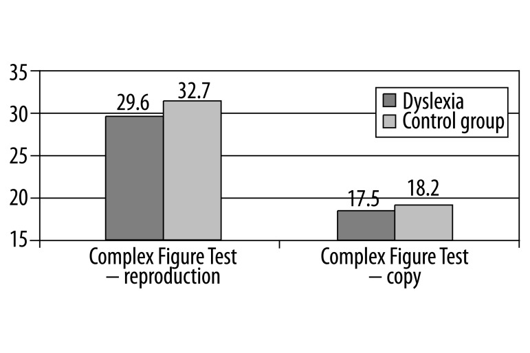
First of all we compared the results obtained by children with dyslexia and children from the control group in the particular tests which measured the level of visuospatial capacity. According to our expectations concerning the Rey-Osterrieth Complex Figure Test, in which a child had to copy a complex figure, demonstrated on the picture – first when looking at it, and then after memorizing it – children with reading difficulties obtained lower results when copying (t=3.8; p=0.000) (Figure 1).
Figure 1.
Comparison of results in the Rey-Osterrieth Complex Figure Test.
When analyzing types of children’s drawings, the most prevailing (34%) was type 4, placing elements side by side without paying attention to important details. The characteristic features of the pictures drawn by children with dyslexia were: a low level of complexity, a tendency for displacing elements or simply omitting them, and the rotations of the components of the copy.
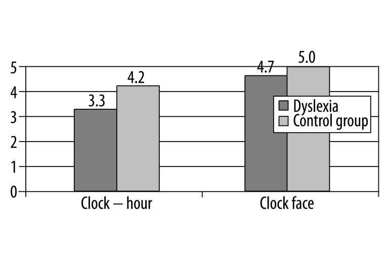
When analyzing the results obtained in the Clock Drawing Test, in which a child had to draw the face of a clock set for a suggested hour, it is necessary to stress the essential differences between the examined groups. The first difference referred to drawing the face of the clock with hands pointing at a particular hour, the second appeared in drawing the face itself and putting figures on it (t=5.0; p=0.000). Children with dyslexia more often set an incorrect time on a correctly drawn face, they also made more mistakes in the face itself (Figure 2).
Figure 2.
Comparison of results in the Clock Drawing Test.
The most common error among children without any deficit was marking the time 10:22 hour instead of 10:27, whereas students with dyslexia apart from setting the incorrect time also placed the figures incorrectly on the face.
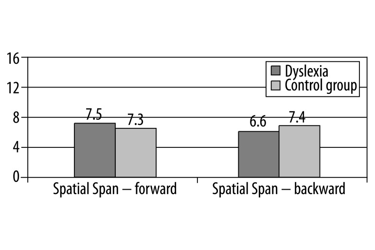
The last test to be applied was the Spatial Span subtest WMS – III which makes use of the ability to maintain a visual-spatial sequence of locations in the working memory of the examined and then its further reconstruction. The examined person is presented with a special arrangements of bricks and after a while he is asked to reconstruct it in the same way and then in the reverse order.
Reconstructing of the sequences of visually presented locations in space turned out to be equally difficult for both examined groups. Children with dyslexia obtained essentially worse results when reconstructing the sequence in the reverse order (t=2.0; p=0.044) (Figure 3).
Figure 3.
Comparison of results in the Spatial Span subtest WMS – III.
Moreover, correlation analyses of the results of particular tests were conducted. The strongest relation was observed between the level of making a copy in the Rey-Osterreth Complex Figure Test and the accuracy in drawing the face of the clock (r=0.35; p=0.003). Those two tasks are highly saturated with the factor connected with graphomotor activity, the disturbance of which is one of the criterial symptoms of dysgraphia.
Discussion
The Rey-Osterrieth Complex Figure Test is the most commonly used in the diagnosis of visual deficit in dyslexia. First of all, it allows one to estimate the perceptional organization of those examined [45]. Type 4 of the drawing, appearing in our research, i.e. placing elements side by side without paying attention to more important details, is, from the perspective of developmental standard, typical of children at the age of 8. Its frequent appearance in the group of 11-year-olds most probably results from an insufficiently formed organization of perception, but it is also connected with a lower graphic capacity among children with developmental dyslexia. A lack of differences in the reproduction making criterion (i.e. reconstructing from memory) together with no essential difference as far as drawing without a delay is concerned, would point to visuospatial orientation deficit rather than disturbances in the memory processes. It appears that the difficulty with making a copy in the Rey-Osterrieth Complex Figure Test may be connected with a lowered level of executive functions and some deficits in the frontal lobe area. As is known, damage to these areas may lead to the smoothness of movement disturbances manifested by a deautomation of motor patterns [1].
In the case of children with specific difficulties in learning but without any obvious damage, the presented symptoms may, however, point to certain difficulties in the functioning of these particular areas. Part of the research also shows the occurrence of essential differences between children with dyslexia and children without deficits as far as drawing from memory is concerned [46]. Children with specific difficulties in learning obtained essentially lower results in this case which proves that there exist problems within the working memory which comprises also the so called visuospatial notes – a system processing spatial information which reaches the brain by means of sight [47].
The way of drawing the clock and types of mistakes made by children with dyslexia point to deficits connected with the right hemisphere which are manifested as attention selectivity deficits and working memory and executive functions’ disturbances. Developmental dyslexia is usually connected with a left-hemisphere deficit because of criteria symptoms such as: phonological proficiency, verbal operating memory. However, poor performance at the Clock Drawing Test is additional proof of the fact that many people with dyslexia experience difficulties in the sphere of visuospatial functions for which language deficit bears no responsibility whatsoever. Eden, Wood and Stein [48] when proposing to include the Clock Drawing Test into the battery of tests diagnosing dyslexia point out that right-hemisphere dysfunction may often appear in children with difficulties in learning to read and write so the test mentioned above may help in a fast and efficient diagnosis.
The high level of difficulty in the Spatial Span subtest WMS – III for all the examined children made the attention factor even more crucial; this can be borne out by the occurrence of essential differences between the examined groups only when trying to reconstruct the sequence in a reverse order. It is believed that direct reconstruction is an accurate measure of working memory in the sphere of visual and visuospatial functions, whereas reversed reconstruction – measures attention. Attention is strictly connected with perception, memory and imagination processes. According to Goldberg [49], paying attention to stimuli which are essential in regarding the target, is connected with the functioning of the frontal lobes, which are responsible for planning.
Conclusions
In recent years, the research into the etiopatogenezis of dyslexia has been dominated by the trend which can be called linguistic. There exists a substantially smaller amount of research concerning visuospatial function deficits in dyslexia when compared to the existing data referring to the linguistic functioning. A substantial part of the obtained material is connected with speech and spelling difficulties, especially with difficulties connected with so called phonological awareness, i.e. ‘an ability to reflect on and manipulate the structure of an utterance as distinct from its meaning’ [50,51]. In the diagnosis of dyslexia special attention is paid to abilities such as: identifying phones in words, spelling, differentiating between similar phones, word finding etc.
The presented research constitutes another piece of evidence showing that children with dyslexia are afflicted with both phonological problems and visuospatial deficits connected with the right posterior parietal cortex. Diagnosis limited only to the linguistic aspect may produce an incomplete picture of the actual pathogenic mechanisms. A clinical picture of dyslexia is influenced by the specific development of both hemispheres [48]. The increasingly popular magnocellular theory of developmental dyslexia presents the neuroanatomic background of visual perception and visuospatial function deficits. While it is true that the disturbance in this sphere does not differentiate dyslexia from other types of learning difficulties, omitting them in the process of diagnosis may lead to a distortion of the picture of a child with developmental dyslexia and thus diminish the chance for effective therapy.
Footnotes
Source of support: Departmental sources
References
- 1.Pąchalska M. Urazy mózgu. Warszawa: Wyd. Naukowe PWN; 2007. Neuropsychologia kliniczna. [in Polish] [Google Scholar]
- 2.Brown JW. A microgenetic approach to time and memory in neropsychology. Acta Neuropsychologia. 2004;2(1):1–12. [Google Scholar]
- 3.Rival C, Olivier I, Ceyte H, Bard C. Age-related differences in the visual processes implied in perception and action: distance and location parameters. J Exp Child Psychol. 2004;87(2):107–24. doi: 10.1016/j.jecp.2003.10.003. [DOI] [PubMed] [Google Scholar]
- 4.Bogdanowicz M, Adryjanek A, Rożyńska M. Poradnik nie tylko dla rodziców. Gdynia: Operon; 2007. Uczeń z dysleksją w domu. [in Polish] [Google Scholar]
- 5.Bakker DJ. Neuropsychological treatment of dyslexia. New York: Oxford University Press; 1990. [Google Scholar]
- 6.Bogdanowicz M. Problem i diagnozowanie. Gdańsk: Wydawnictwo Harmonia; 2003. Ryzyko dysleksji. [in Polish] [Google Scholar]
- 7.Oszwa U. Mathematical, linguistic and visuospatial skills in Polish children with specific learning disabilities in reading and arithmetic. Acta Neuropsychologica. 2006;4(4):286–95. [Google Scholar]
- 8.Dai L, Bellugi U, Chen XN, et al. Is it Williams syndrome? GTF2IRD1 implicated in visual-spatial construction and GTF2I in sociability revealed by high resolution arrays. American Journal of Medical Genetics Part A. 2009;149:302–14. doi: 10.1002/ajmg.a.32652. [DOI] [PMC free article] [PubMed] [Google Scholar]
- 9.Budohoska A, Grabowska A. Dwie półkule jeden mózg. Warszawa: Wiedza Powszechna; 1994. [in Polish] [Google Scholar]
- 10.Kolb B, Whishaw IQ. Fundamentals of Human Neuropsychology. New York: Worth Publishers; 2003. [Google Scholar]
- 11.Kielar-Turska M. Słowo i tekst. Kraków: Wydawnictwo UJ; 1989. Mowa dziecka. [in Polish] [Google Scholar]
- 12.Piaget J, Inhelder B. Psychologia dziecka. Wrocław: Wydawnictwo Siedmioróg; 1989. [in Polish] [Google Scholar]
- 13.Brown JW. Process and the authentic life: Toward a psychology of value. Frankfurt & lancaster: Ontos Verlag; 2005. [Google Scholar]
- 14.Czaplewska E. Rozwój kompetencji w zakresie orientacji przestrzennej u dzieci od 3 do 8 roku życia. Psychologia Rozwojowa. 2002;7(4):109–17. [in Polish] [Google Scholar]
- 15.Czaplewska E, Bogdanowicz K, Kaczorowska-Bray K. An evaluation of visual spatial orientation in preschool children. Acta Neuropsychologica. 2009;7(1):21–32. [Google Scholar]
- 16.Semenza C. Assessing disorders of awareness and representation of body parts. In: Halligan PW, Kischka U, Marshall JC, editors. Handbook of clinical neuropsychology. Oxfored: Oxford Univeristy Press; 2003. pp. 195–213. [Google Scholar]
- 17.Pąchalska M. Procesy poznawcze i emocjonalne. Lublin: Wyd. UMCS; 2008. Rehabilitacja neuropsychologiczna. [in Polish] [Google Scholar]
- 18.Brugger P, Surbeck W, Loester T. Pseudoneglect in representational space: effects of magical ideation. Acta Neuropsychologica. 2007;5(1/2):1–7. [Google Scholar]
- 19.Willcutt EG, Pennington BF, Olson RK, et al. Neuropsychological analyses of comorbidity between reading disability and attention deficit hyperactivity disorder: In search of the common deficit. Developmental Neuropsychology. 2005;27(1):35–78. doi: 10.1207/s15326942dn2701_3. [DOI] [PubMed] [Google Scholar]
- 20.World Health Organization. The ICD-10 classification of mental and behavioural disorders: Diagnostic criteria for research. Geneva: Authors; 1992. [Google Scholar]
- 21.American Psychiatric Association. Diagnostic and Statistical Manual of Mental Disorders. 4th ed. Washington, DC: Authors; 2000. text rev. [Google Scholar]
- 22.Bogdanowicz M. Specyficzne trudności w czytaniu i pisaniu – dysleksja rozwojowa. In: Gałkowski T, Jastrzębowska G, editors. Logopedia – pytania i odpowiedzi Opole. Wydawnictwo Uniwersytetu Opolskiego; 2003. pp. 491–535. [in Polish] [Google Scholar]
- 23.Wadsworth SJ, DeFries JC, Olson RK, Willcutt EG. Colorado longitudinal twin study of reading disability. Annals of Dyslexia. 2007;57:139–60. doi: 10.1007/s11881-007-0009-7. [DOI] [PubMed] [Google Scholar]
- 24.Dick BD, Kaplan BJ, Crawford S. The influence of family history on reading remediation and reading skills in children with dyslexia. Canadian Journal of School Psychology. 2006;21(1–2):106–19. [Google Scholar]
- 25.Pennington BF, McGrath LM, Rosenberg J, et al. Gene X environment interactions in reading disability and attention-deficit/hyperactivity disorder. Developmental Psychology. 2009;45(1):77–89. doi: 10.1037/a0014549. [DOI] [PMC free article] [PubMed] [Google Scholar]
- 26.Galaburda AM, Turco LJ, Ramus F, et al. From genes to behavior in developmental dyslexia. Nature Neuroscience. 2006;9(10):1213–17. doi: 10.1038/nn1772. [DOI] [PubMed] [Google Scholar]
- 27.Habib M. Zaburzenia nabywania zdolności językowych i pisania: najnowsze osiągnięcia neurobiologii. In: Grabowska A, Rymarczyk K, editors. Dysleksja – od badań mózgu do praktyki. Warszawa: Instytutu Biologii Doświadczalnej PAN; 2004. pp. 185–215. [in Polish] [Google Scholar]
- 28.Knight DF, Hynt GW. Neurobiologia dysleksji. In: Reid G, Wearmouth J, editors. Dysleksja Teoria i praktyka. Gdańsk: Gdańskie Wydawnictwo Psychologiczne; 2008. pp. 51–70. [in Polish] [Google Scholar]
- 29.Galaburda AM, Menard MT, Rosen GD. Evidence for aberrant auditory anatomy in developmental dyslexia. The Proceedings of the National Academy of Sciences. 1994;91:8010–13. doi: 10.1073/pnas.91.17.8010. [DOI] [PMC free article] [PubMed] [Google Scholar]
- 30.Eckert MA, Leonard CM, Richards T, et al. Anatomical correlates of dyslexia: Frontal and cerebellar findings. Brain. 2003;126:482–94. doi: 10.1093/brain/awg026. [DOI] [PubMed] [Google Scholar]
- 31.Pernet CR, Poline JB, Demonet JF, Rousselet GA. Brain classification reveals the right cerebellum as the best biomarker of dyslexia. BMC Neuroscience. 2009;10:67–85. doi: 10.1186/1471-2202-10-67. [DOI] [PMC free article] [PubMed] [Google Scholar]
- 32.Everatt J. Procesy wzrokowe. In: Reid G, Wearmouth J, editors. Dysleksja Teoria i praktyka. Gdańsk: Gdańskie Wydawnictwo Psychologiczne; 2008. pp. 121–39. [in Polish] [Google Scholar]
- 33.Snowling MJ. Dyslexia. Oxford: Blackwell Publisher; 2000. [Google Scholar]
- 34.Pavlidis GT. Eye movement differences between dyslexics, normal, and retarded readers while sequentially fixating digits. Am J Optom Physiol Opt. 1985;62(12):820–32. doi: 10.1097/00006324-198512000-00003. [DOI] [PubMed] [Google Scholar]
- 35.Jaśkowski P, Rusiak P. Posterior parietal lobe and developmental dyslexia. Acta Neurobiol Exp (Wars) 2005;65:79–94. doi: 10.55782/ane-2005-1542. [DOI] [PubMed] [Google Scholar]
- 36.Breitmeyer BG, Williams MC. Effects of isoluminant-background color on metacontrast and stroboscopic motion: interactions between sustained (P) and transient (M) channels. Vision Research. 1990;30(7):1069–75. doi: 10.1016/0042-6989(90)90115-2. [DOI] [PubMed] [Google Scholar]
- 37.Livingstone M, Rosen G, Drislane F, Galaburd A. Physiological and anatomical evidence for a magnocellular defect in developmental dyslexia. Proceedings of the National Academy of Sciences. 1991;88:7943–47. doi: 10.1073/pnas.88.18.7943. [DOI] [PMC free article] [PubMed] [Google Scholar]
- 38.Galaburda AM, Livingstone M. Evidence for magnocellular deficit in developmental dyslexia. Annals of the New York Academy of Science. 1993;682:70–82. doi: 10.1111/j.1749-6632.1993.tb22960.x. [DOI] [PubMed] [Google Scholar]
- 39.Stein J. The magnocellular theory of developmental dyslexia. Dyslexia. 2001;7:12–36. doi: 10.1002/dys.186. [DOI] [PubMed] [Google Scholar]
- 40.Bednarek D, Grabowska A. Luminance and chromatic contrast sensitivity in dyslexia: The magnocellular deficit hypothesis revisited. NeuroReport. 2002;13(18):2521–25. doi: 10.1097/00001756-200212200-00028. [DOI] [PubMed] [Google Scholar]
- 41.Matczak A, Piotrowska A, Ciarkowska W. Wechslera dla Dzieci – wersja zmodyfikowana (WISC-R) Warszawa: Pracownia Testów Psychologicznych; 1997. Skala Inteligencji D. [in Polish] [Google Scholar]
- 42.Cohen MJ, Riccio CA, Kibby MY, Edmonds JE. Developmental progression of clock face drawing in children. Child Neuropsychology. 2000;6:64–76. doi: 10.1076/0929-7049(200003)6:1;1-B;FT064. [DOI] [PubMed] [Google Scholar]
- 43.Strupczewska B. Podręcznik. Warszawa: COM PW-Z MEN; 1990. Test Figury Złożonej Rey-Osterrieth. [Google Scholar]
- 44.Wechsler D. Wechsler Memory Scale III. San Antonio, Texas: Psychological Corporation; 1997. [Google Scholar]
- 45.Krasowicz-Kupis G. Diagnozowanie funkcji wzrokowych w dysleksji. In: Krasowicz-Kupis G, editor. Diagnoza dysleksji Najważniejsze problemy. Gdańsk: Wydawnictwo Harmonia; 2003. pp. 225–47. [Google Scholar]
- 46.Mati-Zissi H, Zafiropoulou M. Visuomotor coordination and visuospatial working memory of children with specific reading disabilities: a study using the Rey-Osterrieth Complex Figure. Perceptual and Motor Skills. 2003;97(2):543–46. doi: 10.2466/pms.2003.97.2.543. [DOI] [PubMed] [Google Scholar]
- 47.Baddeley AD. Is working memory stall working? European Psychologist. 2002;7:85–97. [Google Scholar]
- 48.Eden GF, Wood FB, Stein JF. Clock Drawing in developmental dyslexia. J Learn Disabil. 2003;36(3):216–28. doi: 10.1177/002221940303600302. [DOI] [PubMed] [Google Scholar]
- 49.Goldberg E. Frontal lobes and the civilized mind. New York: Oxford Univerity Press; 2001. The executive brain. [Google Scholar]
- 50.Stackhouse J, Wells B. Children’s speech and literacy difficulties: A psycholinguistic framework. London: Whurr; 1997. [Google Scholar]
- 51.Pąchalska M, Moskała M, MacQueen BD, et al. Early neurorehabilitation in a patient with severe traumatic brain injury to the frontal lobes. Med Sci Monit. 2010;16(12):CS157–67. [PubMed] [Google Scholar]


