Abstract
Introduction
Odontoid fractures are the most common upper cervical spine fracture. There are two mechanisms in which odontoid fractures occur, most commonly hyperflexion of the neck resulting in displacement of the dens anteriorly and hyperextension resulting in posterior dens displacement. Type 2 fractures are the most common and are associated with significant non-union rates after treatment. One possible consequence of an odontoid fracture is a synovial cyst, resulting in spinal cord compression, presenting as myelopathy or radiculopathy. Synovial cysts as a result of spinal fracture, usually of the facet joint, are most common in the lumbar region, followed by the thoracic and then cervical region; cervical cysts are rare. Fracture and subsequent cyst formation is thought to be related to hyper-motion or trauma of the spine. This is reinforced by the appearance of spinal synovial cysts most commonly at the level of L4/5; this being the region with the biggest weight-bearing function. The most common site of cervical cyst formation is at the level of C7/T1; this is a transitional joint subjected to unique stress and mechanical forces not present at higher levels. Treatment of a cervical synovial cyst at the level of the odontoid is challenging with little information available in the literature. The majority of cases appear to implement posterior surgical resection of the cyst, with fusion of adjacent cervical vertebrae to stabilise the fracture, resulting in restricted range of movement.
Case presentation
We describe a case concerning a 39-year-old female who presented with uncertain cause of odontoid fracture, resulting in a cystic lesion compressing the upper cervical spinal cord.
Outcome
Minimal invasive surgery of C1/C2 transarticular fusion was successfully performed resulting in significant improvement of neurological symptoms in this patient. At 1-year follow-up, the cyst had resolved without surgical removal and this was confirmed by radiological measures.
Keywords: Transarticular fusion, Odontoid fracture, Cervical cyst, Cystic lesion
Case presentation
A 39-year-old woman presented complaining of numbness in all four limbs, which had been ongoing for 18 months with progressive worsening. A few episodes of bladder and bowel incontinence were reported.
On examination, the patient had a slightly wide based gait, disturbed tandem gait, positive Romberg sign, increased tone and exaggerated reflexes. The patient had reduced sensation to light touch and pinprick in lower limbs to mid thorax. There was absence of sensation to light touch and joint position in upper limbs. Digital rectal examination revealed disturbance of sensation to light touch and pinprick around the anal margin.
Past medical history included congenital acetabular dysplasia, morbid obesity (Body Mass Index >45), and right carpal tunnel syndrome for which a release had been performed, but with no effect. Current drug history included lansoprazole and citalopram. The patient smoked approximately 20 cigarettes per day but was otherwise fit and well.
MRI showed a large cystic lesion behind the vertebral body of C2 resulting in upper cervical cord compression. There was an apparent line running through base of odontoid, and evidence of pseudarthrosis and instability between the odontoid peg tip and the body on CT. This abnormality was thought to be congenital but could have been caused by possible neck trauma occurring approximately 2 years previously.
Diagnostic imaging section
Fig. 1.
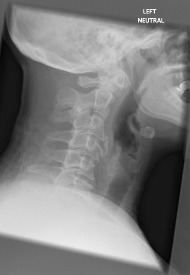
Pre-operative lateral radiograph of the patient displaying odontoid displacement
Fig. 2.
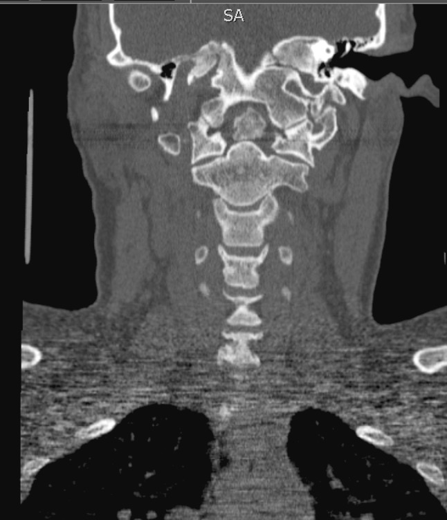
Pre-operative CT section to show asymmetry at the level of C1/C2
Fig. 3.
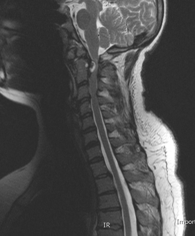
Lateral MRI showing cystic lesion behind vertebral body of C2 pre-operatively
Description of the condition, epidemiology, diagnosis, pathology, differential diagnosis
Treatment of odontoid fractures is dependent upon type of fracture. Stable Type 1 fractures can usually be treated with a hard cervical collar for 6–8 weeks. Type 2 and 3 fractures can be treated by non-surgical or surgical means. A Halo vest would be the non-surgical treatment option of choice. Normally, these cases need extensive fusion with limitation of flexion and extension to allow healing [1–4].
Common surgical procedures for odontoid fractures include internal fixation (odontoid screw fixation) and arthrodesis (posterior fusion); however, superiority of treatments is controversial [5, 6].
In arthrodesis, fusion of bones that form a joint occurs, usually posteriorly, essentially eliminating the joint. The fused joint becomes stable again and provides considerable pain relief to the patients; however, there is loss of flexibility and motion, which is even more considerable in extensive fusion. Fixation of adjacent dens by anteriorly placed screws preserves movement of joints and consequently may be a preferred surgical option.
One possible consequence of an odontoid fracture is a synovial cyst, presenting as myelopathy or radiculopathy. Cervical cysts at C1/C2 level can present due to a range of dens degeneration/trauma, without dens involvement at all, or can themselves be the cause of spinal trauma, for example dens hypoplasia [7]. Where there is dens involvement, treatment is surgical, through one of a number of options including posterior craniocervical fusion [8], decompression and excision of the lesion [9], posterior atlantoaxial arthrodesis [7].
Presentation of cervical cysts alone is most likely related to hypermovement of a spinal joint caused by degeneration, congenital abnormality, inflammation or trauma. Surgical treatment of cysts present without fracture can be effective with hemilaminectomy or full decompressive laminectomy [10].
Rationale for treatment and evidence-based literature
A retrospective study by Kazda et al. [10] compared surgical and non-surgical treatment of type 2 and 3 dens injuries. Patients attending a single centre between 1999 and 2006 were recruited to the study and then followed up for at least 1 year. Out of 32 participants, 15 had undergone surgery and 17 had been managed non-surgically. Non-surgical patients showed restricted range of cervical motion, higher Visual Analogue Scale (VAS) scores and greater fragment displacement, than patients undergoing surgery. Thus it can be concluded that in this cohort of patients, surgery was the superior treatment.
A case report by Lyons et al. [11] summarises treatment of 35 patients with cervical subaxial synovial cysts of unknown origin. All patients underwent posterior surgical resection of their cyst; 21 patients underwent a hemilaminectomy and 14 underwent full decompressive laminectomy. No patients in this report presented with a history of cervical spine trauma or previous surgery at the level of the synovial cyst. No patients had an increase in severity of symptoms post-operatively as measured by the Modified Ranking Scale.
A report by Marbacher et al. [12], described the first case of a synovial cyst arising from pseudarthrosis of a previous dens fracture. The 60-year-old patient presented with symptoms of spinal cord compression. Treatment was surgical removal of the central atlas arch, dens, transverse ligament, tectorial membrane and compressing cyst, followed by a C0–C3 fusion. Two months post-operatively, the patient had recovered completely from spinal cord compression and there was complete resection of the cystic lesion.
The failing of these studies [10–12], is in addressing the issue of reduced mobility arising from posterior fusion of adjacent cervical vertebrae; the patient is left with a permanent functional deficit.
Newer methods, for example, cancellous bone augmentation have been described. In a study by Ruf et al. [13], 17 patients with pseudarthrosis of the dens were operated on between 1991 and 2005. Patients had a hole drilled in the dens, which was then packed with autologous bone graft. Temporary instrumentation of C1/C2 was performed and removed after 3–4 months. Out of 15 patients available for follow-up, 9 (60%) had healing of the pseudarthrosis with preservation of C1/C2 motility. This could consequently be a surgical treatment option, which preserves cervical function.
A case reported by Blondel et al. [14], used a single anterior procedure to stabilise a complicated dens fracture in a 79-year-old male after a bicycle accident. Odontoid screw fixation and C2/C3 fusion were performed. This single procedure achieved stability of the fracture, whilst managing to preserve range of cervical motion in this patient, compared to posterior fusion.
In this case report, transarticular screw fusion rather than occipital cervical fusion was used, to preserve as much neck movement as possible in this relatively young and fit female patient.
Procedure
Posterior C1/C2/C3 decompression and C1/C2 transarticular screw and fusion were performed. The surgeons opted for C1/C2 transarticular fusion in favour of occipital cervical fusion to preserve neck movement, which was about 75%. Occipital plates were positioned pre-emptively should the need for revision of the procedure arise. Pre-operative VAS was 10.
The patient was asked to wear a hard collar post-operatively, which was initially intended to be worn for 3 months.
At 3 months post-operative, CT and MRI showed substantial resolution of cyst and spinal cord to be reasonably free. On examination, wounds from surgery had healed well. The patient had reasonably good gait, however, tandem gait and Romberg’s signs were still positive. Sensation in terms of light touch and pinprick was patchy but noticeably improved. VAS for combination of neck and arm pain was three. Metalwork was stable, however, fusion was not yet completed, so patient was asked to continue to wear collar.
At 9 months follow-up, the patient continued to show substantial improvement. There was still complaint of some upper cervical neck discomfort and numbness in hands and feet, however, reports of pain were now in neck only, with VAS of 6. MRI showed significant resolution of cyst and spinal cord to be free, as at 3 months. Patient was advised to slowly decrease use of hard collar.
Procedure imaging section
Fig. 4.
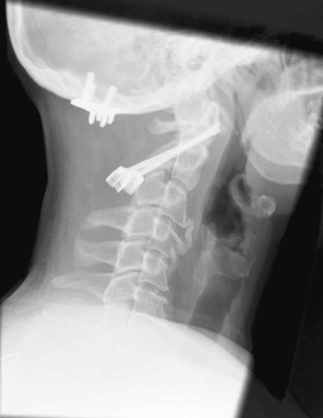
Post-operative lateral radiograph to show accurate positioning of transarticular fusion
Fig. 5.
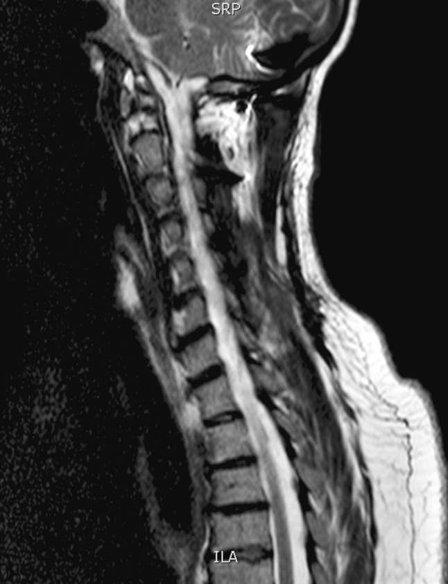
Lateral MRIs to show complete resolution of cyst and free spinal cord at 1-year follow-up
Fig. 6.
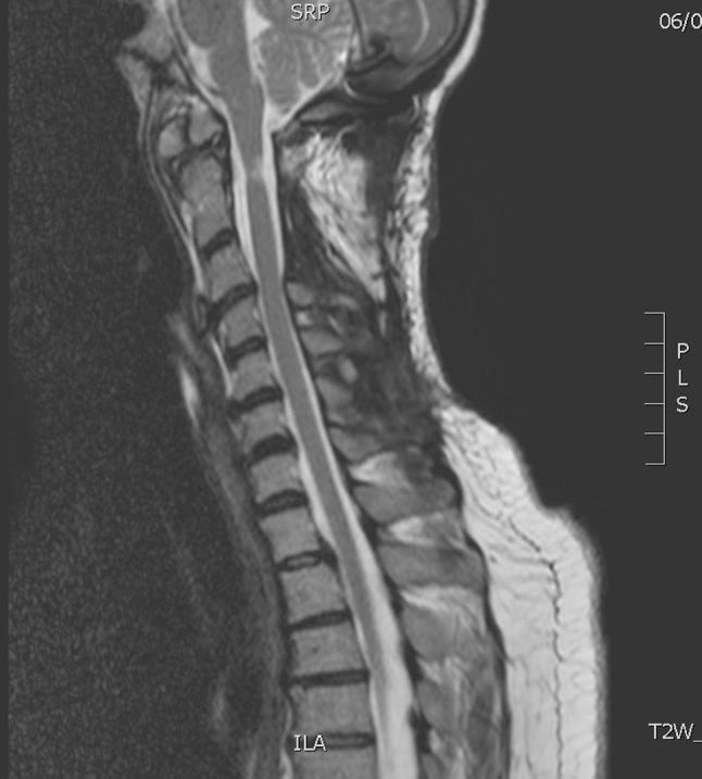
Lateral MRIs to show complete resolution of cyst and free spinal cord at 1-year follow-up
Outcome
MRI scan after 1 year showed complete resolution of the cyst and improvement in space available for the spinal cord at this level and VAS was three. No further surgery was required. Carpal tunnel syndrome was diagnosed bilaterally and referral for left carpal tunnel release was suggested.
This case illustrates an uncertain cause of odontoid fracture, resulting in a cystic lesion compressing the upper cervical spinal cord. Limited surgery of C1/C2 transarticular fusion was successfully performed to avoid use of large constructs and loss of neck motility post-operatively that would result from occipital cervical fusion. The cyst resolved without surgical removal, resulting in improvement of neurological symptoms in this patient.
Acknowledgments
Conflict of interest
None of the authors has any potential conflict of interest.
References
- 1.Anderson LD, D’Alonzo RT. Fractures of the odontoid process of the axis. J Bone Joint Surg Am. 1974;56:1663–1674. [PubMed] [Google Scholar]
- 2.Maak TG, Grauer JN. The contemporary treatment of odontoid injuries. Spine. 2006;31:S53–S60. doi: 10.1097/01.brs.0000217941.55817.52. [DOI] [PubMed] [Google Scholar]
- 3.Ryan MD, Taylor TK. Odontoid fractures in the elderly. J Spinal Disord. 1993;6:397–401. doi: 10.1097/00002517-199306050-00005. [DOI] [PubMed] [Google Scholar]
- 4.Van Gompel J, Morris J, Kasperbauer J, Grander D, Krauss W. Cystic deterioration of the C1–2 articulation: clinical implications and treatment outcomes. J Neurosurg Spine. 2011;14:437–443. doi: 10.3171/2010.12.SPINE10302. [DOI] [PubMed] [Google Scholar]
- 5.Denaro V, Papalia R, Di Martino A, Denaro L, Maffuli N. The best surgical treatment for type II fractures of the dens is still controversial. Clin Orthop Relat Res. 2010;469:742–750. doi: 10.1007/s11999-010-1677-x. [DOI] [PMC free article] [PubMed] [Google Scholar]
- 6.Riesner HJ, Blattert TR, Katscher S, Josten C. Are there therapy algorithms in isolated and combined atlas fractures? Z Orthop Unfall. 2009;147:472–480. doi: 10.1055/s-0029-1185621. [DOI] [PubMed] [Google Scholar]
- 7.Ito T, Hayashi M, Ogino T. Retrodental synovial cyst which disappeared after posterior C1–C2 fusion: a case report. J Orthop Surg. 2000;8:83–87. doi: 10.1177/230949900000800115. [DOI] [PubMed] [Google Scholar]
- 8.Chang H, Park JB, Kim KW. Synovial cyst of the transverse ligament of the atlas in a patient with os odontoideum and atlantoaxial instability. Spine. 2000;25:741–744. doi: 10.1097/00007632-200003150-00016. [DOI] [PubMed] [Google Scholar]
- 9.Jabre A, Shahbabian S, Keller JT. Synovial cyst of the cervical spine. Neurosurgery. 1987;20:316–318. doi: 10.1227/00006123-198702000-00020. [DOI] [PubMed] [Google Scholar]
- 10.Kazda S, Kocis J, Wendsche P, Jarkovskã J. Comparative clinical study assessing C2 dens injuries treatment outcomes. Rozhl Chir. 2009;88:554–558. [PubMed] [Google Scholar]
- 11.Lyons MK, Birch BD, Krauss WE, Patel NP, Nottmeier EW, Boucher OK. Subaxial cervical synovial cysts: report of 35 histologically confirmed surgically treated cases and review of the literatures. Spine. 2011;35(20):E1285–E1289. doi: 10.1097/BRS.0b013e31820709a8. [DOI] [PubMed] [Google Scholar]
- 12.Marbacher S, Lukes A, Vajtai I, Ozdoba C. Surgical approach for synovial cyst of the atlantoaxial joint: a case report and review of the literature. Spine. 2009;34:E528–E533. doi: 10.1097/BRS.0b013e3181ab22c3. [DOI] [PubMed] [Google Scholar]
- 13.Ruf M, Welk T, Mãller M, Merk HR, Harms J. Ventral cancellous bone augmentation of the dens and temporary instrumentation C1/C2 as a function-preserving option in the treatment of dens pseudoarthrosis. J Spinal Disord Tech. 2010;23:285–292. doi: 10.1097/BSD.0b013e3181aac6ff. [DOI] [PubMed] [Google Scholar]
- 14.Blondel B, Metellus P, Fuentes S, Dutertre G, Dufour H. Single anterior procedure for stabilization of a three-part fracture of the axis (odontoid dens and hangman fracture): case report. Spine. 2009;34:E255–E257. doi: 10.1097/BRS.0b013e318195ab2d. [DOI] [PubMed] [Google Scholar]


