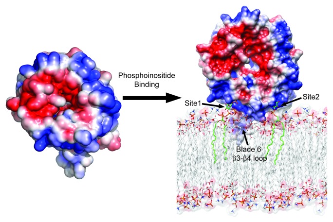
Figure 1. Structural model for phosphatidylinositol phosphate binding and membrane targeting by Atg18 family proteins. Electrostatic potential surface of free and membrane bound forms of Kluyveromyces lactis Hsv2 with saturating blue and red at ± 3 kT/e. The lipid molecules bound at site 1 and site 2 are shown as green stick models.

An official website of the United States government
Here's how you know
Official websites use .gov
A
.gov website belongs to an official
government organization in the United States.
Secure .gov websites use HTTPS
A lock (
) or https:// means you've safely
connected to the .gov website. Share sensitive
information only on official, secure websites.
