Abstract
To determine whether isotype matched immunoglobulin (Ig; Ig′κ/Ig′λ) ratios had prognostic significance in patients with intact Ig multiple myeloma (MM). Novel immunoassays measuring serum concentrations of the Ig heavy chain/light chain (HLC) subsets IgGκ, IgGλ, IgAκ and IgAλ were compared with monoclonal protein (‘M-spike') quantification by serum protein electrophoresis, β2-microglobulin (β2-M), albumin, serum free light chain (FLC) and cytogenetic markers in relation to outcome in 339 MM patients. Abnormal IgGκ/IgGλ and IgAκ/IgAλ ratios present in the respective tumor isotypes at clinical presentation were predictive of shorter progression-free survival (PFS) (hazard ratio (HR) 1.9; P=0.0002), predominantly due to the suppression of the uninvolved (polyclonal) Ig of the same isotype as the tumor (HR 1.8; P=0.002). No significant associations were observed between PFS and M-spike concentrations, suppression of non-tumor Igs of different isotypes or FLC κ/λ ratios. β2-M and HLC ratios were independently prognostic (P=0.045 and P=0.001). A staging system using β2-M and extreme HLC ratios (<0.01 or >200) had greater prognostic value than the widely used ISS staging system (HR 1.7; P=0.00002 vs HR 1.3; P=0.017). These results suggest that HLC ratios may have a role in clinical management of MM.
Keywords: multiple myeloma, immunoglobulin, prognostic factors
Introduction
International guidelines recommend serum protein electrophoresis and serum free light chain (FLC) immunoassays with derived kappa/lambda (κ/λ) ratios to screening for monoclonal gammopathies.1, 2 Compared with the absolute FLC concentration, the use of the κ/λ ratio is a more sensitive marker of monoclonal FLC production as it incorporates suppression of the non-tumor (uninvolved) FLC in its calculation.3 In addition, baseline FLC κ/λ ratios have been reported to be prognostic in multiple myeloma (MM),4, 5 in contrast to densitometric measurements of intact immunoglobulin (Ig) M-spike.6, 7 The added utility of FLC κ/λ ratios has prompted interest in the analysis of κ/λ ratios of intact Igs, now possible due to the availability of immunoassays for the intact Ig subsets; IgGκ, IgGλ, IgAκ and IgAλ (heavy chain/light chain (HLC) immunoassays).8, 9 These molecules are then measured in pairs to produce ratios of tumor-produced (involved) Ig to polyclonal (uninvolved) Ig concentrations, for example, IgGκ/IgGλ. Preliminary evaluations suggest that HLC κ/λ ratios are diagnostically sensitive in MM and AL (amyloid light chain) amyloidosis, can be used to monitor both diseases10, 11 and in some instances may be the only useful marker of disease progression.12 One report also proposes HLC ratios may be prognostic in monoclonal gammopathies of undetermined significance.13 Of particular interest is the ability of the HLC assays to quantify suppression of the uninvolved, polyclonal Igs of the same isotype as the tumor. To date, this has not been possible, either by immunoassays or by electrophoretic methods.
The purpose of the current study was to determine whether HLC measurements, including Ig isotype-specific suppression, had prognostic significance in patients with intact Ig MM. The relationship between HLC measurements and other prognostic indicators such as M-spike, β2-microglobulin (β2-M), albumin, FLCs and cytogenetic abnormalities was also examined.
Patients and methods
Patients and serum samples
Presentation serum samples from patients recruited to the Inter Groupe Français du Myelome 2005-01MM trial were studied, excluding those with FLC-only disease. A total of 339 patients were evaluated, comprising 245 IgG (166 IgGκ, 79 IgGλ) and 94 IgA (60 IgAκ, 34 IgAλ) isotypes. All samples were taken at the time of initial clinical presentation and patients were monitored for progression-free survival (PFS) and overall survival (OS). In accordance with the trial protocol, patients received bortezomib (Velcade, Millennium Pharmaceuticals, Cambridge, MA, USA) and dexamethasone or VAD (vincristine, doxorubicin and dexamethasone) as induction therapy plus or minus DCEP (dexamethasone, cyclophosphamide, etoposide and cisplatin), followed by high-dose melphalan with a stem cell autograft as first line therapy.14 Serum samples were stored at −80 °C until analysis; insufficient samples were available for some measurements (Table 1). A panel of 129 and 138 healthy blood donors was used to generate IgG and IgA HLC reference intervals, respectively (Table 1).
Table 1. Concentrations of serum immunoglobulins and free light chains in normal subjects and 339 patients with multiple myeloma, showing median values together with (95% ranges).
| Normals | IgGκ myeloma (n=166) | IgGλ myeloma (n=79) | IgAκ myeloma (n=60) | IgAλ myeloma (n=34) | |
|---|---|---|---|---|---|
| Monoclonal Ig by SPEP (g/l) | Not applicable | 40.5 (8.0–90) | 46.0 (10.9–85) | 34.0 (11.4–89) | 28.0 (3.46–62) |
| Total IgG (g/l) | 10.9a (7.05–17.45) | 45.7 (12.5–105) | 53.8 (6.21–104) | 3.43 (0.85–8.78) | 3.9 (1.53–7.83) |
| IgGκ (g/l) | 6.82b (4.01–9.77) | 33 (5.6–92) | 0.41 (0.12–5.84) | Not recorded | Not recorded |
| IgGλ (g/l) | 3.83b (1.98–5.71) | 0.33 (0.072–2.03) | 35.5 (4.8–91) | Not recorded | Not recorded |
| IgGκ/IgGλ ratio | 1.87b (1.09–2.60) | 93.5 (5.13–864) | 0.018 (0.001–0.878) | Not recorded | Not recorded |
| Total IgA (g/l) | 2.39a (0.87–5.76) | 0.31 (0.06–2.61) | 0.28 (0.07–2.33) | 39.3 (9.6–107.6) | 32.8 (1.8–65.5) |
| IgAκ (g/l) | 1.19c (0.47–2.82) | Not recorded | Not recorded | 36.7 (5.36–110) | 0.3 (0.018–2.17) |
| IgAλ (g/l) | 0.98c (0.36–1.98) | Not recorded | Not recorded | 0.072 (0.017–1.13) | 30.1 (1.95–50) |
| IgAκ/IgAλ ratio | 1.27c (0.80–2.04) | Not recorded | Not recorded | 462 (11.2–6020) | 0.01 (0.018–0.255) |
| Total IgM (g/l) | 1.34a (0.46–3.86) | 0.22 (0.04–1.61) | 0.21 (0.037–0.93) | 0.18 (0.04–0.66) | 0.21 (0.056–8.77) |
| FLC-κ (mg/l) | 7.30d (3.3–19.4) | 175 (9.66–5645) | 7.14 (0.40–37.9) | 183 (7.99–12 165) | 6.53 (1.27–36.1) |
| FLC-λ (mg/l) | 12.40d (5.7–26.3) | 5.43 (0.50–17.0) | 315 (9.62–7015) | 2.24 (0.18–143) | 220 (17.4–22 683) |
| FLC ratio | 0.60d (0.31–1.2) | 60.1 (1.8–2237) | 0.02 (0.0–1.05) | 68.7 (0.98–10 298) | 0.02 (0.0001–0.56) |
Laboratory methods
Total IgG, IgA and IgM (Siemens Dade Behring, Munich, Germany), IgGκ, IgGλ, IgAκ, IgAλ (Hevylite, Binding Site, Birmingham, UK) and serum FLC using polyclonal sheep antisera (Freelite, Binding Site, expressed as either κ/λ or λ/κ when calculating tertiles) were analyzed on a Dade Behring BNII nephelometer (Siemens AG, Munich, Germany). HLC normal ranges and ratios (either IgGκ/IgGλ or IgAκ/IgAλ) were obtained from analysis of blood donor samples. The degree of systemic humoral immuno-suppression was determined from concentrations of non-tumor Igs (that is, IgG and IgM in IgA patients and IgA and IgM in IgG patients). Reference ranges and medians for IgG, IgA and IgM were obtained from a general adult population.15 All other serum measurements were made in centers in France at the time of sample collection: β2-M and albumin (Siemens Dade Behring) by nephelometry (results of which were used to stage MM patients16), M-spike by serum protein electrophoresis densitometry (Sebia, Paris, France) and the cytogenetic markers Del:13, t4:14 and Del:17p by fluorescence in situ hybridization.17
Statistical analysis
Associations between different assay methods and different markers were tested using Pearson's correlation analysis. Differences in PFS and OS between patient groups were investigated using Kaplan–Meier survival curves with the Mantel–Cox/log rank test used to indicate significance. A Cox proportional hazards model was utilized to compare the association of all variables with PFS. All statistical analyses were performed using SPSS version 18 (Chicago, IL, USA).
Ethics permission
The study was approved by the relevant national health authority agency and the Ethics Committee of the University of Nantes.
Results
The serum concentrations of IgG and IgA HLC in IgG and IgA MM patients were determined, along with serum FLCs (Table 1). For individual patients, a high degree of correlation existed between serum M-spike, involved HLC Ig and total involved Ig concentrations. Pearson's correlations comprised: M-spike vs total involved Ig, 0.87, P=9 × 10⁻⁵; M-spike vs involved HLC, 0.80, P<10−10; involved HLC vs total involved Ig, 0.87, P<10−10. However, there was no relationship between the concentration of involved HLC and isotype matched FLC (IgGκ vs FLC, −0.15, P=0.06; IgGλ vs FLCλ, −0.06, P=0.59; IgAκ vs FLCκ, −0.01, P=0.95; and IgAλ vs FLCλ, −0.37, P=0.03).
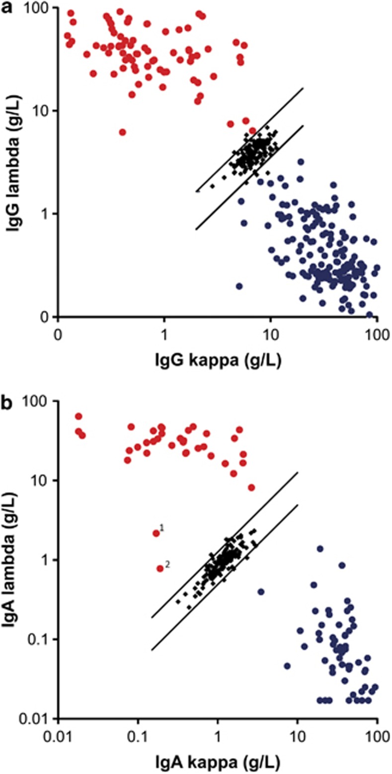
Figures 1a and b summarize IgG or IgA HLC measurements for all patients and normal samples using HLC Ig′κ/Ig′λ dot plots. All IgG MM patients had IgG HLC ratios outside the 95% confidence limits of the normal ranges; the same was true for IgA MM HLC ratios.
Figure 1.
Serum HLC concentrations in patients with multiple myeloma. (a) 166 IgGκ (blue circles) and 79 IgGλ (red circles). (b) 60 IgAκ (blue circles) and 34 IgAλ (red circles). Individual results from blood donor samples and 95% confidence limits (diagonal lines) are shown. Concentrations of lambda FLCs in samples 1 and 2 in Figure 2b were 103 000 and 8500 mg/l, respectively.
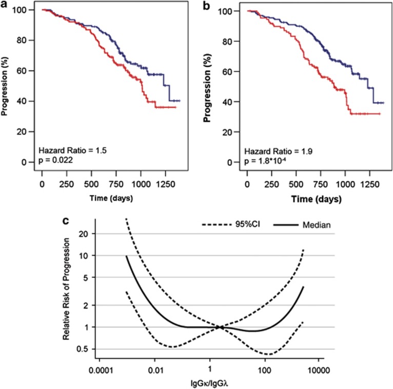
During the study period, 125 patients (37%) had disease progression and 46 patients (14%) died. When patients were categorized according to baseline M-spike concentrations above or below the median values, no significant differences in PFS (P=0.14) or OS (P=0.46) were observed. Similarly, there was no significant difference in outcome when patients were categorized into tertiles of M-spike concentration (PFS: P=0.07 and OS: P=0.4). Nephelometric measurements of total IgG or IgA also failed to demonstrate significant association with outcome. In contrast, there was a significant correlation between HLC κ/λ ratios and outcome. Figure 2a compares the PFS for patients with HLC ratios above or below median concentrations (using κ/λ ratios for IgGκ and IgAκ tumors and λ/κ ratios for IgGλ and IgAλ tumors). The difference in PFS between the two arms (HLC ratio > or <median) was statistically significant (P=0.022). This significance increased when more extreme ratios were considered (>0.01 to <200 compared with <0.01 or >200). Using this stratification, ∼1/3rd of the patients (n=116) had more extreme HLC ratios associated with shorter PFS (hazard ratio (HR) 1.9; P=0.0002: Figure 2b). Using the same HLC ratio stratification for OS, there was a tendency towards significant difference between the two groups (P=0.08), although there were only 14% deaths at this stage of the trial.
Figure 2.
Kaplan–Meier analysis of HLC ratios in relation to clinical outcome: (a) HLC ratios above (red, n=163) or below (blue, n=162) median values vs PFS. (b) HLC ratios with values of >0.01 to <200 (blue, n=209) vs more extreme values (<0.01 or >200: red, n=116), in relation to PFS. (c) Relationship between the HLC IgG ratios and PFS showing that more extreme ratios (<0.01 or >200) are associated with shorter PFS (P<0.001). Median (solid line) and 95% confidence limits (broken lines) are shown.
When IgG and IgA MM patients were analyzed separately, increasingly abnormal HLC κ/λ ratios were associated with shortened PFS in IgG patients (Figure 2c, P<0.001) but not in IgA patients (P=0.32). For IgG patients, the risk of progression rapidly increased with the extent of the ratio abnormality (Figure 2c), thereby providing support for risk stratification using these more extreme ratios.
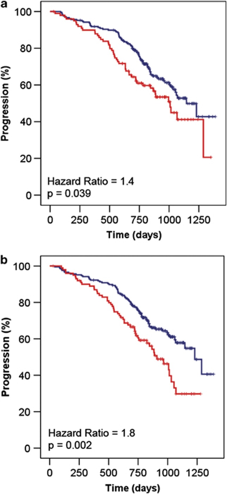
The contribution of involved and uninvolved HLC to the risk of progression was examined. Figure 3a shows PFS for involved HLC levels when comparing IgG and IgA patients who had concentrations in the top tertile with the rest (HR 1.4; P=0.039). However, the association between suppressed levels of the uninvolved HLC was more significantly associated with adverse PFS (Figure 3b; HR 1.8; P=0.002). Thus, suppression of the uninvolved HLC concentrations accounted for the majority of the association between HLC κ/λ ratios and PFS.
Figure 3.
Kaplan–Meier analysis of HLC concentrations in relation to clinical outcome: comparison of PFS for the lower 2/3rds (blue, n=209) vs upper 1/3rd (red, n=116) for: (a) involved HLCs and (b) uninvolved HLCs.
No significant association was observed between PFS and concentrations of the non-tumor isotype above or below median values in IgA patients, IgG and IgM correlations with PFS were not significant (P=0.169 and P=0.477), and in IgG patients, IgA and IgM correlations were not significant (P=0.952 and P=0.977).
Table 2 shows the correlations between PFS and the various measured parameters using univariate and multivariate Cox regression analysis. HLC κ/λ ratios and β2-M were individually more significant than other markers, including albumin, FLC κ/λ ratios and cytogenetic tests (Del:13, t4-14 and Del:17p). Multivariate analysis identified HLC κ/λ ratios and β2-M as being significantly correlated with outcome; albumin, FLC ratios and cytogenetic tests made no contribution.
Table 2. Significance of markers for progression-free survival assessed by univariate and multivariate Cox regression analysis.
| Covariates | Univariate analysis | Multivariate analysis (n=242) |
|---|---|---|
| Del:13 | P=0.03a (n=283) | P=0.546 |
| t4-14 | P=0.05a (n=252) | P=0.515 |
| Del:17p | P=0.08 (n=277) | P=0.457 |
| β2-M >5.5 mg/l | P=0.51 (n=308) | P=0.407 |
| β2-M >3.5 mg/l | P=0.001a (n=308) | P=0.045a |
| Albumin <35 g/l | P=0.153 (n=302) | P=0.828 |
| FLC tertiles | P=0.589 (n=307) | P=0.689 |
| Monoclonal Ig tertilesb | P=0.16 (n=300) | P=0.748 |
| HLC ratios of <200 to >0.01 vs more extreme values | P=0.017a (n=308) | P=0.001a |
Abbreviations: β2-M, β2-microglobulin; FLC, serum free light chain; HLC, immunoglobulin heavy/light chain; Ig, immunoglobulin.
n=number of results available for the analysis.
Significant result at P<0.05.
Monoclonal immunoglobulins as measured by SPEP densitometry.
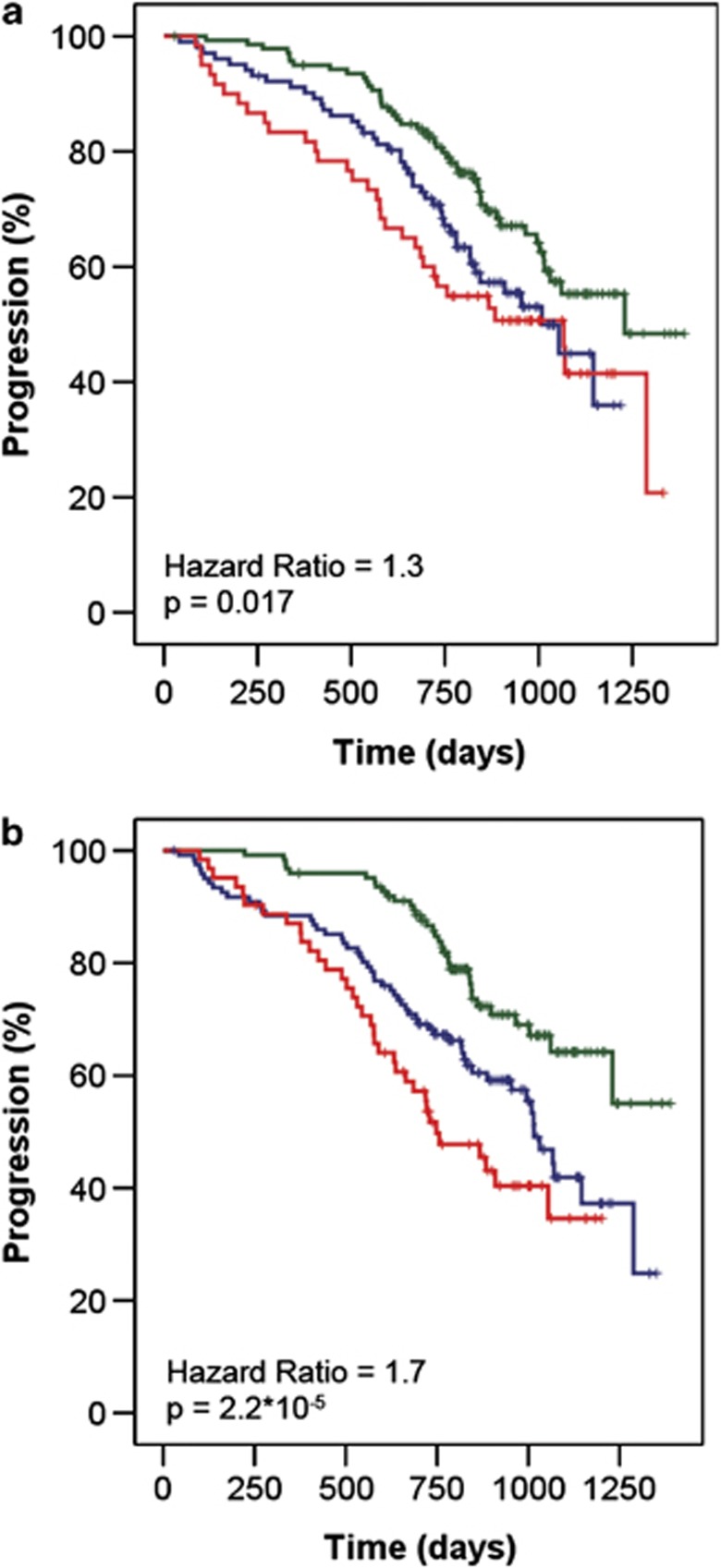
Figure 4a shows the correlation between PFS and the ISS stages for all patients (P=0.017). Figure 4b shows results of a staging system based upon three categories using β2-M and HLC ratios: Stage 1, normal values; Stage 2, either β2-M >3.5 mg/l or extreme HLC ratios (κ/λ <0.01 or >200); and Stage 3, β2-M >3.5 mg/l and extreme HLC ratios (κ/λ <0.01 or >200). Using this model, Stage 3 was more significantly associated with shorter PFS than ISS stage 3 disease (P=0.000002).
Figure 4.
Kaplan–Meier analysis of PFS based upon (a) the ISS that utilizes elevated serum β2-M and low albumin concentrations. Stage I (green, n=140) β2-M <3.5 mg/l and albumin ⩾35 g/l, stage II (blue, n=102) not stage I or II, stage III (red, n=60) β2-M >5.5 mg/l; (b) a risk stratification based upon elevated serum β2-M and more extreme HLC κ/λ ratios (within or outside of 0.01–200) as a replacement for albumin. Low risk (green, n=124) β2-M <3.5 mg/l and a less extreme HLC ratio (0.01–200), intermediate risk (blue, n=122) either β2-M >3.5 mg/l or an extreme HLC ratio (<0.01 or >200), high risk (red, n=62) both β2-M >3.5 mg/l and an extreme HLC ratio (<0.01 or >200).
Discussion
Here, for the first time we show a correlation between HLC ratio measurements and PFS in MM at diagnosis. In common with previous reports there was no association between baseline M-spike16 measurements and outcome. Similarly, suppression of the non-tumor associated Ig′s were of little prognostic use.
The prognostic utility of HLC ratios is largely due to the relative suppression of the polyclonal, uninvolved HLC concentrations. This is the first report of this phenomenon in MM and is supported by similar observations in monoclonal gammopathies of undetermined significance patients.13 Such HLC isotype-specific suppression of polyclonal Igs suggests that bone marrow micro-environment niches may be affected, selectively, by growth of IgG- or IgA-producing tumor cells.
Although uninvolved HLC suppression is the main component of the HLC prognostic utility (HR 1.8; P=0.002), there is also a weak correlation with the involved HLC concentrations (HR 1.4; P=0.039), such that a combination (in the form of the HLC κ/λ ratio) provides the most significantly correlation (HR 1.9; P=0.0002). Curiously, HLC κ/λ ratios had a greater prognostic power in IgG MM than in IgA MM. This is likely to be a reflection of the different number of MM patients (245 IgG compared with 94 IgA) analyzed for each isotype. Alternatively, this could be a reflection of a subtle difference between IgA and IgG MM and larger studies are required to investigate these results further.
We propose that the HLC κ/λ ratio is more prognostic than the serum M-spike level or isotype-specific suppression as the ratio is unaffected by two mechanisms that influence serum measurements of monoclonal Igs. First, variations in hematocrit and plasma volume in MM cause Ig serum concentrations to change by 50% or more, independently of alterations in tumor production rates.18 As both involved and uninvolved HLC measurements are affected equally, the Ig′κ/Ig′λ ratios compensate for these processes, with better reflection of tumor production rates. Second, serum IgG molecules are removed from the circulation by a concentration-dependent process, so that measurements do not reliably relate to tumor production. IgG Fc receptors located on nucleated cells recycle IgG many times, extending the half-life to 21 days at normal serum concentrations. At high IgG concentrations, IgG Fc receptors are saturated, causing the excess IgG to be catabolised; consequently, the overall half-life of IgG lies somewhere between 3 days (for the component that is rapidly catabolised) and 21 days (for the component that is recycled). As the half-lives of polyclonal IgG and monoclonal IgG are affected equally, IgG HLC ratios are unaffected by changes in IgG half-life and may be a more accurate reflection of tumor production than M-spike concentration.
HLC tests measure the tumor-produced Ig more accurately than total Ig measurements, as they use separate immunoassays for Ig′κ and Ig′λ molecules. By comparison, total Ig immunoassays for IgG and IgA include all the polyclonal, non-tumor Igs along with the monoclonal component. Furthermore, traditional, M-spike serum protein electrophoresis measurements by densitometry are limited by co-migrating proteins, such as transferrin being included in any measurements, —a particular concern for fast-migrating IgA M-spikes.
In recent years, cytogenetic abnormalities have been identified as important prognostic factors in MM. Three well known variants were measured in this study; partial or complete deletion of chromosome 13 (Del:13);19 the specific Ig heavy-chain translocation t4:14;20 and mono-allelic deletions of the p53 locus—17p13.21 Although all three markers correlated with PFS associations they were of less prognostic significance than HLC ratios (Table 2).
The impact of HLC ratios was particularly apparent using multivariate analysis, where HLC ratios and β2-M concentrations were the only significant independent variables for identifying patients with reduced PFS. Several previous studies have shown serum polyclonal Freelite immunoassay FLC κ/λ ratios to be predictive of survival.4, 5, 22 This was not observed in this cohort presumably as patients with light chain MM were excluded. Previously, the prognostic utility of serum FLC ratios in predicting OS has been described. In this cohort we had insufficient follow-up data to perform this analysis and recognize this as a study limitation. Other limitations of this study include relatively small patient numbers for IgA MM compared with IgG MM, on which the main correlations were based. As mentioned above, survival data are lacking as the trial is at a relatively early stage of maturity, with only 14% mortality at the time of analysis. Furthermore, additional studies are required to assess the utility of the HLC measurement in patient monitoring and predicting survival after maximal treatment response.
As shown in this study, HLC ratios are predictive of outcome, which may be useful in disease staging at presentation and in assessing the depth of response. These results need confirmation in other plasma cell dyscrasias (including IgM HLC ratios) and, in particular, it would be of interest to investigate monoclonal gammopathy of undetermined significance, smoldering MM,23 Waldenstrom's macroglobulinemia and B-cell chronic lymphocytic leukemia.
In summary, we have provided evidence that serum ratios of IgGκ/IgGλ and IgAκ/IgAλ correlate with disease outcome in MM. In contrast, total IgG and IgA measurements were of little prognostic utility. Furthermore, HLC κ/λ ratios were the most significant parameter for outcome prediction in a multivariate analysis that included β2-M, albumin and cytogenetic abnormalities. Clearly, HLC measurements provide additional information to standard laboratory tests, which may be of considerable importance in MM patient management.
Acknowledgments
We thank Roger Holder, Department of Clinical Studies, University of Birmingham, and Dr Mathew Maurer Mayo Clinic, Rochester for their statistical advice.
ARB wrote the manuscript and developed the study hypothesis; SJH analyzed the data and co-authored the manuscript; SJH and NJF developed the assays and generated the data; J-LH, CM, PM, MA and HA-L provided the clinical samples and analysis and revised the manuscript.
ARB is a shareholder in Binding Site group, Ltd, while SJH and NJF are employees. J-LH, CM, PM, MA and HA-L declared no conflict of interest.
References
- Dispenzieri A, Kyle R, Merlini G, Miguel JS, Ludwig H, Hajek R, on behalf of the International Myeloma Working Group et al. International Myeloma Working Group guidelines for serum-free light chain analysis in multiple myeloma and related disorders. Leukemia. 2008;23:215–224. doi: 10.1038/leu.2008.307. [DOI] [PubMed] [Google Scholar]
- Anderson CL, Chaudhury C, Kim J, Bronson CL, Wani MA, Mohanty S, et al. Perspective-FcRn transports albumin: relevance to immunology and medicine. Trends Immunol. 2006;27:343–348. doi: 10.1016/j.it.2006.05.004. [DOI] [PubMed] [Google Scholar]
- Drayson M, Tang LX, Drew R, Mead GP, Carr-Smith H, Bradwell AR. Serum free light-chain measurements for identifying and monitoring patients with nonsecretory multiple myeloma. Blood. 2001;97:2900–2902. doi: 10.1182/blood.v97.9.2900. [DOI] [PubMed] [Google Scholar]
- Kyrtsonis MC, Vassilakopoulos TP, Kafasi N, Sachanas S, Tzenou T, Papadogiannis A, et al. Prognostic value of serum free light chain ratio at diagnosis in multiple myeloma. Br J Haematol. 2007;137:240–243. doi: 10.1111/j.1365-2141.2007.06561.x. [DOI] [PubMed] [Google Scholar]
- Snozek CL, Katzmann JA, Kyle RA, Dispenzieri A, Larson DR, Therneau TM, et al. Prognostic value of the serum free light chain ratio in newly diagnosed myeloma: proposed incorporation into the international staging system. Leukemia. 2008;22:1933–1937. doi: 10.1038/leu.2008.171. [DOI] [PMC free article] [PubMed] [Google Scholar]
- Durie BG, Jacobson J, Barlogie B, Crowley J. Magnitude of response with myeloma frontline therapy does not predict outcome: importance of time to progression in southwest oncology group chemotherapy trials. J Clin Oncol. 2004;22:1857–1863. doi: 10.1200/JCO.2004.05.111. [DOI] [PubMed] [Google Scholar]
- Dingli D, Pacheco JM, Nowakowski GS, Kumar SK, Dispenzieri A, Hayman AR, et al. Relationship between depth of response and outcome in multiple myeloma. J Clin Oncol. 2007;25:4933–4937. doi: 10.1200/JCO.2007.11.7879. [DOI] [PubMed] [Google Scholar]
- Bradwell AR, Harding SJ, Fourrier NJ, Wallis GLF, Drayson M, Carr-Smith H, et al. Assessment of monoclonal gammopathies by nephelometric measurement of individual immunoglobulin kappa/lambda ratios. Clin Chem. 2009;55:1646–1655. doi: 10.1373/clinchem.2009.123828. [DOI] [PubMed] [Google Scholar]
- Keren DF. Heavy/Light-chain analysis of monoclonal gammopathies. Clin Chem. 2009;55:1606–1608. doi: 10.1373/clinchem.2009.132753. [DOI] [PubMed] [Google Scholar]
- Ludwig H, Harding S, Bradley C, Milosavljevich D, Drayson M, Morgan G, et al. Abnormal serum IgA kappa/IgA lambda ratios at maximum response predict poor progression free survival in myeloma patients Blood 2009114(abstract 4879). [Google Scholar]
- Wechalekar A, Harding S, Lachmann H, Gillmore JD, Wassef NL, Thomas M, et al. Serum immunoglobulin heavy/light chain ratios (Hevylite) in patients with systemic AL amyloidosis Amyloid 201017(abstract 186). [Google Scholar]
- Donato L, Zeldenrust S, Murray D, Katzmann J. A 71 year old woman with multiple myeloma status after stem cell transplantation. Clin Chem. 2011;57:1645–1649. doi: 10.1373/clinchem.2011.163766. [DOI] [PubMed] [Google Scholar]
- Katzmann J, Clark R, Dispenzieri A, Kyle R, Landgren O, Bradwell AR, et al. Isotype-specific heavy/light chain (HLC) suppression as a predictor of myeloma development in monoclonal gammopathy of undetermined significance (MGUS) Blood 2009114(abstract 1788). [Google Scholar]
- Harousseau JL, Mathiot C, Attal M, Marit D, Caillot C, Hullin T, et al. Bortezomib/dexamethasone versus VAD as induction prior to autologous stem cell transplantation (ASCT) in previously untreated multiple myeloma (MM): updated data from IFM 2005/1 trial J Clin Oncol 200826(abstract 8505). [Google Scholar]
- Gonzalez-Quintela A, Alende R, Gude F, Campos J, Rey J, Meijide LM, et al. Serum levels of immunoglobulins (IgG IgA, IgM) in a general adult population and their relationship with alcohol consumption, smoking and common metabolic abnormalities. Clin Exp Imm. 2007;151:42–50. doi: 10.1111/j.1365-2249.2007.03545.x. [DOI] [PMC free article] [PubMed] [Google Scholar]
- Greipp PR, San Miguel J, Durie BG, Crowley JJ, Barlogie B, Blade J, et al. International staging system for multiple myeloma. J Clin Oncol. 2005;23:3412–3420. doi: 10.1200/JCO.2005.04.242. [DOI] [PubMed] [Google Scholar]
- Facon T, Avet-Loiseau H, Guillerm G, Moreau P, Geneviève F, Zandecki M, et al. Chromosome 13 abnormalities identified by FISH analysis and serum β2-microglobulin produce a powerful myeloma staging system for patients receiving high-dose therapy. Blood. 2001;97:1566–1571. doi: 10.1182/blood.v97.6.1566. [DOI] [PubMed] [Google Scholar]
- Alexanian R. Blood volume in monoclonal gammopathy. Blood. 1977;49:301–307. [PubMed] [Google Scholar]
- Fonseca R, Harrington D, Oken MM, Dewald GW, Bailey RJ, Van Wier SA, et al. Biological and prognostic significance of interphase fluorescence in situ hybridization detection of chromosome 13 abnormalities (_13) in multiple myeloma: an Eastern Cooperative Oncology Group Study. Cancer Res. 2002;62:715–720. [PubMed] [Google Scholar]
- Moreau P, Facon T, Leleu X, Morineau N, Huyghe P, Harousseau JL, et al. Recurrent 14q32 translocations determine the prognosis of multiple myeloma, especially in patients receiving intensive chemotherapy. Blood. 2002;100:1579–1583. doi: 10.1182/blood-2002-03-0749. [DOI] [PubMed] [Google Scholar]
- Gertz MA, Lacy MQ, Dispenzieri A, Greipp PR, Litzow MR, Henderson KJ, et al. Clinical implications of t(11;14)(q13;q32), t(4;14)(p16.3;q32), and -17p13 in myeloma patients treated with high-dose therapy. Blood. 2005;106:2837–2840. doi: 10.1182/blood-2005-04-1411. [DOI] [PMC free article] [PubMed] [Google Scholar]
- van Rhee F, Bolejack V, Hollmig K, Pineda-Roman M, Anaissie E, Epstein J, et al. High serum-free light chain levels and their rapid reduction in response to therapy define an aggressive multiple myeloma subtype with poor prognosis. Blood. 2007;110:827–832. doi: 10.1182/blood-2007-01-067728. [DOI] [PMC free article] [PubMed] [Google Scholar]
- Pérez-Persona E, Vidriales MB, Mateo G, García-Sanz R, Mateos MV, García de Coca A, et al. New criteria to identify risk of progression in monoclonal gammopathy of uncertain significance and smoldering multiple myeloma based on multiparameter flow cytometry analysis of bone marrow plasma cells. Blood. 2007;110:2586–2592. doi: 10.1182/blood-2007-05-088443. [DOI] [PubMed] [Google Scholar]
- Katzmann JA, Clark RJ, Abraham RS, Bryant S, Lymp JF, Bradwell AR, et al. Serum reference intervals and diagnostic ranges for free kappa and lambda immunoglobulin light chains: relative sensitivity for detection of monoclonal light chains. Clin Chem. 2002;28:1437–1444. [PubMed] [Google Scholar]


