Abstract
BACKGROUND: Melanoma is a relatively immunogenic tumor, in which infiltration of melanoma cells by T lymphocytes is associated with a better clinical prognosis. We hypothesized that radiation-induced cell death may provide additional stimulation of an anti-tumor immune response in the setting of anti-CTLA-4 treatment. METHODS: In a pilot melanoma patient, we prospectively tested this hypothesis. We treated the patient with two cycles of ipilimumab, followed by stereotactic ablative radiotherapy to two of seven hepatic metastases, and two additional cycles of ipilimumab. RESULTS: Subsequent positron emission tomography-computed tomography scan indicated that all metastases, including unirradiated liver lesions and an unirradiated axillary lesion, had completely resolved, consistent with a complete response by RECIST. CONCLUSION: The use of radiotherapy in combination with targeted immunotherapy as a noninvasive in vivo tumor vaccine strategy appears to be a promising method of enhancing the induction of systemic immune responses and anti-tumor effect.
Case Report
Here, we report a 57-year-old male patient diagnosed with stage IIA cutaneous melanoma in November 2007 following biopsy of a mole on his left posterior arm that revealed melanoma, ulcerated, with a Breslow thickness of 1.75 mm. He underwent a wide local excision of his primary lesion and left axillary sentinel node biopsy in December 2007. Because initial pathologic review was read as positive for isolated tumor cells in the intracapsular region, he subsequently underwent a completion left axillary dissection in December 2007. Twenty-two axillary lymph nodes were removed and found to be negative, with one lymph node containing benign capsular nevus cells. He was therefore staged as IIA [per American Joint Committee on Cancer (AJCC) designation of isolated tumor cells as node-negative at the time] and did not receive any adjuvant treatment.
The patient remained disease-free until May 2010, when he developed a new 6- to 8-mm subcutaneous nodule within the melanoma scar. Punch biopsy confirmed locally recurrent melanoma (satellitosis), and he underwent wide local excision of the satellite metastasis. Pathology revealed residual/recurrent melanoma as well as a 4-mm microsatellite focus of melanoma within the subcutaneous mass, at least 1.5 cm from the main tumor mass. Metastatic workup with positron emission tomography (PET)-computed tomography (CT) in June 2010 was negative for distant disease. He then underwent adjuvant radiotherapy to the site of recurrence in the left posterior arm, receiving 50.4 Gy in 20 fractions followed by adjuvant systemic therapy with 1 month of high-dose interferon completed in August 2010. Repeat PET-CT in January 2011 showed no evidence of disease.
In May 2011, the patient noted a new subcutaneous nodule in the left axillary region, and fine-needle aspiration (FNA) confirmed in-transit melanoma metastasis. PET-CT in May 2011 revealed two new sub-centimeter hypermetabolic foci within the liver in addition to the axillary metastasis. The left axillary in-transit metastasis was excised in May 2011, and CT-guided liver biopsy confirmed metastatic melanoma.
We hypothesized that radiation-induced tumor cell death may provide further stimulation of an anti-tumor immune response in the setting of treatment with anti-CTLA-4, and we discussed prospectively testing this hypothesis by combining these two clinically indicated and standard-of-care treatments in this patient. The patient agreed and was started on ipilimumab at 3 mg per kilogram of body weight every 3 weeks for a total of four doses, with radiation planned to start after two doses. After receiving the first two doses of ipilimumab, repeat PET-CT scan in August 2011, performed for radiotherapy treatment planning purposes, showed progression of liver metastases, with enlargement of the two previously noted lesions, as well as development of five new hypermetabolic foci in the liver. As planned, the patient received stereotactic radiotherapy to two of the liver metastases to 54 Gy in three fractions. During administration of cycle 3 of ipilimumab, he noted the rapid development of a new 5-cm palpable subcutaneous nodule on his left upper arm, which was confirmed by fine-needle aspiration to represent another in-transit melanoma recurrence. He received cycle 4 of ipilimumab in September 2011.
One week later, the patient was admitted to the hospital with nausea and vomiting and was found to have hypophysitis with an undetectable cortisol level. He was started on systemic prednisone with immediate improvement of nausea and vomiting and continued on a slow prednisone taper for the next several months.
In December 2011, follow-up PET-CT scan revealed no FDG uptake in the areas of previously noted liver lesions. There was persistent focal uptake in the left upper arm at the site of the previously biopsied in-transit melanoma metastasis. The decision was made to observe the left arm lesion for potential treatment response and not to pursue surgical excision, as it was unlikely to change his clinical outcome, and to continue to follow him closely.
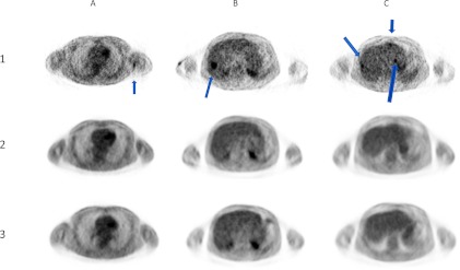
The patient was seen again in late February 2012, at which time his left arm lesion had resolved completely. PET-CT scan at that time revealed complete resolution of the hypermetabolic lesion in his left arm and continued complete resolution of all of the hyper-metabolic liver lesions. He was seen again in August 2012 with an unremarkable physical exam. PET-CT scan at that time again showed no evidence of metabolically active malignancy and continued complete resolution of the previously hypermetabolic left arm lesion and all of the hypermetabolic liver lesions, at 1-year post-radiation (Figure 1). The patient continues on a slow steroid taper for resolving hypophysitis.
Figure 1.
PET scans performed before treatment (row 1), at 6 months after treatment (row 2), and at 12 months after treatment (row 3). Column A (arrow) shows untreated axillary lesion that was no longer apparent at 6 and 12 months after treatment. Column B (arrow) shows one of two treated hepatic lesions, which showed complete response to treatment at 6 and 12 months after treatment. Column C (arrows) shows three of five untreated hepatic lesions, which also showed complete response to treatment at 6 and 12 months after treatment.
Discussion
Our patient experienced a systemic complete response to ipilimumab and local radiotherapy following high-dose radiotherapy to two of seven metastatic liver lesions after having experienced disease progression on ipilimumab alone. This is the second report to date of a patient with metastatic melanoma who had an “abscopal” response to radiotherapy with ipilimumab and the first report of a patient who achieved a complete response with this combined treatment [1].
Melanoma is a relatively immunogenic tumor. It is therefore an immune responsive disease in which infiltration of melanoma lesions by T lymphocytes is associated with a better clinical prognosis [2]. However, engagement of the T cell antigen receptor by itself is not sufficient for full T cell activation. A second costimulatory signal is required for induction of interleukin-2 (IL-2) production, proliferation, and differentiation to affect function of naïve T cells. Abundant recent data indicate that the primary source of this costimulation is mediated by engagement of CD28 on the T cell surface by members of the B7 family on the antigen-presenting cell [3]. Expression of B7 has been shown to be limited to specialized cells of the hematopoietic lineage, including dendritic cells, activated macrophages, and activated B cells. CTLA-4 is an inhibitory receptor, and inhibiting CTLA-4 thereby stimulates the immune response [4]. CTLA-knockout mice appear to have spontaneously activated T cells evident at 1 week after birth, followed by rampant lymphoproliferation and lymphadenopathy. Since thymocyte differentiation and selection proceed normally in CTLA-4-deficient mice, the rampant T cell expansion indicates that CTLA-4 plays a critical role in downregulating T cell responses in the periphery [5]. When used as part of a tumor vaccine strategy, anti-CTLA-4 antibodies may generate anti-tumor response by two possible mechanisms: 1) CTLA-4 blockade on tumor-specific effector T cells, resulting in clonal activation and expansion, and 2) CTLA-4-targeted depletion of tumor-induced Tregs, which could inhibit immune responses to tumor antigens [1,6,7]. Radiation also causes a dose-dependent reduction in Treg proliferation, which parallels the induction in apoptosis in Tregs, and can affect both the phenotype and suppression function in Tregs [8]. Therefore, the combined effects of both ipilimumab and radiation on Tregs may be important determinants of the efficacy of this combination therapy.
The concept of using radiotherapy as a component of a noninvasive tumor vaccine strategy is a well-established concept, with promising translational potential as tumor vaccine strategies and immunotherapies have become available, and is the subject of completed (NCT00185965: CpG + radiotherapy for lymphoma) and ongoing clinical trials, including NCT00861614: ipilimumab + radiotherapy for metastatic prostate cancer; NCT01421017: Toll-like receptor 7 agonist + radiotherapy for metastatic breast cancer; NCT01401062: anti-transforming growth factor-β + radiotherapy for metastatic breast cancer [9]. Preclinical studies show that local radiotherapy induces tumor cell death and release of tumor antigens and increases antigen presentation, which can promote an effective immune response [10,11]. An effective radiotherapy and immunotherapeutic combination, therefore, would produce an endogenous in situ vaccination against the tumor, which unlike most other vaccine strategies is entirely noninvasive and does not require intratumoral injections.
The “abscopal” effect, or what could more accurately be termed an “in vivo vaccination” effect, has previously been demonstrated in pre-clinical models. Demaria et al. showed decreased pulmonary metastases and improved survival in mice treated with radiotherapy in the context of CTLA-4 blockade [11]. The effect of dose, fractionation, and timing of the “abscopal” effect is controversial [10]. Several pre-clinical studies suggest that hypofractionation is optimal, while other studies suggest that conventional fractionation is more effective at promoting an “abscopal” effect [10].
The first clinical report of an “abscopal” effect in a patient receiving ipilimumab with concurrent radiotherapy was recently reported by Postow et al., in which they describe a patient with metastatic melanoma with a paraspinal mass, splenic lesions, and right hilar lymphadenopathy [1]. This patient was treated palliatively for back pain caused by the paraspinal mass, to 28.5 Gy in three fractions. She continued to receive ipilimumab. Five months after radiotherapy, all lesions were found to have regressed on CT scan, and 10 months after radiotherapy, lesions were stable with continued presence of minimal disease.
Our case differs from the one described by Postow et al. in several ways. First, our patient was asymptomatic at the time of radiotherapy and was prospectively treated with ipilimumab, not only because he had progressive metastatic disease, but also because we hypothesized that it may provide an endogenous vaccination, which could potentially amplify the systemic anti-melanoma T cell response in the setting of CTLA-4 blockade. Second, our patient achieved a complete response with no evidence of disease on CT or PET scan, although our follow-up interval (12 months) is still relatively short. Third, given the location of his metastatic disease, we were able to treat with radiotherapy to a higher dose per fraction (54 Gy in three fractions vs 28.5 Gy in three fractions). This may have resulted in an increased systemic immune response as suggested in the preclinical work by Dewan et al., with limited data suggesting a dose-response relationship [10].
While delayed response to ipilimumab alone has been reported, the historical overall response rate to ipilimumab in stage IV melanoma is <10%, with a complete response rate of <2% [7]. The patient's left axillary tumor demonstrated likely pseudoprogression, given the rapid growth followed by rapid resolution, observed in approximately 10% of ipilimumab responses. Finally, the patient also experienced autoimmune hypophysitis, a well-described adverse effect of ipilimumab, and he has responded well to a gradual and ongoing prednisone taper [12].
At the Stanford Cancer Institute, we are currently enrolling patients on a study in which patients with metastatic melanoma are treated concurrently with ipilimumab and radiotherapy, with the goal of enhancing the induction of systemic anti-melanoma immune responses that will inhibit growth and kill melanoma cells in sites of established metastases outside of the radiotherapy field more effectively than with anti-CTLA-4 alone, with minimal or no additional toxicity. The use of targeted immunotherapy in combination with radiation therapy is becoming an area of increasing interest and clinical investigation in melanoma. Seung et al. recently reported results from a phase 1 study combining stereotactic body radiotherapy and IL-2 in patients with metastatic melanoma or metastatic renal cell carcinoma and found that 8 of 12 patients had a complete response (CR) or partial response (PR) by RECIST in unirradiated lesions after treatment, with a significantly greater frequency of proliferating CD4 cells with an early activated effector memory phenotype in the peripheral blood of responders versus nonresponders [13]. This in vivo tumor vaccine strategy has potential clinical applicability for the treatment of a variety of tumor types, not only with anti-CTLA-4 or IL-2 but also with other immunotherapies currently in development, such as anti-CD137, anti-PD-1, and anti-PD-L1.
References
- 1.Postow MA, Callahan MK, Barker CA, Yamada Y, Yuan J, Kitano S, Mu Z, Rasalan T, Adamow M, Ritter E, et al. Immunologic correlates of the abscopal effect in a patient with melanoma. N Engl J Med. 2012;366(10):925–931. doi: 10.1056/NEJMoa1112824. [DOI] [PMC free article] [PubMed] [Google Scholar]
- 2.Clemente CG, Mihm MC, Jr, Bufalino R, Zurrida S, Collini P, Cascinelli N. Prognostic value of tumor infiltrating lymphocytes in the vertical growth phase of primary cutaneous melanoma. Cancer. 1996;77(7):1303–1310. doi: 10.1002/(SICI)1097-0142(19960401)77:7<1303::AID-CNCR12>3.0.CO;2-5. [DOI] [PubMed] [Google Scholar]
- 3.Lenschow DJ, Walunas TL, Bluestone JA. CD28/B7 system of T cell costimulation. Annu Rev Immunol. 1996;14:233–258. doi: 10.1146/annurev.immunol.14.1.233. [DOI] [PubMed] [Google Scholar]
- 4.Walunas TL, Lenschow DJ, Bakker CY, Linsley PS, Freeman GJ, Green JM, Thompson CB, Bluestone JA. CTLA-4 can function as a negative regulator of T cell activation. Immunity. 1994;1(5):405–413. doi: 10.1016/1074-7613(94)90071-x. [DOI] [PubMed] [Google Scholar]
- 5.Krummel MF, Sullivan TJ, Allison JP. Superantigen responses and co-stimulation: CD28 and CTLA-4 have opposing effects on T cell expansion in vitro and in vivo. Int Immunol. 1996;8(4):519–523. doi: 10.1093/intimm/8.4.519. [DOI] [PubMed] [Google Scholar]
- 6.Gabriel EM, Lattime EC. Anti-CTL-associated antigen 4: are regulatory T cells a target? Clin Cancer Res. 2007;13(3):785–788. doi: 10.1158/1078-0432.CCR-06-2820. [DOI] [PubMed] [Google Scholar]
- 7.Hodi FS, O'Day SJ, McDermott DF, Weber RW, Sosman JA, Haanen JB, Gonzalez R, Robert C, Schadendorf D, Hassel JC, et al. Improved survival with ipilimumab in patients with metastatic melanoma. N Engl J Med. 2010;363(8):711–723. doi: 10.1056/NEJMoa1003466. [DOI] [PMC free article] [PubMed] [Google Scholar]
- 8.Cao M, Cabrera R, Xu Y, Liu C, Nelson D. Gamma irradiation alters the phenotype and function of CD4-CD25- regulatory T cells. Cell Biol Int. 2009;33(5):565–571. doi: 10.1016/j.cellbi.2009.02.007. [DOI] [PMC free article] [PubMed] [Google Scholar]
- 9.Milas L, Hunter N, Withers HR. Concomitant immunity to pulmonary metastases of a murine fibrosarcoma: influence of removal of primary tumor by radiation or surgery, of active specific immunization and treatment with Corynebacterium granulosum. Int J Radiat Oncol Biol Phys. 1976;1(11–12):1171–1178. doi: 10.1016/0360-3016(76)90090-0. [DOI] [PubMed] [Google Scholar]
- 10.Dewan MZ, Galloway AE, Kawashima N, Dewyngaert JK, Babb JS, Formenti SC, Demaria S. Fractionated but not single-dose radiotherapy induces an immune-mediated abscopal effect when combined with anti- CTLA-4 antibody. Clin Cancer Res. 2009;15(17):5379–5388. doi: 10.1158/1078-0432.CCR-09-0265. [DOI] [PMC free article] [PubMed] [Google Scholar]
- 11.Demaria S, Kawashima N, Yang AM, Devitt ML, Babb JS, Allison JP, Formenti SC. Immune-mediated inhibition of metastases after treatment with local radiation and CTLA-4 blockade in a mouse model of breast cancer. Clin Cancer Res. 2005;11(2 pt 1):728–734. [PubMed] [Google Scholar]
- 12.Yang JC, Hughes M, Kammula U, Royal R, Sherry RM, Topalian SL, Suri KB, Levy C, Allen T, Mavroukakis S, et al. Ipilimumab (anti-CTLA4 antibody) causes regression of metastatic renal cell cancer associated with enteritis and hypophysitis. J Immunother. 2007;30(8):825–830. doi: 10.1097/CJI.0b013e318156e47e. [DOI] [PMC free article] [PubMed] [Google Scholar]
- 13.Seung SK, Curti BD, Crittenden M, Walker E, Coffey T, Siebert JC, Miller W, Payne R, Glenn L, Bageac A, et al. Phase 1 study of stereotactic body radiotherapy and interleukin-2—tumor and immunological responses. Sci Transl Med. 2012;4(137):137ra74. doi: 10.1126/scitranslmed.3003649. [DOI] [PubMed] [Google Scholar]


