Fig. 8.

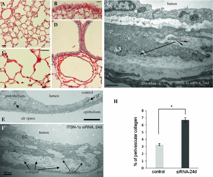
Chronic ITSN-1s deficiency leads to dense perivascular collagen depositions Representative micrographs of Picrosirius Red-stained lung sections of control (A) and siRNAITSN-treated mice for 24d (B–D), to assess collagen deposition. Short septal walls remnants were often noticed with collagen bundles at the tip (C, arrows). Bars 20 μm. Electron micrographs show segments of the alveolar septal wall in a control microvessel (E), as well as in a postcapillary venule (10–15 μm diameter) (F), and a precapillary arteriole (20–25 μm diameter) (G) of ITSN-1s deficient mice. Collagen fibrils (G, arrows), and fibrilar material (proteoglicans, elastic fibers, etc.), accumulated in the basement membrane between the ECs, smc and fibroblast. j1—interendothelial junction, j2—epithelial junction, EC—endothelial cell, pvs—perivascular space, smc—smooth muscle cell. Bar 500 nm. H Quantification of the amount of collagen encircling middle-sized lung vessels in control and ITSN-1s chronic inhibition mice. Results are representative for 3 different experiments, with 3 mice/experimental condition. Collagen layers were measured in 20–25 randomly chosen high power fields comprising 25 medium-sized blood vessels for each experimental condition. Data are expressed as mean ± SEM, *P < 0.05
