Abstract
Benign fibrous histiocytomas of the skin sometimes extend into the deeper dermis with higher cellularity and show more aggressive clinical courses in comparison with typical dermatofibromas. They may occur either as a true neoplasm or in a reactive process. We describe a case of fibrous histiocytoma which was triggered by tuberculin skin test.
Keywords: dermatofibroma, fibrous histiocytoma, tuberculin, tumor
Case
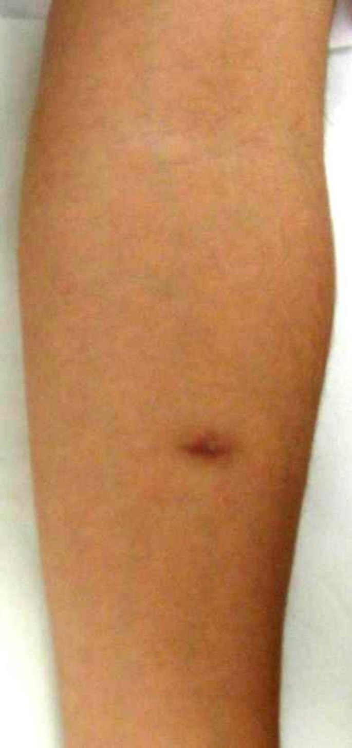
A 25-year-old male complained of a nodule which developed at the tuberculin injected site on the forearm. Four months before, he was administered 0.1 ml tuberculin test containing 0.25 µg/0.5 mL purified protein derivative (PPD). The tuberculin reaction was positive (15x15 mm of erythematous induration) at 48 h. A physical examination revealed an 8-mm sized, brownish firm nodule on the flexor aspect of the left forearm [Fig. 1]. Histological examination revealed fibrous proliferation in the dermis extending into the deep dermis with a hypercellularity. The proliferated fibroblasts were immunoreactive for factor XIIIa, HLA-DR, alpha-smooth muscle actin (α-SMA) and CD68, but negative for CD34. The tumor cells did not exhibit atypical mitotic figures, pleomorphism, or nuclear atypia. The overlying epidermis was not elongated, and there was no increase of basal pigmentation in the overlying epidermis.
Figure 1.

Clinical appearance of a nodule on the site of tuberculin injection.
In this case, fibrous histiocytoma occurred on the tuberculin-innoculated site 4 months after the tuberculin skin test. Results of immunohistochemistry showed that the majority of tumor cells were positively stained with HLA-DR, factor XIIIa, α-SMA and CD68. A superficial lesion of fibrous histiocytoma is regarded as dermatofibroma. Some believe that dermatofibroma is the result of an abortive immunoreactive process mediated by dermal dendritic cells, while others believe that dermatofibroma is a benign neoplastic process.[1] Previous studies have shown that dermatofibromas originate from fibroblasts, histiocytes, and perivascular cells. In vitro studies have shown that cultured monocyte-derived dendritic cells are transformed to cells sharing many of the characteristics of dermatofibroma.[1]
The tuberculin reaction is the delayedtype hypersensitivity reaction to PPD of tuberculin. Previous studies of histological changes of tuberculin reaction showed that all individuals did not show uniform patterns and large variations are recognized depending on time. Acute phase reaction is classified into three types, i.e. the perivascular dermatitis type, the basal spongiotic dermatitis type, and the erythema multiforme type.[2] Local cytokine expression on peripheral blood showed that interferon-γ (IFN-γ) reached a peak at 48 h following PPD injection mainly on CD3+ T-cells, while tumor necrosis factor-α (TNF-α), interleukin-1 (IL-1) and IL-6 were enhanced mainly on CD68+ macrophages/monocytes throughout the periods of 7 days.[3] PPD mediates dendritic cell maturation which is reflected by surface expression of CD83 and MHC II.[4] The histological features of palisading granuloma with central degenerative changes were observed in biopsies taken 4 months and 1 year after PPD injection.[2] Also, granuloma annulare-like changes were induced at the site of old intracutaneous tuberculin injections 6 months later.[5] Thus, histiocytes are activated at the late phase reaction, and may lead to the hyperproliferation in the present case.
To our knowledge, this is the first case of fibrous histiocytoma following tuberculin test. Although the occurrence of fibrous histiocytoma at the site of tuberculin injection may be a mere coincidence, the tuberculin injection may have played a part in the development of fibrous tumors. Fibrous tumors may be included in the differential diagnosis for tumors which occur at the site of tuberculin injection site.
References
- Aiba S, Tagami H. Phorbol 12-myristate 13-acetate can transform monocyte-derived dendritic cells to different cell types similar to those found in dermatofibroma. J Cutan Pathol. 1998;25:65–71. doi: 10.1111/j.1600-0560.1998.tb01692.x. [DOI] [PubMed] [Google Scholar]
- Kuramoto Y, Tagami H. Histopathologic pattern analysis of human intracutaneous tuberculin reaction. Am J Dermatopathol. 1989;11:329–337. doi: 10.1097/00000372-198908000-00006. [DOI] [PubMed] [Google Scholar]
- Chu CQ, Field M, Andrew E, Haskard D, Feldmann M, Maini RN. Detection of cytokines at the site of tuberculin-induced delayed-type hypersensitivity in man. Clin Exp Immunol. 1992;90:522–529. doi: 10.1111/j.1365-2249.1992.tb05877.x. [DOI] [PMC free article] [PubMed] [Google Scholar]
- Bagheri K, Delirezh N, Moazzeni SM. PPD extract induces the maturation of human monocyte-derived dendritic cells. Immunopharmacol Immunotoxicol. 2008;30:91–104. doi: 10.1080/08923970701812654. [DOI] [PubMed] [Google Scholar]
- Fisher I. An unusual late histologic change seen in the intracutaneous tuberculin reaction. J Invest Dermatol. 1954;23:233–235. doi: 10.1038/jid.1954.103. [DOI] [PubMed] [Google Scholar]


