Abstract
There are few observation papers regarding the natural history of an aneurysm. We report on a case of a completely occluded middle cerebral artery (MCA) aneurysm. A 47-year-old female patient presented with a headache and was diagnosed with rupture of a right MCA aneurysm. Due to a high risk of direct neck clipping, she received conservative treatment after craniotomy and wrapping of her aneurysm. The patient's condition showed improvement, with complete occlusion of the aneurysm and considerable reduction of the aneurysm in size after approximately three years. This is a rare case of an aneurysm of MCA that showed spontaneous resolution. Finally, on the angiogram, characteristics of an aneurysm to occlude spontaneously will be presumed based on literature reviews.
Keywords: Middle cerebral artery aneurysm, Spontaneous remission, Angiography
INTRODUCTION
Many studies on unruptured aneurysms have reported a prevalence of approximately 3.6 to 6% and a bleeding rate of approximately 2.3 to 0.3%/year.1),18) For the giant aneurysm (≥ 25 mm), a rupture rate of 6% has been reported for one year since the first diagnosis, and based on the natural history, it was considered bad due to high lethality.1),15),20) However, with technological developments in the fields of surgery and anesthetics since the 1970s-1980s, the mortality and morbidity associated with the operation have shown a rapid decrease. In addition, advances in diagnostic technology have made it possible to find and treat an unruptured aneurysm that has already been diagnosed prior to the rupture.8-10) Accordingly, most studies on unruptured aneurysms have reported symptoms that appear prior to the rupture of an aneurysm, and have discussed correct diagnostic procedures, various treatments and results.
The last path of the natural history of an unruptured aneurysm concerns death or disability caused by the rupture. However, few studies on unruptured aneurysms that have disappeared by spontaneous cure have been reported. Due to medical ethics, conduct of such clinical studies is actually impossible. We report on a large right middle cerebral artery (MCA) aneurysm, which induced subarachnoid hemorrhage (SAH) after rupture and showed spontaneous occlusion after a wrapping operation. The characteristics and causes of the aneurysm and possible spontaneous cure are examined based on cerebral angiogram with literature reviews.
CASE REPORT
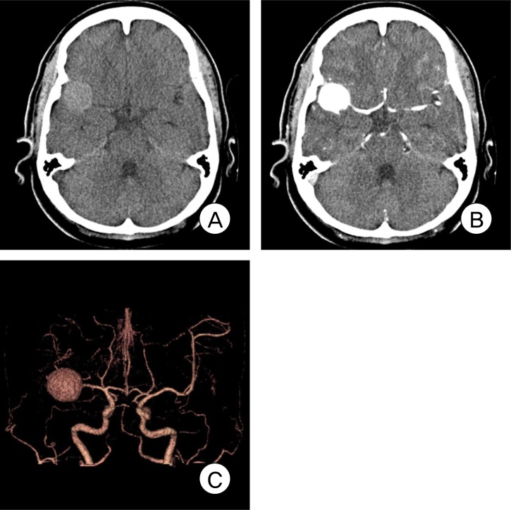
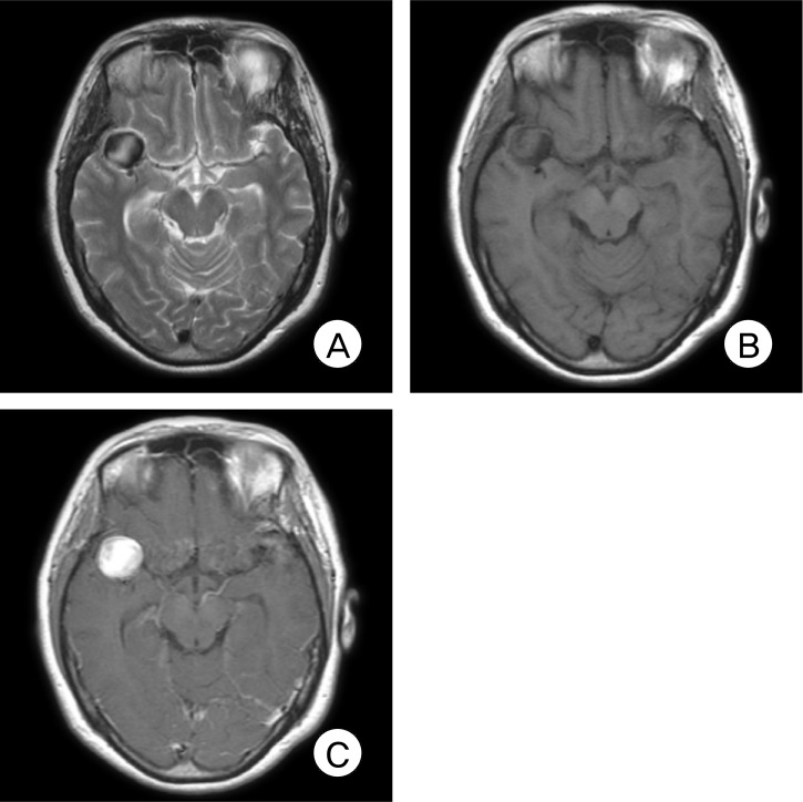
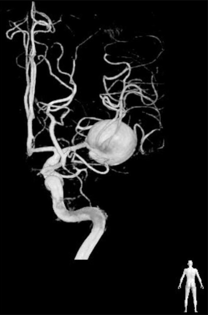
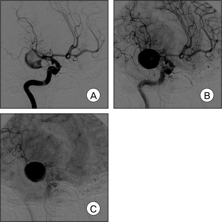
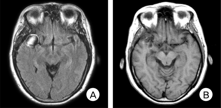
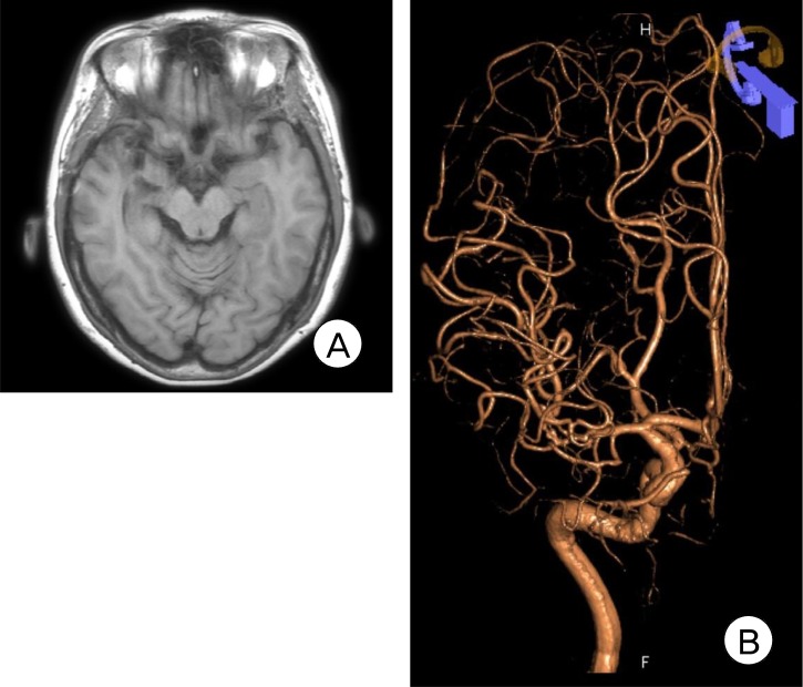
A 46-year-old female patient visited the hospital, complaining of a headache. No abnormal findings were observed on the first neurologic examination, while other medical examinations also showed normal findings. Findings on brain computed tomography (CT) examination showed a slightly high-density intra axial mass lesion measuring approximately 2.5 cm lateral sides on the right sylvian fissure. It showed no edema around the mass, iso-density and strong enhancement without calcification inside (Fig. 1A, B). According to findings on a three-dimensional CT angiogram (3-D CTA) examination, the mass was diagnosed as a saccular aneurysm in the right MCA bifurcation (Fig. 1C). T2 weighted images from a brain magnetic resonance image (MRI) examination showed a high signal in the aneurysm and a heterogeneous peripheral low signal. At the back of the aneurysm sac, M2 branches were advanced and deviated toward the medial side. A T1 weighted image showed a low signal in a part close to the Circle of Willis and an iso signal far from it. Like the CT image, it showed a slightly low signal with dense contrast enhancement and difference in filling in the aneurysm sac (Fig. 2A, B, C). In addition, SAH was not found on the CT examination; however, a small amount of SAH was noted around the aneurysm and along the sylvian fissure. In the right internal carotid artery (ICA) angiogram (three-dimension subtraction image) posterior to anterior view, an aneurysm measuring 21.6 × 20.9 mm in the right MCA bifurcation was observed in a lateral direction. It formed a broad neck along an inferior branch of M2 and several perforating branches were observed around the neck (Fig. 3). It is specific in the angiogram that the filling of the aneurysm in the early arterial phage was not partially performed. Filling of the aneurysm in the later arterial phage was completed, without excretion up to the capillary phage. This is referred to as a slow filling and delayed excretion (Fig. 4A, B, C). The patient underwent surgery after approximately ten days. Direct clipping in the operation field was difficult; therefore, the operation was completed after wrapping the ruptured portion using surgical glue. The patient was discharged from the hospital without complications. According to findings on the MRI examination performed approximately months after discharge, a target sign that appeared as a high signal, and a low signal in a slightly inner portion and a high signal again in a central portion, was seen around the aneurysm on the T1 image (Fig. 5A). An examination after 19 months showed that the size of the aneurysm had decreased considerably (Fig. 5B). Finally, according to findings on the angiogram and MRI examination (which were performed after three years and eight months), there was no trace of the mass in the sylvian fissure due to the considerable reduction in the size of the aneurysm and almost everything in the M2 branch was intact (Fig. 6A, B).
Fig. 1.
(A) Axial computed tomography (CT) scan shows a slightly high-density intra axial mass lesion measuring approximately 2.5 cm, along the peripheral side of the right sylvian fissure. No edema was observed around the mass. There was no calcification either. (B) After contrast enhancement, the mass lesion showed significant enhancement. (C) Three-dimensional CT angiography reconstruction showing a saccular aneurysm in the right middle cerebral artery (MCA) bifurcation.
Fig. 2.
(A) T2 weighted images from a brain magnetic resonance image (MRI) examination show a high signal in the aneurysm sac and a heterogeneous, peripheral low signal. (B) T1 weighted image shows a low signal in a portion close to the Circle of Willis and an iso signal far from it. (C) After contrast enhancement, strong improvement with a filling difference in the aneurysm sac is observed.
Fig. 3.
Right internal carotid artery angiogram (three-dimensional subtraction image) posterior to anterior view. An aneurysm measuring 21.6 × 20.9 mm in the right MCA bifurcation is observed in a lateral direction, and an aneurysm of the right MCA bifurcation formed a broad neck along an inferior branch of M2. Several perforating branches are observed around the neck.
Fig. 4.
Right internal carotid artery angiogram. Slow filling and delayed excretion pattern are noted (A, B and C).
Fig. 5.
(A) MRI obtained eight months later. A target sign that appeared as a high signal, a low signal in the slightly inner portion and a high signal again in the central portion, is seen on the T1 image around the aneurysm. (B) After 19 months, the size of the aneurysm shows a considerable decrease.
Fig. 6.
(A) MRI obtained three years and eight months later, showing considerable reduction in the size of the aneurysm and no trace of the mass in the sylvian fissure. (B) Three-dimensional angiography reveals an occlusion of the aneurysm and intact M2 branches.
DISCUSSION
Occlusion of an aneurysm due to spontaneous thrombosis was first reported in 1955.2) The risk of a ruptured aneurysm became known in the mid 1970s, however, due to high mortality and morbidity associated with the operation, administration of an active treatment for ruptured aneurysm was difficult. There have been some reports on spontaneous occlusion of the aneurysm. They have been widely spread by Yasargil since the mid-1970s, which led to an increase in the success rate of operation and a decrease in complications from anesthesia. It also led to a rapid decrease in mortality and morbidity from the operation.21)
However, according to the development of diagnostic technologies, including CT, MRI, and other methods, the number of cases of diagnosed unruptured aneurysm has shown a continuous increase, and treatment of unruptured aneurysm has recently become a controversial issue. In addition, questions arose regarding the bleeding rate in the natural history of an unruptured aneurysm, or whether to treat the aneurysm or monitor its progress by comparing the bleeding rate, and mortality and morbidity of the operation. Likewise, a randomized controlled study is the most ideal approach to understanding the natural history of a ruptured aneurysm. However, limitations posed by medical ethics make such an approach impossible.
Many papers have reported a high bleeding rate in the natural history of an unruptured giant aneurysm, while several case reports on spontaneously cured aneurysms have been published. Loevnet et al.12) reported a large basilar artery aneurysm found nine months after a seven-year-old boy received a trauma, which showed complete disappearance after six years. Morón et al.14) reported on a traumatic posterior cerebral artery aneurysm of a six-year-old boy, which disappeared completely after five years. Other cases of spontaneous occlusion of a traumatic aneurysm have been reported, insisting that less than 15% of traumatic aneurysms were cured spontaneously.15) Krapt et al.11) suggested that a congenital, unruptured giant aneurysm in a nine-month-old female infant was completely occluded and absorbed, based on three-year examination. There have also been papers on infants who could not undergo an operation or spontaneous occlusion of a traumatic aneurysm, however, few reports on the natural history of an un-ruptured aneurysm have been published. Vasconcellos et al.3) proposed that 25% (five cases) of 20 cases showed courses of spontaneous occlusion during observation of giant carotid cavernous aneurysms of the internal carotid artery with a relatively low risk of bleeding. The possibility of spontaneous occlusion was relatively high in cases of improvement of initial symptoms, such as retrobulbar pain or migraine during courses of carotid cavernous giant aneurysms.
A factor to analogize spontaneous occlusion of an unruptured aneurysm is the volume-to-orifice ratio.20)29) Through analysis of the results for the ratio of the size (mm2) of the aneurysm neck to the aneurysm volume (mm3) from studies on 21 aneurysms, a value greater than 25 mm indicates the potential for thrombosis, and a value greater than 28 mm indicates a strong possibility of thrombosis.
Thus, it is assumed that as the volume-to-orifice ratio grows larger, thrombosis appears more frequently by inducement of red blood cell aggregation thrombosis due to blood stasis in the aneurysm. Accordingly, the pathophysiology of thrombosis is not clear, however, it has received the most support. The volume-to-orifice ratio calculated in our case was 28 mm, and the results of angiography showed partial dye filling in the aneurysm in the early arterial phage, maximum filling in the late arterial phage, and slow filling delayed excretion in the capillary phage. Accordingly, it appears to be attributed to blood stasis in the aneurysm. The other study insisted on the possibility that thrombosis could cause intermittent vasospasm or dehydration caused by non-ionic contrast media while reporting cases of aneurysm that has caused thrombosis after angiography. However, it was impossible to demonstrate the correct mechanism.7)
Partial thrombosis of a giant aneurysm from 48 to 76% has been reported, however, sources demonstrating complete thrombosis are scarce.6) Through observation reports on 22 cases of giant intracranial aneurysms, Whittle and et al.19) overthrew the conventional assumption that partial thrombosis in an aneurysm can reduce hemorrhage risk, by demonstrating that there was no difference in incidence of SAH in 12 cases of thrombosis and non-thrombosis. They also insisted that mortality generated after SAH, and mortality and morbidity before and after surgery did not differ. As a result, they reported that partial thrombosis of the aneurysm did not reduce the risk of SAH and only aneurysms with total thrombosis reducing the risk of SAH. They also argued that partial thrombosis did not influence procedures or methods for treatment of giant aneurysms.
Due to the dynamic and reversible courses of thrombosis of aneurysms, observation of a course over a long period of time is necessary. In addition, its generation and resorption are performed repeatedly, and complications such as thromboembolic infarct can occur.13)
Whittle et al. insisted that wrapping of a ruptured aneurysm had an effect on small aneurysms, but that wide-based, large, or giant types of MCA aneurysms are associated with a high risk of rebleeding and fatal outcomes. Wrapping was generally known as an inadequate method of treating an aneurysm.5),16) Accordingly, it is impossible for wrapping to stop rebleeding of the aneurysm.5),17) Deshmukh et al.4) reported that according to angiographic follow-up studies on 34 patients who underwent wrapping of an aneurysm for an average of 3.4 years, there was no change in the size and shape of the aneurysm. Despite reports on rupture caused by wrapping or a change in size, there was no basis for occlusion of the aneurysm after wrapping, as manifested in our case.
CONCLUSION
The Authors report on a case of spontaneous occlusion and absorption of a saccular aneurysm of MCA bifurcation. It was assumed that in the pathophysiology of spontaneous occlusion of an aneurysm, sluggish flow in the aneurysm may be the cause and the volume-to-orifice ratio of the angiography, slow filling, and delayed excretion through prediction of slow flow may manifest. In cases of inoperable aneurysm, the volume-to-orifice ratio was greater than 28 mm. Patients who manifested slow filling and delayed excretion had no other symptoms or complications. They propose observation as one method of treatment.
However, long-term observation is more critical for an aneurysm that does not involve a stationary lesion and one that has dynamic courses. Conduct of further studies on the characteristics and types of aneurysms that will undergo spontaneous occlusion is needed.
References
- 1.International Study of Unruptured Intracranial Aneurysms Investigators. Unruptured intracranial aneurysms-risk of rupture and risks of surgical intervention. N Engl J Med. 1998 Dec;339(24):1725–1733. doi: 10.1056/NEJM199812103392401. [DOI] [PubMed] [Google Scholar]
- 2.De Lange SA. [A case of spontaneous thrombosis of a cerebral arteriovenous aneurysm] Ned Tijdschr Geneeskd. 1955 Mar;99(13):944–946. Dutch. [PubMed] [Google Scholar]
- 3.Vasconcellos LP, Flores JAC, Conti MLM, Veiga JCE, Lancellotti CLP. Spontaneous thrombosis of internal carotid artery: a natural history of giant carotid cavernous aneurysms. Arq Neuropsiquiatr. 2009 Jun;67(2A):278–283. doi: 10.1590/s0004-282x2009000200020. [DOI] [PubMed] [Google Scholar]
- 4.Deshmukh VR, Kakarla UK, Figueiredo EG, Zabramski JM, Spetzler RF. Long-term clinical and angiographic follow-up of unclippable wrapped intracranial aneurysms. Neurosurgery. 2006 Mar;58(3):434–442. doi: 10.1227/01.NEU.0000199158.02619.99. discussion 434-42. [DOI] [PubMed] [Google Scholar]
- 5.Fujiwara S, Fujii K, Nishio S, Fukui M. Long-term results of wrapping of intracranial ruptured aneurysms. Acta Neurochir (Wien) 1990;103(1-2):27–29. doi: 10.1007/BF01420188. [DOI] [PubMed] [Google Scholar]
- 6.Gerber S, Dormont D, Sahel M, Grob R, Foncin JF, Marsault C. Complete spontaneous thrombosis of a giant intracranial aneurysm. Neuroradiology. 1994 May;36(4):316–317. doi: 10.1007/BF00593270. [DOI] [PubMed] [Google Scholar]
- 7.Hay KL, Bull BS. Factors influencing the activation of platelets by nonionic contrast medium. J Vasc Interv Radiol. 1996 May-Jun;7(3):401–407. doi: 10.1016/s1051-0443(96)72879-8. [DOI] [PubMed] [Google Scholar]
- 8.Heiskanen O. Risks of surgery for unruptured intracranial aneurysms. J Neurosurg. 1986 Oct;65(4):451–453. doi: 10.3171/jns.1986.65.4.0451. [DOI] [PubMed] [Google Scholar]
- 9.Inagawa T. Surgical treatment of multiple intracranial aneurysms. Acta Neurochir (Wien) 1991;108(1-2):22–29. doi: 10.1007/BF01407662. [DOI] [PubMed] [Google Scholar]
- 10.King JT, Jr, Berlin JA, Flamm ES. Morbidity and mortality from elective surgery for asymptomatic, unruptured, intracranial aneurysms: a meta-analysis. J Neurosurg. 1994 Dec;81(6):837–842. doi: 10.3171/jns.1994.81.6.0837. [DOI] [PubMed] [Google Scholar]
- 11.Krapf H, Schoning M, Petersen D, Kuker W. Complete asymptomatic thrombosis and resorption of a congenital giant intracranial aneurysm. J Neurosurg. 2002 Jul;97(1):184–189. doi: 10.3171/jns.2002.97.1.0184. [DOI] [PubMed] [Google Scholar]
- 12.Loevner LA, Ting TY, Hurst RW, Goldberg HI, Schut L. Spontaneous thrombosis of a basilar artery traumatic aneurysm in a child. AJNR Am J Neuroradiol. 1998 Feb;19(2):386–388. [PMC free article] [PubMed] [Google Scholar]
- 13.Maruyama M, Asai T, Kuriyama Y, Sawada T, Ogata J, Nishimura T, et al. Positive platelet scintigram of a vertebral aneurysm presenting thromboembolic transient ischemic attacks. Stroke. 1989 May;20(5):687–690. doi: 10.1161/01.str.20.5.687. [DOI] [PubMed] [Google Scholar]
- 14.Moron F, Benndorf G, Akpek S, Dempsy R, Strother CM. Spontaneous thrombosis of a traumatic posterior cerebral artery aneurysm in a child. AJNR Am J Neuroradiol. 2005 Jan;26(1):58–60. [PMC free article] [PubMed] [Google Scholar]
- 15.Nakstad P. Spontaneous occlusion of traumatic pericallosal aneurysm and pericallosal artery. Neuroradiology. 1987;29(3):312. doi: 10.1007/BF00451778. [DOI] [PubMed] [Google Scholar]
- 16.Sato K, Fujiwara S, Kameyama M, Ogawa A, Yoshimoto T, Suzuki J. Follow-up study on ruptured aneurysms treated by wrapping. Neurol Med Chir (Tokyo) 1990;30(10):734–737. doi: 10.2176/nmc.30.734. [DOI] [PubMed] [Google Scholar]
- 17.Todd NV, Tocher JL, Jones PA, Miller JD. Outcome following aneurysm wrapping: a 10-year follow-up review of clipped and wrapped aneurysms. J Neurosurg. 1989 Jun;70(6):841–846. doi: 10.3171/jns.1989.70.6.0841. [DOI] [PubMed] [Google Scholar]
- 18.Wardlaw JM, White PM. The detection and management of unruptured intracranial aneurysms. Brain. 2000 Feb;123(Pt 2):205–221. doi: 10.1093/brain/123.2.205. [DOI] [PubMed] [Google Scholar]
- 19.Whittle IR, Dorsch NW, Besser M. Spontaneous thrombosis in giant intracranial aneurysms. J Neurol Neurosurg Psychiatry. 1982 Nov;45(11):1040–1047. doi: 10.1136/jnnp.45.11.1040. [DOI] [PMC free article] [PubMed] [Google Scholar]
- 20.Wiebers DO, Whisnant JP, Huston J, 3rd, Meissner I, Brown RD, Jr, Piepgras DG, et al. Unruptured intracranial aneurysms: natural history, clinical outcome, and risks of surgical and endovascular treatment. Lancet. 2003 Jul;362(9378):103–110. doi: 10.1016/s0140-6736(03)13860-3. [DOI] [PubMed] [Google Scholar]
- 21.Yasargil MG, Vise WM, Bader DC. Technical adjuncts in neurosurgery. Surg Neurol. 1977;8(5):331–336. [PubMed] [Google Scholar]


