Abstract
Many recombinant therapeutic proteins are purified from Escherichia coli. While expression in E. coli is easily achieved, some disadvantages such as protein aggregation, formation of inclusion bodies, and contamination of purified proteins with the lipopolysaccharides arise. Lipopolysaccharides have to be removed to prevent inflammatory responses in patients. Use of the Gram-positive Bacillus anthracis as an expression host offers a solution to circumvent these problems. Using the multiple protease-deficient strain BH460, we expressed a fusion of the N-terminal 254 amino acids of anthrax lethal factor (LFn), the N-terminal 389 amino acids of diphtheria toxin (DT389) and human transforming growth factor alpha (TGFα). The resulting fusion protein was constitutively expressed and successfully secreted by B. anthracis into the culture supernatant. Purification was achieved by anion exchange chromatography and proteolytic cleavage removed LFn from the desired fusion protein (DT389 fused to TGFα). The fusion protein showed the intended specific cytotoxicity to epidermal growth factor receptor-expressing human head and neck cancer cells. Final analyses showed low levels of lipopolysaccharides, originating most likely from contamination during the purification process. Thus, the fusion to LFn for protein secretion and expression in B. anthracis BH460 provides an elegant tool to obtain high levels of lipopolysaccharide-free recombinant protein.
Keywords: tumor therapy, immunotoxin, tobacco etch virus protease, diphtheria toxin, lipopolysaccharide, endotoxin1
Introduction
Bacillus anthracis is a Gram-positive bacterial pathogen that is well known for its secreted toxin, anthrax toxin. The toxin is a well-studied, three-protein exotoxin consisting of protective antigen (PA), edema factor (EF), and lethal factor (LF). PA is a receptor-binding component which acts to deliver the enzymatic moieties LF and EF to the cytosol of eukaryotic cells. The three toxin proteins are encoded on the plasmid pXO1 and all three are secreted with high efficiency, enabling their isolation in high purity from culture supernatants [1]. PA binds to either of its receptors (capillary morphogenesis gene 2 or tumor endothelial marker 8) on the cell surface and is internalized after proteolytic activation and oligomerization. Up to four EF and LF molecules bind to the PA oligomer and are co-internalized into endosomal compartments. Acidification in the endosomes results in membrane integration of the PA oligomer and pore formation. EF and LF are pulled through the PA pore and act on their cytosolic substrates.
The N-terminal 254 amino acids of LF (LFn) are sufficient for effective binding to the PA oligomer and delivery into the cytosol. Successful transport of other proteins fused to LFn was demonstrated for Pseudomonas exotoxin A catalytic domain, diphtheria toxin A chain, shiga toxin catalytic domain [2], as well as the reporter beta-lactamase [3]. Fusions of LFn to potent protein toxins, e.g. Pseudomonas exotoxin A catalytic domain, led to design of efficient anti-tumor drugs that are being studied in preclinical analyses [4]. Tumor-targeting of these modified LF proteins was achieved by mutation of the protease-cleavage site of PA to require activation by tumor-enriched cell-surface proteases, e.g. matrix metalloproteases or urokinase plasminogen activator [5].
The LFn fusion to potent enzymes in combination with tumor-selective PA is one example of a targeted protein toxin for anti-tumor therapy. Several similar compounds have been described containing highly potent bacterial and plant toxins that act catalytically in the cytosol of targeted cells to kill the cells (reviewed in [6]). Most commonly, specificity has been sought by linking these toxins chemically or genetically to antibodies that bind to cell surface materials enriched on tumor cells. These proteins, initially termed “immunotoxins” and now more generally described as “targeted toxins” (TT), have been under development for decades [7]. The only FDA-approved TT to date is denileukin diftitox, containing diphtheria toxin lacking its receptor-binding domain and genetically fused to human interleukin-2.
Production of TTs is problematic and expensive. Most TTs are produced in E. coli, since they are toxic for eukaryotic expression systems. This results in heavy contamination of proteins with lipopolysaccharides that have to be removed before use of the proteins in animal experiments or patients. Furthermore, expression in E. coli commonly results in inclusion body formation. Isolation of denatured proteins from inclusion bodies requires unfolding and refolding of the proteins during the purification process, which presents another source of protein losses. Slower protein expression reduces inclusion body formation, however, purification is more problematic since many cellular proteins are associated with the purified protein and further purification steps may be necessary.
The objective of this study was the expression of a TT in the protease-deficient B. anthracis strain BH460 [1]. In order to achieve secretion from the bacteria, LFn was fused to a TT consisting of the N-terminal 389 amino acids of diphtheria toxin (DT389) and human transforming growth factor alpha (TGFα). The fusion protein was purified from the supernatant of the growth medium, LFn was removed from the desired fusion protein (DT389 fused to TGFα) by proteolytic cleavage. The fusion protein was further purified and analyzed.
Materials and Methods
Expression and Purification of LFn-DT-TGF in B. anthracis BH460
The plasmid encoding LFn-TEV-DT389-TGFα (LFn-DT-TGF) was isolated from Dam and Dcm methylation-deficient E. coli strain SCS110 and transformed into B. anthracis strain BH460 [1] by electroporation. BH460 is a sporulation-deficient, virulence plasmid-cured strain with six proteases deleted. Transformant colonies were grown overnight on LB-agar plates supplemented with 10 μg/ml kanamycin. Several colonies were resuspended in 7 L FA growth and protein expression medium [8] with 10 μg/ml kanamycin and grown at 37 °C for 12 h. For the analysis of different growth conditions, cells were incubated in 5 mL FA medium for 4, 6, 8, and 10 h at either 30 or 37 °C. Samples growing at 37 °C were additionally supplemented with the protease inhibitor 4-(2-Aminoethyl) benzenesulfonyl fluoride hydrochloride (AEBSF, US Biological, MA; final concentration 167 μM). LFn-DT-TGF was secreted in the supernatant and purified by hydrophobic interaction chromatography and subsequently by anion exchange chromatography as described earlier [1]. Details on the purification process are described in the supplemental data. Removal of LFn from the fusion protein LFn-DT-TGF in order to generate DT389-TGFα (DT-TGF) was achieved by tobacco etch virus (TEV) protease cleavage. TEV protease was purified as described [9].
Following TEV protease digestion, the entire sample was diluted 1:2 with water to reduce the phosphate concentration and loaded on a ceramic hydroxyapatite column and purified as described [1]. An additional anion exchange chromatography step was introduced by loading the samples on a MonoQ column for purification with an Äkta chromatography system. The DT-TGF-containing fractions were analyzed by SDS-PAGE, dialyzed as described above, concentrated (Amicon Ultrafiltration devices, 30 kDa molecular weight cutoff, Millipore, Billerica, MA), filter sterilized, and stored in aliquots at −80 °C. DT-TGF was analyzed by electrospray ionization mass spectrometry (HP/Agilent 1100 MSD, Hewlett Packard, Palo Alto, CA) at the NIDDK core facility, Bethesda, MD) to confirm that the mass matched the mass calculated from its sequence.
Cell culture and Cytotoxicity of DT-TGF
Cytotoxicity experiments were performed on HN6 cells (human head and neck cancer cell line) [10]. Dose-response curves for DT-TGF were obtained by incubation on 10,000 HN6 cells / well in 96-well plates (0.2–64.3 nM) for 6 h and a further 42 h after washing the cells twice with medium. Cell survival was determined by using the oxidative indicator 3-(4,5-Dimethylthiazol-2-yl)-2,5-diphenyltetrazolium bromide (MTT, Sigma Aldrich, St. Louis, MO). Details on cell culture and cytotoxicity assay setup are described in detail in the supplemental data. The relative cell survival, designated as survival index (SI), was calculated after blank subtraction (wells without cells) as the percentage of living cells in treated wells in relation to untreated cells (cells without toxin). The 50 % survival index (SI50, 50 % cell survival in comparison to untreated controls) was determined by a nonlinear regression curve fit (least square fit with “log(inhibitor) versus normalized response”) using GraphPad Prism 5.02.
Tumor necrosis factor-α release assay for lipopolysaccharide content
Tumor necrosis factor-α (TNF-α) release was studied on RAW264.7 cells (mouse leukemic monocyte macrophages). Cells were maintained as described above for HN6 cells. Cells were seeded in 6-well plates (1 × 10⁶ cells in 2 mL medium) and incubated at 37 °C overnight before addition of DT-TGF (10 μg/mL), edema factor purified from E. coli (10 μg/mL), or lipopolysaccharide (LPS, 10 pg/mL) in 2 mL fresh medium. After 18 h incubation at 37 °C, the supernatant was removed, cleared by centrifugation and subjected to TNF-α detection by the Mouse TNF-α ELISA Kit (Invitrogen, Grand Island, NY) according to the manufacturer’s instructions. The statistical significance of different TNF-α concentrations was determined by a non-paired t-test using GraphPad Prism 5.02 and a one-tailed significance of p ≤ 0.05 was interpreted as being statistically significant.
Results
Expression of LFn-DT-TGF
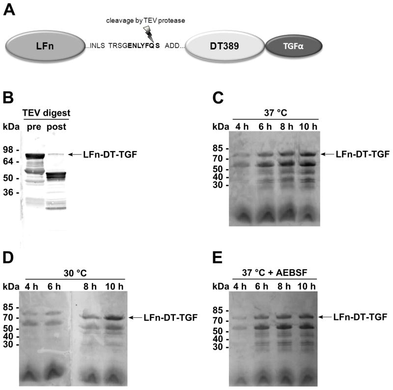
The fusion of LFn to DT-TGF through a TEV protease cleavage site linking LFn and DT389 was designed to allow purification of DT-TGF from the supernatant of B. anthracis cultures (Figure 1A). Cleavage of LFn-DT-TGF (79.8 kDa) by TEV protease for release of DT-TGF (49.2 kDa) was verified after overnight incubation of an LFn-DT-TGF-containing culture supernatant (Figure 1B). DT-TGF was detected by Western blotting using an anti-diphtheria toxin antibody.
Figure 1. Schematic structure of LFn-DT-TGF and expression and cleavage analyses.

(A) The N-terminal 254 amino acids of LF are fused via a linker containing a TEV protease cleavage site to the 389 N-terminal amino acids of diphtheria toxin and TGFα. (B) TEV protease cleavage of LFn-DT-TGF in the supernatant of B. anthracis BH460 (10-fold concentrated). Immunodetection by anti-diphtheria toxin. (C-E) Expression of LFn-DT-TGF in B. anthracis strain BH460 at 30 or 37 °C and in the presence of 167 μM protease inhibitor AEBSF. Supernatants were collected after 4–10 h, 10-fold concentrated and detected by SDS-PAGE and subsequent Coomassie staining.
Different expression parameters were tested, e.g. 37 °C, 30 °C, and 37 °C in the presence of the protease inhibitor AEBSF. As expected, expression at 37 °C results in the highest amount of LFn-DT-TGF (Figure 1C). However, the protein was either already cleaved during the expression or secreted as incompletely synthesized protein, as shown by a further protein band of approximately 55 kDa. The expression at 30 °C (Figure 1D) and the supplementation of the growth medium with AEBSF did not increase the amount of full length LFn-DT-TGF (Figure 1E).
Purification of LFn-DT-TGF
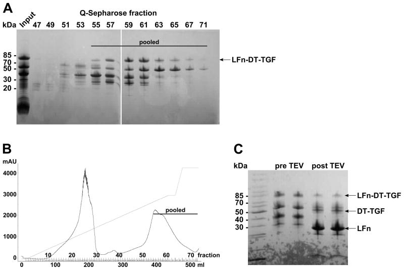
LFn-DT-TGF secreted in the supernatant of the BH460 culture was concentrated by hydrophobic interaction chromatography, applied to a Q-Sepharose anion exchange column. Gradient elution of the column resulted in enrichment of LFn-DT-TGF (Figure 2A). Pooling of the fractions containing LFn-DT-TGF (fractions 55–72) removed impurities from the BH460 expression culture from the sample (Figure 2B). The yield of partially purified LFn-DT-TGF after anion exchange chromatography was determined as 33.7 mg/L growth medium with a total quantity of 236 mg. Purified proteins were dialyzed and concentrated before the TEV protease digestion was performed on LFn-DT-TGF. The proteolytic treatment successfully removed LFn from DT-TGF (Figure 2C). Prematurely cleaved or incompletely synthesized fusion proteins (protein bands at 55, 40 and 30 kDa) were cleaved as well, since all these bands were no longer detected after TEV protease digestion. It can thus be assumed that all of these fusion proteins contained full length LFn but incomplete DT-TGF. This assumption is corroborated by the high intensity band for LFn, of 29.5 kDa (identified by mass spectrometry, data not shown). The protein band for DT-TGF with a molecular mass of 49.2 kDa was only observed following the cleavage.
Figure 2. Purification of LFn-DT-TGF.

(A) Q-Sepharose anion exchange chromatography elution fractions analyzed by SDS-PAGE and Coomassie staining. (B) Elution profile of LFn-DT-TGF purification. (C) TEV protease cleavage (overnight at 4 °C) of pooled LFn-DT-TGF fractions analyzed by SDS-PAGE and Coomassie staining.
Purification of DT-TGF
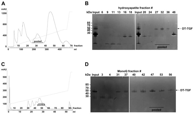
The TEV protease-digested sample was applied to ceramic hydroxyapatite chromatography and DT-TGF was successfully separated from LFn and other impurities (Figure 3A). This purification step proved to be highly efficient for the purification of the protein, as demonstrated by the elution profile of the hydroxyapatite chromatography step. The analysis of the fractions containing DT-TGF (pooled fractions 26–37) by SDS-PAGE showed a second band below DT-TGF with a slightly lower molecular mass (Figure 3B). In order to remove this potential degradation product, high resolution anion chromatography (MonoQ column) was applied as the final purification step (Figure 3C) and achieved the separation of full length DT-TGF (Figure 3D). A considerable amount of the full-size DT-TGF was lost due to contamination by the smaller fragment in fractions preceding pooled fractions 45–53. Following the purification step the protein concentration of DT-TGF was determined to be 0.9 mg/mL and the total amount of purified DT-TGF was 5.4 mg. Based on the volume of the growth medium (7 L) the yield of the purification was calculated to be 0.8 mg/L after all purification steps.
Figure 3. Purification of DT-TGF after TEV cleavage.

(A) Elution profile of DT-TGF hydroxyapatite chromatography purification. (B) Elution fractions were analyzed by SDS-PAGE and Coomassie staining. Pooled fractions were dialyzed and submitted to a further purification step. (C) Elution profile of DT-TGF purification by MonoQ anion exchange chromatography. (D) Elution fractions were analyzed by SDS-PAGE and Coomassie staining. Pooled fractions were dialyzed and stored at −20 °C.
Identification of DT-TGF
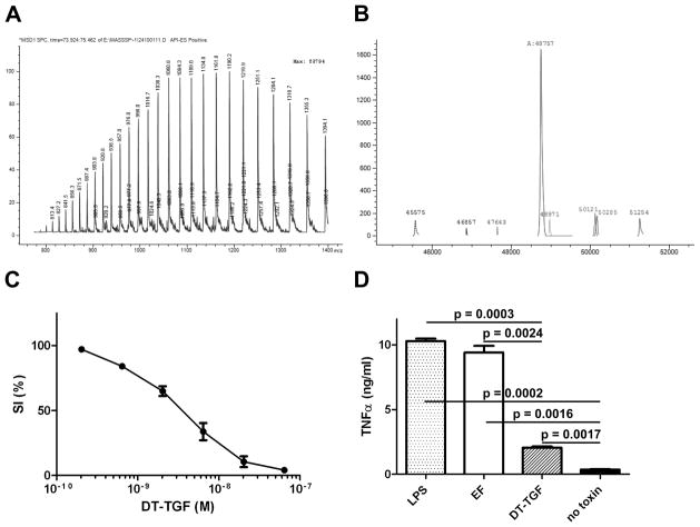
The mass spectra obtained after analysis of DT-TGF corresponded with the expectations and the low background signal demonstrates the purity of the sample (Figure 4A). The mass identification for the protein resulted in a main fragment with 48,757 Da (Figure 4B). This result differs by only 3.4 Da from the calculated mass of 48,460.4 Da, a difference that is within the usual variance we have observed for many other proteins examined by this method.
Figure 4. Analysis of purified DT-TGF.

(A and B) Electron spray ionization mass spectrometry analysis of DT-TGF. (A) Mass spectrum for DT-TGF (m/z ratio on x axis and relative abundance on x axis). (B) Deconvolution of the data in A to obtain intact mass of DT-TGF. (C) Cytotoxicity of DT-TGF on HN6 human head and neck cancer cells. HN6 cells (10,000/well) were exposed to different concentrations of DT-TGF for 48 h and viable cells were quantitated in an MTT assay. Relative survival was calculated as the percentage of living cells after treatment in relation to untreated cells. Error bars indicate S.E.M. of two independent experiments performed in triplicate. (D) TNF-α release assay. 1 × 10⁶ RAW264.7 cells were grown overnight and incubated with 10 μg/mL DT-TGF or anthrax toxin edema factor (EF, purified from E. coli) or 10 pg/mL LPS for 18 h at 37 °C. TNF-α in the supernatants was quantitated by TNF-α ELISA.
Functional Analysis of DT-TGF
Cytotoxicity analyses of DT-TGF employed the human head and neck cancer cell line HN6, which express epidermal growth factor receptor, the target receptor of TGFα. TGFα within DT-TGF acts as targeting moiety to direct the fusion protein to the receptor and initiate internalization. A 48-h toxin exposure resulted in dose-dependent cytotoxicity with a SI50 value of 3.40 ± 0.04 nM (Figure 4C). This result indicates that the purified DT-TGF is fully functional and exerts a very high cytotoxic effect on this model tumor cell line. The expression of DT-TGF in B. anthracis avoids contamination of the protein with LPS. Purified DT-TGF was analyzed in a TNF-α release assay to detect any inflammatory responses of macrophages to the presence of LPS. DT-TGF, edema factor purified from E. coli, and a positive control containing 10 pg/mL LPS were incubated on mouse macrophages and the supernatant of the cell culture was analyzed for TNF-α release (Figure 4D). DT-TGF, EF, and LPS did not induce cell killing in RAW cells in an experiment performed with the identical concentrations and incubation time as used for the TNF-α release assay (data not shown). RAW cells do not express epidermal growth factor receptor (the target receptor for DT-TGF) and EF is not delivered into the cells unless anthrax protective antigen is present. The analyses showed very high TNF-α concentrations in both the edema factor sample and a LPS positive control with TNF-α concentrations of 10.3 ± 0.2 ng/mL and 9.4 ± 0.5 ng/mL, respectively. The TNF-α concentration of the sample treated with DT-TGF was significantly lower than any of these samples (2.1 ± 0.2 ng/mL TNF-α, p ≤ 0.0016). However, even though DT-TGF resulted in a 4.5-fold lower TNF-α release from the macrophages, a TNF-α release was induced, since the untreated cells released only 0.4 ± 0.1 ng/mL TNF-α.
Discussion
The fusion of the targeted fusion toxin DT-TGF to LFn resulted in the successful expression of LFn-DT-TGF and its secretion to the growth medium of the protease-deficient B. anthracis strain BH460. The fusion protein LFn-DT-TGF was under the control of the PA promoter to achieve high protein expression [11]. This system proved to be a very efficient recombinant expression system for the three proteins of anthrax toxins. An advantage of the described expression system is the constitutive expression of the recombinant protein. No induction of protein expression is necessary, thus avoiding the need to monitor bacterial growth in the expression phase. However, the DT-TGF-coding DNA sequence was not optimized for expression in B. anthracis and a host-specific optimization towards a higher A/ T content could result in a further significant increase of DT-TGF yield. Codon optimization is expected to yield a drastic increase and Gao et al. observed a more than 4-fold increase in the yield of glucose oxidase expressed in Pichia pastoris upon codon optimization [12].
The expression in B. anthracis is furthermore very advantageous since B. anthracis has a very efficient secretion system for its toxins. By fusing DT-TGF to LFn and the PA signal sequence the resulting fusion protein was secreted efficiently and high yields of the protein were observed in the supernatant. Secretion of the recombinant protein provides a dramatic advantage for the purification of the protein. Contamination by other proteins was very low and allowed for high purity of the recombinant protein already after the first purification step. The presence of several proteins of a smaller size is attributed to degradation of the protein during the expression and purification (Figure 2C).
However, this expression system depends on use of the non-virulent plasmid-cured strain BH460, lacking expression of six extracellular proteases [1]. Otherwise the strong proteolytic activity in the supernatant results in rapid degradation of secreted proteins. Similar approaches were developed in recent years to provide extracellular protease-deficient strains of Bacillus subtilis [13], and in E. coli for a cell free translation system [14]. The Gram-positive protein expression systems became more prominent in recent years due to the high yield of expressed protein in genetically improved strains and the avoidance of LPS contaminations [15]. However, remaining proteases secreted to the supernatant of growing BH460 may still be limiting the yields of intact LFn-DT-TGF. Our studies revealed the presence of a 55-kDa fragment of LFn-DT-TGF in all growth conditions tested. This fragment was detected less efficiently by the polyclonal antibody directed against diphtheria toxin, indicating that it probably contains only a portion of the DT389 sequence (Figure 1B). While expression at 30 °C gave a higher ratio of full length to fragmented LFn-DT-TGF, the absolute amount of LFn-DT-TGF was higher upon expression at 37 °C. It was observed that proteolytic activity during the purification process reduced the amount of purified protein. While the supernatant of the growth culture showed mainly two protein bands of 80 and 55 kDa, further bands of 40 and 30 kDa were detected before the TEV protease digest, while the intensity of the band resembling full length LFn-DT-TGF with 80 kDa is weaker. Characterization of the types of trace proteases remaining may allow their inhibition in future work to improve the process.
The TEV protease digestion proved to be very efficient, consistent with its wide use for removal of tags added to facilitate affinity purification [16]. The TEV protease cleavage site was chosen according to a study by Kapust et al. [17]. The overnight digest left only a small fraction of the full length protein uncleaved (Figure 1B and 2C). The major band on the Coomassie-stained gel after the TEV protease digest was the 29-kDa fragment of LFn. All of the four major protein bands identified before the TEV protease digest were cleaved by the TEV protease and produced LFn and different DT-TGF fragments. The two-step purification of DT-TGF following the TEV protease was necessary only because of the presence of DT-TGF fragments having chromatographic properties very similar to those of the full size protein. Other contaminating proteins are not detectable at this stage of the purification. Since the instability of DT-TGF during expression and purification decreases the final yield, a careful analysis of the generated protein fragments either by mass spectrometry or N-terminal sequencing would help to identify cleavage sites that could be mutated in order to generate a more stable second generation fusion protein.
One of the main advantages of protein expression in B. anthracis is the lack of LPS contaminations with the purified protein. This advantage of Gram-positive bacteria has being identified before [18]. The analysis of LPS contaminations of the purified protein DT-TGF revealed significantly lower levels of LPS in comparison to a protein isolated from Gram-negative E. coli (Figure 4B). The LPS-induced TNF-α release in the DT-TGF sample most likely originated from buffers, column resins and other materials of the purification procedure with considerable Gram-negative bacterial contaminations. In order to further reduce the LPS content of the purified DT-TGF, the purification needs to be performed with materials reserved exclusively for the use with samples originating from LPS-free samples. Other groups reported much higher levels of LPS-induced TNF-α release. Lichtman et al. stimulated cells with higher concentrations of LPS (0.1–100 ng/ml LPS compared to 0.01 ng/ml in our experiment), and assayed at a later time (48 h vs the 18 h used in our study), which can explain the much higher TNF-α concentrations observed (approximately 250 ng/ml, vs the 10 pg/ml we found) [19]. The TNF-α release assay was used as an alternative for the limulus amebocyte assay to monitor the more relevant induction of an inflammatory response to LPS contaminations. The conditions used for the TNF-α release did not induce any cell killing or growth arrest in the macrophages. Thus, the observed TNF-α release is not a secondary effect of the toxins but a primary response to the LPS contamination of the samples.
The model protein drug DT-TGF, chosen for the expression and purification in the B. anthracis system, exerted a high cytotoxicity towards the epidermal growth factor receptor-expressing cell line HN6. This head and neck cancer cell line is a suitable model to show the anti-tumor activity of the tumor-targeted toxin in cell culture experiments [20]. The observed SI50 value of 3.4 nM is well in accordance with other epidermal growth factor receptor-targeted toxins and shows the expected high activity [21]. Further studies analyzing the suitability of DT-TGF in a tumor mouse model should be performed to demonstrate the efficacy of DT-TGF in tumor therapy. In conclusion, the expression system described here is a very effective combination of an extracellular protease-deficient B. anthracis host system in combination with a high protein level secreted LF-based recombinant protein.
Supplementary Material
Highlights.
Non-infectious and protease-deficient B. anthracis protein expression system
Successful expression and purification of a tumor-targeted fusion protein drug
Very low endotoxin contamination of purified protein
Efficient protein secretion simplifies purification
Functional anti-tumor fusion protein purified
Acknowledgments
We thank D. Eric Anderson (NIDDK) for assistance with ESI-MS, Dr. Thomas Bugge (NIDCR) for providing the cDNA of DT-TGF, and Dr. Andrei Pomerantsev (NIAID) for providing the BH460 strain. This work was supported by the Intramural Research Program of the NIAID.
Footnotes
Abbreviations: DT389, N-terminal 389 amino acids of diphtheria toxin; DT-TGF, DT389-TGFα; EF, edema factor; LF, lethal factor; LFn, lethal factor N-terminus; LFn-DT-TGF, LFn-TEV-DT389-TGFα; LPS, lipopolysaccharide; PA, protective antigen; TEV, tobacco etch virus; TGFα, transforming growth factor alpha; TNF-α, tumor necrosis factor-α; TT, targeted toxin
Publisher's Disclaimer: This is a PDF file of an unedited manuscript that has been accepted for publication. As a service to our customers we are providing this early version of the manuscript. The manuscript will undergo copyediting, typesetting, and review of the resulting proof before it is published in its final citable form. Please note that during the production process errors may be discovered which could affect the content, and all legal disclaimers that apply to the journal pertain.
Reference List
- 1.Pomerantsev AP, Pomerantseva OM, Moayeri M, Fattah R, Tallant C, Leppla SH. A Bacillus anthracis strain deleted for six proteases serves as an effective host for production of recombinant proteins. Protein Expr Purif. 2011;80:80–90. doi: 10.1016/j.pep.2011.05.016. [DOI] [PMC free article] [PubMed] [Google Scholar]
- 2.Arora N, Leppla SH. Fusions of anthrax toxin lethal factor with shiga toxin and diphtheria toxin enzymatic domains are toxic to mammalian cells. Infection and Immunity. 1994;62:4955–4961. doi: 10.1128/iai.62.11.4955-4961.1994. [DOI] [PMC free article] [PubMed] [Google Scholar]
- 3.Hobson JP, Liu S, Rono B, Leppla SH, Bugge TH. Imaging specific cell-surface proteolytic activity in single living cells. Nat Methods. 2006;3:259–261. doi: 10.1038/nmeth862. [DOI] [PubMed] [Google Scholar]
- 4.Abi-Habib RJ, Singh R, Liu S, Bugge TH, Leppla SH, Frankel AE. A urokinase-activated recombinant anthrax toxin is selectively cytotoxic to many human tumor cell types. Mol Cancer Ther. 2006;5:2556–2562. doi: 10.1158/1535-7163.MCT-06-0315. [DOI] [PubMed] [Google Scholar]
- 5.Liu S, Redeye V, Kuremsky JG, Kuhnen M, Molinolo A, Bugge TH, Leppla SH. Intermolecular complementation achieves high-specificity tumor targeting by anthrax toxin. Nat Biotechnol. 2005;23:725–730. doi: 10.1038/nbt1091. [DOI] [PMC free article] [PubMed] [Google Scholar]
- 6.Fuchs H, Bachran C. Targeted tumor therapies at a glance. Curr Drug Targets. 2009;10:89–93. doi: 10.2174/138945009787354557. [DOI] [PubMed] [Google Scholar]
- 7.Hetzel C, Bachran C, Tur MK, Fuchs H, Stocker M. Improved immunotoxins with novel functional elements. Curr Pharm Des. 2009;15:2700–2711. doi: 10.2174/138161209788923930. [DOI] [PubMed] [Google Scholar]
- 8.Rosovitz MJ, Schuck P, Varughese M, Chopra AP, Mehra V, Singh Y, McGinnis LM, Leppla SH. Alanine scanning mutations in domain 4 of anthrax toxin protective antigen reveal residues important for binding to the cellular receptor and to a neutralizing monoclonal antibody. Journal of Biological Chemistry. 2003;278:30936–30944. doi: 10.1074/jbc.M301154200. [DOI] [PubMed] [Google Scholar]
- 9.Tropea JE, Cherry S, Waugh DS. Expression and purification of soluble His(6)-tagged TEV protease. Methods Mol Biol. 2009;498:297–307. doi: 10.1007/978-1-59745-196-3_19. [DOI] [PubMed] [Google Scholar]
- 10.Yeudall WA, Crawford RY, Ensley JF, Robbins KC. MTS1/CDK4I is altered in cell lines derived from primary and metastatic oral squamous cell carcinoma. Carcinogenesis. 1994;15:2683–2686. doi: 10.1093/carcin/15.12.2683. [DOI] [PubMed] [Google Scholar]
- 11.Park S, Leppla SH. Optimized production and purification of Bacillus anthracis lethal factor. Protein Expr Purif. 2000;18:293–302. doi: 10.1006/prep.2000.1208. [DOI] [PubMed] [Google Scholar]
- 12.Gao Z, Li Z, Zhang Y, Huang H, Li M, Zhou L, Tang Y, Yao B, Zhang W. High-level expression of the Penicillium notatum glucose oxidase gene in Pichia pastoris using codon optimization. Biotechnol Lett. 2012;34:507–514. doi: 10.1007/s10529-011-0790-6. [DOI] [PubMed] [Google Scholar]
- 13.Wu XC, Lee W, Tran L, Wong SL. Engineering a Bacillus subtilis expression-secretion system with a strain deficient in six extracellular proteases. Journal of Bacteriology. 1991;173:4952–4958. doi: 10.1128/jb.173.16.4952-4958.1991. [DOI] [PMC free article] [PubMed] [Google Scholar]
- 14.Jiang X, Oohira K, Iwasaki Y, Nakano H, Ichihara S, Yamane T. Reduction of protein degradation by use of protease-deficient mutants in cell-free protein synthesis system of Escherichia coli. J Biosci Bioeng. 2002;93:151–156. doi: 10.1263/jbb.93.151. [DOI] [PubMed] [Google Scholar]
- 15.Biedendieck R, Borgmeier C, Bunk B, Stammen S, Scherling C, Meinhardt F, Wittmann C, Jahn D. Systems biology of recombinant protein production using Bacillus megaterium. Methods Enzymol. 2011;500:165–195. doi: 10.1016/B978-0-12-385118-5.00010-4. [DOI] [PubMed] [Google Scholar]
- 16.Sun C, Liang J, Shi R, Gao X, Zhang R, Hong F, Yuan Q, Wang S. Tobacco etch virus protease retains its activity in various buffers and in the presence of diverse additives. Protein Expr Purif. 2012;82:226–231. doi: 10.1016/j.pep.2012.01.005. [DOI] [PubMed] [Google Scholar]
- 17.Kapust RB, Tozser J, Copeland TD, Waugh DS. The P1′ specificity of tobacco etch virus protease. Biochem Biophys Res Commun. 2002;294:949–955. doi: 10.1016/S0006-291X(02)00574-0. [DOI] [PubMed] [Google Scholar]
- 18.Ilk N, Schumi CT, Bohle B, Egelseer EM, Sleytr UB. Expression of an endotoxin-free S-layer/allergen fusion protein in gram-positive Bacillus subtilis 1012 for the potential application as vaccines for immunotherapy of atopic allergy. Microb Cell Fact. 2011;10:6. doi: 10.1186/1475-2859-10-6. [DOI] [PMC free article] [PubMed] [Google Scholar]
- 19.Lichtman SN, Wang J, Lemasters JJ. LPS receptor CD14 participates in release of TNF-alpha in RAW 264.7 and peritoneal cells but not in kupffer cells. Am J Physiol. 1998;275:G39–46. doi: 10.1152/ajpgi.1998.275.1.G39. [DOI] [PubMed] [Google Scholar]
- 20.Yoo GH, Subramanian G, Piechocki MP, Ensley JF, Kucuk O, Tulunay OE, Lonardo F, Kim H, Won J, Stevens T, Lin HS. Effect of docetaxel on the surgical tumor microenvironment of head and neck cancer in murine models. Arch Otolaryngol Head Neck Surg. 2008;134:735–742. doi: 10.1001/archotol.134.7.735. [DOI] [PubMed] [Google Scholar]
- 21.Bachran C, Schneider S, Riese SB, Bachran D, Urban R, Schellmann N, Zahn C, Sutherland M, Fuchs H. A lysine-free mutant of epidermal growth factor as targeting moiety of a targeted toxin. Life Sci. 2011;88:226–232. doi: 10.1016/j.lfs.2010.11.012. [DOI] [PubMed] [Google Scholar]
Associated Data
This section collects any data citations, data availability statements, or supplementary materials included in this article.


