
Fig. 5. sgH/gL xpressed endogenously inhibits cell-cell fusion in a dose dependent manner.
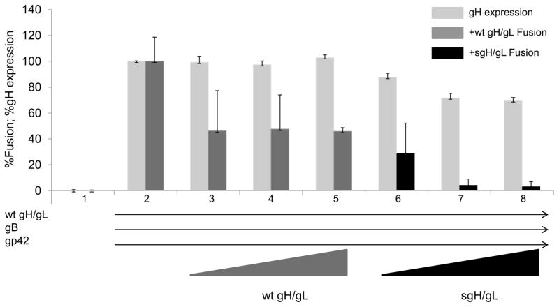
All CHO-K1 target cells were transiently co-transfected with HLA-DR and T7 polymerase. Lane 1: CHO-K1 effector cells were transfected with vector alone. Lane 2–8: All effector cells contain wild type gH (0.5 μg), excess gL (2.5 μg), gB (0.5 μg) and gp42 (2 μg). Lanes 3–5: Effector cells contain additional wild type gH (0.5, 1.0 and 1.5 μg respectively). Lanes 6–8: Effector cells contain additional sgH (0.5, 1.0 and 1.5 μg). Cells were detached twenty four hours post-tranfection and target cells were overlaid onto effector cells. Fusion activity was accessed for wt gH/gL (dark gray bars) and sgH/gL (black bars) eighteen to twenty four hours post overlay by the addition of passive lysis buffer followed by the addition of luciferase substrate. Luciferase activity was measured with a Perkin-Elmer Victor plate reader. gH expression (light gray bars) was determined by cELISA using anti-gH antibody E1D1. Data shown are representative results of three independent experiments. Error bars represent standard deviations for the normalized values.