Abstract
Sour and salt taste interactions are not well understood in the peripheral gustatory system. Therefore, we investigated the interaction of acetic acid and NaCl on taste processing by rat chorda tympani neurons. We recorded multi-unit responses from the severed chorda tympani nerve (CT) and single-cell responses from intact narrowly tuned and broadly tuned salt-sensitive neurons in the geniculate ganglion simultaneously with stimulus-evoked summated potentials to signal when the stimulus contacted the lingual epithelium. Artificial saliva served as the rinse and solvent for all stimuli [0.3 M NH4Cl, 0.5 M sucrose, 0.1 M NaCl, 0.01 M citric acid, 0.02 M quinine hydrochloride (QHCl), 0.1 M KCl, 0.003–0.1 M acetic acid, and 0.003–0.1 M acetic acid mixed with 0.1 M NaCl]. We used benzamil to assess NaCl responses mediated by the epithelial sodium channel (ENaC). The CT nerve responses to acetic acid/NaCl mixtures were less than those predicted by summing the component responses. Single-unit analyses revealed that acetic acid activated acid-generalist neurons exclusively in a concentration-dependent manner: increasing acid concentration increased response frequency and decreased response latency in a parallel fashion. Acetic acid suppressed NaCl responses in ENaC-dependent NaCl-specialist neurons, whereas acetic acid-NaCl mixtures were additive in acid-generalist neurons. These data suggest that acetic acid attenuates sodium responses in ENaC-expressing-taste cells in contact with NaCl-specialist neurons, whereas acetic acid-NaCl mixtures activate distinct receptor/cellular mechanisms on taste cells in contact with acid-generalist neurons. We speculate that NaCl-specialist neurons are in contact with type I cells, whereas acid-generalist neurons are in contact with type III cells in fungiform taste buds.
Keywords: chorda tympani, electrophysiology, epithelial sodium channel, geniculate ganglion
much is known about taste bud receptor proteins that initiate signal transduction and how individual peripheral neurons code information unique to each of the five basic taste qualities. Little is known, however, about how the peripheral nervous system processes information about two or more basic stimuli when presented together in a mixture. The purpose of this study was to investigate how the peripheral nervous system deciphers information about mixtures of NaCl and acetic acid in rat chorda tympani (CT) neurons.
In rodents, two salt transduction pathways have been identified. The first is selective for the sodium cation detected by an epithelial sodium channel (ENaC) located on the apical membrane of taste bud receptor cells (Heck et al. 1984). The second pathway is cation nonselective, but the transduction mechanism is not well understood. Lyall et al. (2004) proposed that this pathway involved an apically located variant of the transient receptor potential family of ion channels, TRPV1t, but this was recently disputed with the use of both whole nerve and single-unit recordings from rat CT neurons (Breza and Contreras 2012).
Taste bud cells with ENaC communicate with afferent neurons that respond selectively to sodium (and lithium) salts, and these responses are attenuated with ENaC antagonists such as amiloride (Lundy and Contreras 1999; Ninomiya and Funakoshi 1988; Rehnberg et al. 1993) or benzamil (Breza and Contreras 2012; Breza et al. 2010) and are thereby deemed amiloride/benzamil sensitive. In contrast, taste bud cells with the nonspecific cation channel communicate with broadly tuned afferent neurons that respond to NaCl, KCl, CaCl2, NH4Cl, and other basic taste stimuli, especially acids (Boudreau 1983; Breza and Contreras 2012; Breza et al. 2010; Lundy and Contreras 1999; Ninomiya and Funakoshi 1988). This latter neuron type is virtually unaffected by amiloride or benzamil and are thereby deemed amiloride/benzamil insensitive.
The mechanisms underlying sour-taste transduction of natural, organic acids (i.e., citric and acetic) in fungiform taste buds are not firmly established but appear to necessitate, at least in part, decreased intracellular pH (Huang et al. 2008; Lyall et al. 2001) and the expression of a PKD2L1 channel on the cell membrane (Horio et al. 2011; Huang et al. 2006; Ishimaru et al. 2006). Citric acid and acetic acid are weak carboxylic acids and gain entry into cells by passive-lipophilic diffusion and dissociate, thereby decreasing intracellular pH (Huang et al. 2008; Lyall et al. 2001). This mechanism may explain why there is a poor correlation between the acidity (pH) of a solution and the intensity of the sour sensation. In fact, acetic acid evokes much larger CT nerve signals than HCl at the same pH, and responses to equimolar concentrations of HCl, citric acid, and acetic acid are similar (Lyall et al. 2001).
On the basis of whole nerve experiments in rats and in hamsters, sour stimuli decrease the sodium-response magnitude in the amiloride/benzamil-sensitive pathway (sodium selective), whereas the response to the sour/salty mixture in the amiloride/benzamil-insensitive pathway (cation nonselective) is additive (Formaker et al. 2012; Lyall et al. 2002), equal to the sum of the component responses. The mechanism of suppression on the benzamil-sensitive pathway may involve decreased sodium passage through apical ENaC as has been shown in heterologous expression systems (Chalfant et al. 1999). In the present study we tested whether acids also decrease sodium responses in NaCl-specialist neurons, which are presumably in contact with ENaC-expressing taste receptor cells. In contrast, the notion that sour/salty mixtures activate the amiloride/benzamil-insensitive pathway with magnitudes that are similar to summation of the individual sour and salty responses indicates that acid and salt transduction in this pathway originate from distinct receptor cell populations.
In general, responses of CT neurons to salts and acids in neurons utilizing the nonselective cation pathway are increased with increasing temperature and are most robust when the adapted temperature approximates body temperature (Breza et al. 2006; Lundy and Contreras 1997, 1999; Lyall et al. 2004). Use of artificial saliva as the solution rinse and solvent also increases single-unit responses to the basic taste stimuli (Breza et al. 2010; Matsuo and Yamamoto 1992), and the combination between artificial saliva and elevated temperature optimizes whole nerve and single-unit responses (Breza and Contreras 2012; Breza et al. 2010; Lawhern et al. 2011). To our knowledge, neither whole nerve nor single-unit experiments with weak acids or acid/salt mixtures have been conducted under experimental conditions that approximate normal physiological conditions (adaptation of the tongue with 35°C artificial saliva). Accordingly, we recorded 5-s responses to taste stimuli representing the basic taste qualities, a concentration series of acetic acid and acetic acid-NaCl mixtures, from the severed CT nerve and from intact NaCl-specialist and acid-generalist neurons in the rat geniculate ganglion under highly controlled conditions. Phasic responses (i.e., 1 s after stimulus onset) to acetic acid, NaCl, and mixtures of acetic acid and NaCl were measured to assess the effect of acetic acid-NaCl mixtures on NaCl responses in NaCl-specialist and acid-generalist pathways during a behaviorally relevant timeframe. We chose acetic acid because it is a weak acid and is an effective stimulus in the whole CT nerve at pH values higher than those of equimolar concentrations of HCl or citric acid (Lyall et al. 2001). Benzamil was used to distinguish NaCl responses mediated by ENaC from those mediated by the nonselective pathway. On the basis of whole nerve studies with ENaC blockers, we hypothesized that acetic acid would attenuate NaCl responses in NaCl specialists, whereas responses to acetic-NaCl mixtures in acid generalists would be additive, equal to the sum of the component responses.
METHODS
Animals and Surgery
All animal procedures were approved by the Institutional Animal Care and Use Committee. Adult male Sprague-Dawley rats (Charles River Laboratories; n = 19) weighing 399–675 g were housed individually in plastic cages in a temperature-controlled colony room on a 12:12-h light-dark cycle with lights on at 0700. Rats were anesthetized with urethane (1.7 g/kg body wt), tracheostomized, and secured in a nontraumatic head holder. For CT preparations (n = 8), rats were placed in a custom-made brass head holder that could be rotated for the nerve dissection. The whole CT nerve was exposed by a mandibular approach, transected proximally, and desheathed for recording. For single-unit experiments (n = 11), rats were placed in a stereotaxic instrument with blunt ear bars. The geniculate ganglion was exposed with a dorsal approach (Breza and Contreras 2012; Breza et al. 2006, 2007, 2010; Lawhern et al. 2011; Lundy and Contreras 1999). Each rat's tongue was gently extended and held in place by a suture attached to its ventral surface. Body temperature was maintained at 37°C with a custom-made (Paul Hendrick, Florida State University) heating pad and temperature controller.
Recording Techniques
Electrogustogram.
The electrogustogram (EGG) was recorded in vivo with Ag-AgCl electrodes by means of nonpolarizing saline-agar-filled capillary pipettes (diameter 100 μm, 0.15 M NaCl, 0.5% agar). The active electrode (amplifier's negative lead) was placed near the receptive field, and an indifferent electrode (amplifier's positive lead) made contact with muscle on the neck. The amplifier's electrode lead with negative polarity was used as the active electrode (viewing purposes only) so that an increase in conductance (voltage drop) from electrolyte solutions (NaCl, QHCl, KCl, NH4Cl) resulted in upward deflections (Breza and Contreras 2012; Breza et al. 2010). The signal was amplified (DC ×50; A-M Systems, Sequim, WA), digitized with waveform hardware and software (Spike2; Cambridge Electronic Design, Cambridge, UK), and stored on a computer for off-line analysis.
CT nerve.
The CT nerve was cut near its entrance into the tympanic bulla and draped over a platinum wire hook (amplifier's positive lead), and the entire cavity was then filled with high-quality paraffin oil (VWR International) to isolate the signal from ground and maintain nerve integrity. An indifferent electrode (amplifier's negative lead) was attached to the skin overlying the cranium with a stainless steel wound clip. Neural activity was differentially amplified (AC ×10,000; A-M Systems; bandpass 300–5,000 Hz), observed with an oscilloscope, integrated using the root mean square (RMS) method (200-ms time constant), digitized and stored as described above.
Geniculate ganglion.
Low-impedance (1.1–1.9 MΩ) glass-insulated tungsten microelectrodes (tip diameter 1 μm) were mounted on a stereotaxic micromanipulator (Siskiyou Design Instruments, Grants Pass, OR) and advanced downward from the dorsal surface of the ganglion. Unit/few-unit activity (2 of 17 recordings had 2 neurons each, all other recordings were isolated single units) was recorded extracellularly (criteria 3:1 signal-to-noise ratio). An indifferent electrode (amplifier's negative lead) was attached to the skin overlying the cranium with a stainless steel wound clip. Neural activity was amplified (AC ×10,000; A-M Systems; bandpass 300–5,000 Hz), observed with an oscilloscope, digitized, and stored as described above.
Stimulus Delivery and Stimulation Protocols
Solutions were presented to the tongue at a constant flow rate (50 μl/s; dead volume 25 μl; see Breza and Contreras 2012 for more details) and temperature (35 ± 0.3°C) with the use of an air-pressurized 32-channel commercial fluid delivery system (16 channels for the present study) and heated perfusion cube (OctaFlow; ALA Scientific Instruments, Farmingdale, NY), respectively. All solutions and pharmacological reagents were reagent grade and were purchased from VWR International or Sigma Aldrich. Artificial saliva (0.015 M NaCl, 0.022 M KCl, 0.003 M CaCl2, and 0.0006 MgCl2; pH 5.8 ± 0.2) served as the rinse solution and solvent for all stimuli. We tested CT nerve responses to 5-s applications of 0.3 M NH4Cl, 0.5 M NaCl, 0.5 M sucrose, 0.1 M NaCl, 0.01 M citric acid, 0.02 M QHCl, 0.1 M KCl, 0.003–0.1 M acetic acid, and 0.003–0.1 M acetic acid mixed with 0.1 M NaCl. The tongue was pretreated for 60 s with 1 μM benzamil mixed in artificial saliva and then stimulated with 0.1 M NaCl (without benzamil) to assess the effect of benzamil pretreatment on NaCl responses mediated via apical ENaCs. Because benzamil is light sensitive, we wrapped the reservoir containing this compound in aluminum foil throughout the duration of the experiment. Stock solutions of benzamil (5 mM) were stored at −20°C.
We tested each single neuron's response to 5 s of stimulation with the following stimuli: 0.5 M sucrose, 0.1 M NaCl, 0.01 M citric acid, 0.02 M QHCl, 0.1 M KCl, 0.003–0.1 M acetic acid, and 0.003–0.1 M acetic acid mixed with 0.1 M NaCl. For single units, the protocol for taste stimulus delivery, benzamil pretreatment, and 0.1 M NaCl recovery was essentially the same as for CT protocols except each stimulus was presented 3–4 times and averaged. The standard stimuli (0.5 M sucrose, 0.1 M NaCl, 0.01 M citric acid, 0.02 M QHCl, 0.1 M KCl, and 0.3 M NH4Cl) were applied twice at the beginning and twice at the end of the protocol to validate the stability of the recording.
Measurement of Solution pH
The pH of each solution was measured with a pH meter (Mettler Toledo FE20). The results of solution pH are shown in Table 1.
Table 1.
Solution pH of taste stimuli
| Solution | pH |
|---|---|
| Artificial saliva | 5.8 |
| 0.3 M NH4Cl | 5.15 |
| 0.5 M sucrose | 5.29 |
| 0.1 M NaCl | 5.36 |
| 0.01 M citric acid | 2.60 |
| 0.02 M QHCl | 6.14 |
| 0.1 M KCl | 5.66 |
| 0.003 M acetic acid | 3.55 |
| 0.01 M acetic acid | 3.33 |
| 0.03 M acetic acid | 3.16 |
| 0.1 M acetic acid | 2.82 |
| 0.003 M acetic acid + 0.1 M NaCl | 3.54 |
| 0.01 M acetic acid + 0.1 M NaCl | 3.30 |
| 0.03 M acetic acid + 0.1 M NaCl | 3.13 |
| 0.1 M acetic acid + 0.1 M NaCl | 2.80 |
QHCl, quinine hydrochloride.
Data Analysis
CT nerve.
A 5-s period of baseline activity immediately prior to each stimulus was used to calculate the area under the curve for the integrated (RMS) 5-s response during stimulation by way of vertical cursors in Spike2. The EGG was used to mark the time when the stimulus first contacts the lingual epithelium and start of analysis as described previously (Breza and Contreras 2012; Breza et al. 2010; Lawhern et al. 2011). Consistent responses (criteria ± 10%) to standards (0.3 M NH4Cl and 0.5 M NaCl) at the beginning and end of the protocol were indicators of nerve integrity and recording stability (see Figs. 1 and 2). Whole nerve responses were normalized to the average response to 0.3 M NH4Cl at the beginning and end of the protocol.
Fig. 1.
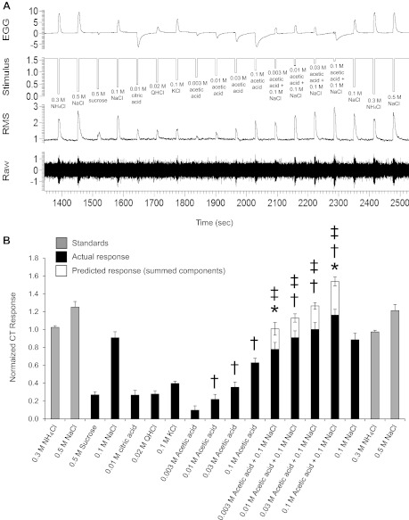
Raw electrophysiological traces from the chorda tympani (CT) nerve (A) and average responses (B, n = 7) to 0.3 M NH4Cl, 0.5 M NaCl, 0.5 M sucrose, 0.1 M NaCl, 0.01 M citric acid, 0.02 M quinine hydrochloride (QHCl), 0.1 M KCl, 0.003–0.1 M acetic acid, 0.003–0.1 M acetic acid mixed with 0.1 M NaCl, and predicted responses (sum of component responses to 0.003–0.1 M acetic acid and 0.1 M NaCl). Data are normalized to the average 0.3 M NH4Cl response. *Significantly different from 0.1 M NaCl alone; †significantly different from the preceding concentration; ‡significantly different from their predicted responses at the same concentration. EGG, electrogustogram; RMS, root mean square.
Fig. 2.
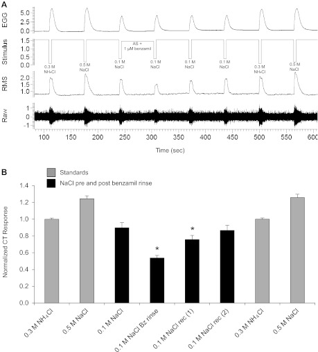
Raw electrophysiological traces from the CT nerve (A) and average responses (B, n = 7) to 0.3 M NH4Cl, 0.5 M NaCl, and 0.1 M NaCl before and after pretreatment with 1 μM benzamil. Data are normalized to the average 0.3 M NH4Cl response. *Significant differences in 0.1 M NaCl responses post benzamil treatment. AS, artificial saliva; rec, recovery.
Geniculate ganglion.
Spike templates were formed by amplitude and waveform shape. Spontaneous firing rate for each neuron was calculated as the average number of spikes/100 ms during the 5 s immediately before each stimulus. The EGG was used to mark the time when the stimulus first contacts the lingual epithelium and start of analysis. Response frequency was calculated as the difference between the spontaneous firing rate immediately before stimulation and the average number of spikes/100 ms occurring during a full 5-s period of chemical stimulation. Multiple applications of the same stimulus (baseline and stimulus) were averaged within each cell to produce an average response to that chemical stimulus (Breza and Contreras 2012; Breza et al. 2010; Lawhern et al. 2011). We used a hierarchal cluster analysis [Pearson's product-moment correlation coefficient (r − 1) and the average-linking method between subjects (Statistica, StatSoft, Tulsa, OK)] to categorize neurons by their average responses to 0.5 M sucrose, 0.1 M NaCl, 0.01 M citric acid, 0.02 M QHCl, 0.1 M KCl, 0.3 M NH4Cl, 0.003–0.1 M acetic acid, 0.003–0.1 M acetic acid + 0.1 M NaCl, 0.1 M NaCl immediately following pretreatment with 1 μM benzamil, and 0.1 M NaCl recovery responses following benzamil application.
Responses to the 0.5 M sucrose, 0.1 M NaCl, 0.01 M citric acid, 0.02 M QHCl, 0.1 M KCl, and 0.3 M NH4Cl were used to determine the breadth of tuning (H) for each neuron, calculated as H = −K Σ pi log pi, where K is a scaling constant (1.285 for 6 stimuli) and pi is the proportion of the response to individual stimuli to which the neuron responded against the total responses to all the stimuli (Smith and Travers 1979). H values range from 0 to 1; 0 corresponds to neurons that responded to only one stimulus, and 1 corresponds to neurons that responded equally to all the stimuli. Thus H values provide a quantitative measure of breadth of tuning. Because the entropy measure is unable to deal with negative proportions, absolute values were used for inhibitory responses; inhibitory responses were generally infrequent and of low magnitude. Taking the absolute value of inhibitory responses was the original method of dealing with inhibitory responses (Travers and Smith 1979) as discussed in Smith and Travers (1979), and we have also recently adopted this method (Breza and Contreras 2012). The results of entropy were compared with the noise-to-signal ratio (N/S) as described previously (Breza and Contreras 2012; Spector and Travers 2005). Briefly, the N/S was calculated as the product of the response to the second-best stimulus and the reciprocal of the response to the best stimulus (N/S = 2nd best stimulus × 1/best stimulus). Effect size, using Glass's d method; d = (x̄2 − x̄1)/SD x̄1, was calculated for each neuron's average response to a chemical stimulus over the full 5-s period and was used to indicate the response magnitude for each neuron and as a neuron group (Breza and Contreras 2012). By convention, effect sizes of 0.2, 0.5, and 0.8–∞ are used to indicate small, medium, and large effects (Cohen 1992) and are therefore used here to indicate the size of the effect (difference in response from baseline). The results from the N/S and the Glass's d method were compared with the breadth of tuning results shown in Table 2.
Table 2.
Breadth of tuning, noise-to-signal ratios, and effect sizes to 5-s applications with five or six standard stimuli
| Glass's d |
||||||||
|---|---|---|---|---|---|---|---|---|
| Neuron Type | H, 5 stimuli | N/S, 2 stimuli | Sucrose | NaCl | Citric acid | QHCl | KCl | NH4Cl |
| NaCl specialist | 0.46 ± 0.07 | 0.23 ± 0.09 | 1.30 ± 0.50 | 17.31 ± 5.61 | 0.31 ± 0.12 | 0.28 ± 0.12 | 1.13 ± 0.24 | Omitted |
| Acid generalist | 0.89 ± 0.01 | 0.76 ± 0.07 | 0.91 ± 0.16 | 2.61 ± 0.44 | 2.90 ± 0.72 | 0.92 ± 0.72 | 1.47 ± 0.29 | Omitted |
| Glass's d |
||||||||
|---|---|---|---|---|---|---|---|---|
| Neuron Type | H, 5 stimuli | N/S, 2 stimuli | Sucrose | NaCl | Citric acid | QHCl | KCl | NH4Cl |
| NaCl specialist | 0.60 ± 0.06 | 0.42 ± 0.10 | 1.30 ± 0.50 | 17.31 ± 5.61 | 0.31 ± 0.12 | 0.28 ± 0.12 | 1.13 ± 0.24 | 4.07 ± 1.10 |
| Acid generalist | 0.83 ± 0.02 | 0.53 ± 0.06 | 0.91 ± 0.16 | 2.61 ± 0.44 | 2.90 ± 0.72 | 0.92 ± 0.72 | 1.47 ± 0.29 | 7.05 ± 1.33 |
Values are mean ± SE. Breadth of tuning is indicated by H values for each neuron group, whereas noise-to-signal ratios are indicated by N/S values, and effect sizes are indicated by Glass's d for each of the standard stimuli.
For a detailed analysis of spike rate, we examined each group's average response (spikes/100 ms relative to baseline) to each stimulus in the test protocol and the summed responses to NaCl and acetic acid. Effect sizes (Glass's d method) for each 100-ms bin following stimulus onset were calculated for each neuron's average response to a chemical stimulus and were averaged within each neuron group to estimate response latency and response magnitude (peak response) as described previously in greater detail (Breza and Contreras 2012). In this study, we used a medium (0.5) effect size to determine response latency. The effect size needed to stay at or above 0.5 for at least five 100-ms bins for response latency to be determined as described previously (Breza and Contreras 2012).
Statistics.
Further statistical analyses were conducted with appropriate analysis of variance (ANOVA; Statistica) or t-test within and between subjects. One-way repeated-measures (RM) ANOVA was used to evaluate the effect of all test stimuli and NaCl recovery following benzamil pretreatment on integrated CT nerve responses and single units. Two-way RM ANOVA was used to compare the average spikes/100 ms relative to baseline (1-s intervals) for NaCl with or without benzamil pretreatment within neuron type (stimulus × time). Spontaneous firing rate before each stimulus was averaged for each neuron, and average baseline firing rate for each neuron group was then evaluated with an independent t-test. Significant main effects or interactions (P < 0.05) of ANOVAs were further examined with Fisher's least significant difference method. Graphic and tabular data are means ± SE.
RESULTS
Chorda Tympani: Standard Taste Stimuli, Acetic Acid, and Taste Mixtures
Shown in Fig. 1A are the typical responses from one CT nerve recording and, in Fig. 1B, the average responses from seven male rats to 5-s applications of the standard taste stimuli, 0.3 M NH4Cl, and to an acetic acid and acetic acid + 0.1 M NaCl concentration series. The polarity of the EGG waveform was positive for ionic solutions (NaCl, KCl, QHCl, NH4Cl) and negative for nonionic solutions (sucrose, citric, and acetic acid), and the magnitude of EGG response increased with acetic acid and NaCl concentration. Regardless, chemical stimulation triggered an immediate EGG response that occurred at approximately the same time for all stimuli.
The CT nerve responses varied with taste stimulus [F(17,102) = 83.55, P < 0.001] and acetic acid concentration. The average response to 0.1 M NaCl was significantly greater than the response to sucrose, citric acid, QHCl, and KCl (all P values <0.001) and to all acetic acid concentrations (all P values <0.001). The response to 0.01 M citric acid was similar to the responses to 0.01 and 0.03 M acetic acid, but the response to 0.1 M acetic acid was greater than the response to 0.01 M citric acid (P < 0.001) despite similarities in solution pH (Table 1). The CT nerve responses increased with acetic acid concentration alone (all P values <0.05) and mixed with 0.1 M NaCl (P values < 0.05).
To investigate mixture effects, we compared the actual responses to the acetic acid-NaCl mixtures with the sum of the component responses, because the mixture responses differed significantly from the responses to acetic acid alone (all P values <0.001). The responses to acetic acid-NaCl mixtures were less than the sum of the component responses at each concentration (P values <0.001). The responses to 0.003, 0.01, 0.03, and 0.1 M acetic acid-NaCl were 22, 19, 21, and 24% less than predicted, respectively. The overall suppressive effect across all concentrations was 21%. The responses to 0.5 M NaCl and 0.3 M NH4Cl at the beginning and end of the protocol did not differ statistically, demonstrating response stability over the recording interval.
Chorda Tympani: Effects of Benzamil Pretreatment
Shown in Fig. 2A are the typical responses from one CT nerve recording and, in Fig. 2B, the average responses of seven male rats to short-duration 5-s applications of 0.1 M NaCl with and without 1 μM benzamil pretreatment, as well as the average responses to 0.3 M NH4Cl and 0.5 M NaCl at the beginning and end of the protocol. The responses differed across stimuli [F(7,42) = 60.51, P < 0.001]. Benzamil pretreatment reduced the response to NaCl by 40% (P < 0.001) during the 5-s stimulation period and by 16% 60 s after returning to the artificial saliva rinse (P < 0.01). Full recovery occurred after 2 min of artificial saliva rinse (P = 0.49). The responses to 0.3 M NH4Cl and 0.5 M NaCl remained stable from the beginning to the end of the protocol.
Geniculate Ganglion: Basic Firing Characteristics
The results from the hierarchical cluster analysis are shown in the dendrogram of Fig. 3. Analysis of agglomeration by means of a scree plot (data not shown) indicated that an abrupt upward deflection occurred at 0.42, which separated the 21 neurons into 2 main groups of 13 NaCl specialists and 8 acid generalists. Figure 4 shows that the NaCl-specialist neuron was completely unresponsive to acetic acid at all concentrations but responded well to the acetic acid-NaCl mixture. Figure 4 shows that benzamil pretreatment reduced spike rate and increased response latency to NaCl. In contrast, the acid-generalist neuron responded in a concentration-dependent manner to acetic acid alone and to the mixture with NaCl, and NaCl responses were unaffected by benzamil pretreatment. Figure 4 also shows that the spontaneous firing rate during the artificial saliva rinse was much higher for the acid-generalist neuron. In fact, the average spontaneous firing rate differed significantly (T = −2.13, P = 0.05) between the NaCl specialists (mean = 0.23 ± 0.01 spikes/100 ms) and the acid generalists (mean = 0.41 ± 0.02 spikes/100 ms). This result is consistent with our recent findings (Breza and Contreras 2012; Breza et al. 2010; Lawhern et al. 2011).
Fig. 3.
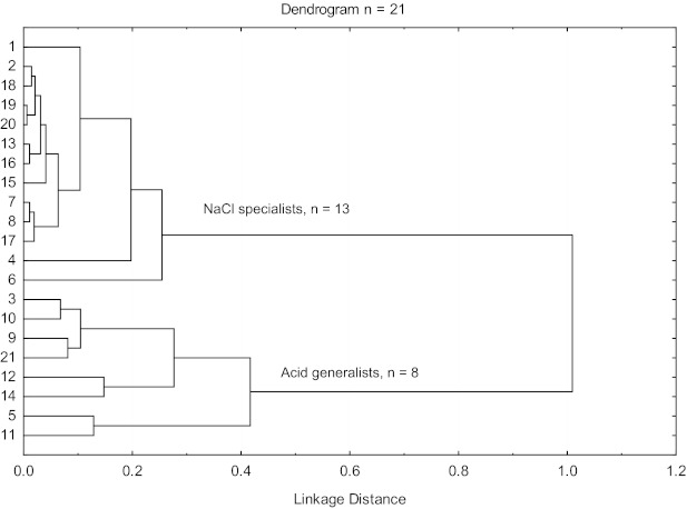
Dendrogram of the results from the hierarchical cluster analysis.
Fig. 4.
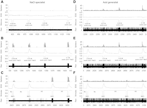
Raw electrophysiological traces from a NaCl-specialist neuron (A–C) and acid-generalist neuron (D–F) to 0.003–0.1 M acetic acid, 0.003–0.1 M acetic acid mixed with 0.1 M NaCl, and 0.1 M NaCl with and without benzamil pretreatment.
NaCl Specialists and Acid Generalists: Average 5-s Responses
NaCl specialists.
Shown in Fig. 5 are the average 5-s responses by the NaCl-specialist and acid-generalist neuron groups. The two groups responded differently across stimuli [F(15, 180) = 41.86, P < 0.001]. The NaCl specialists were narrowly tuned, responding more to 0.1 M NaCl than to any other single stimulus (all P values <0.001). The breadth of tuning and the N/S were low when 0.3 M NH4Cl was excluded from the analyses (see Table 2). Without 0.3 M NH4Cl, the effect size for 0.1 M NaCl was 17 times greater than for other basic taste stimuli and KCl (Table 2). Including 0.3 M NH4Cl in the analyses increased the breadth of tuning and N/S, and the effect size analysis revealed that the response to 0.1 M NaCl was four times greater than to 0.3 M NH4Cl. Benzamil pretreatment decreased the spike rate to 0.1 M NaCl by 76% (P < 0.001); spike rate was fully restored by the third NaCl presentation.
Fig. 5.
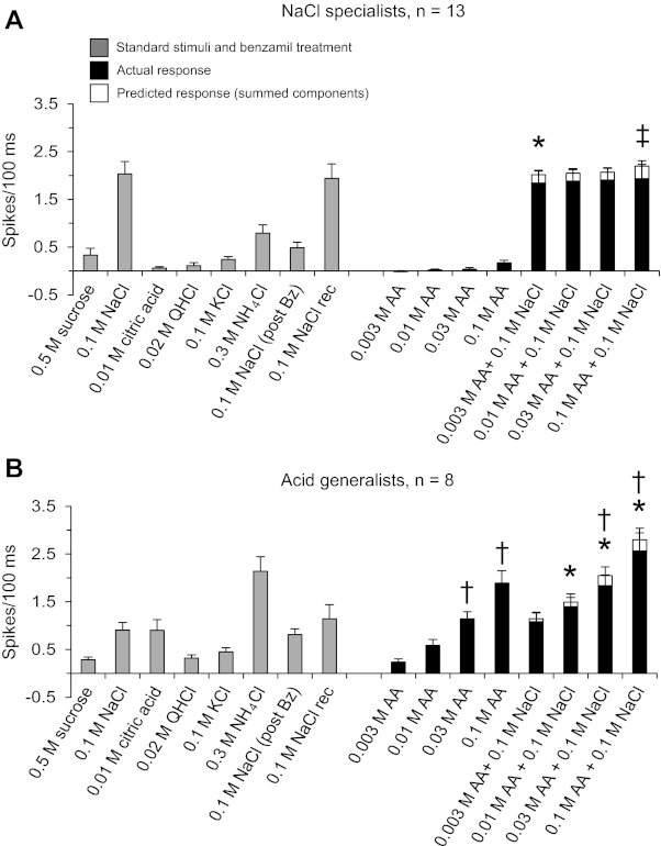
Average responses to all test stimuli (5-s presentations relative to baseline) from NaCl specialists (A) and acid generalists (B). *Significantly different from 0.1 M NaCl alone; †significantly different from the preceding concentration; ‡significantly different from their predicted responses at the same concentration. AA, acetic acid; Bz, benzamil.
The NaCl-specialist group was largely unresponsive to all acetic acid concentrations. The response to 0.003 M acetic acid + NaCl (P < 0.05) was significantly less than the response to NaCl alone, but responses to acetic acid-NaCl mixtures at all other concentrations were equal to the response to NaCl alone. Nevertheless, we found that the actual responses to the mixtures were significantly smaller than the responses to the summed components [F(8, 96) = 3.21, P < 0.01]. The response to the 0.1 M acetic-NaCl mixture was significantly less than the response to the summed components (P < 0.01) and marginally less for the other three comparisons (0.003, 0.01, 0.03 M; P = 0.055, P = 0.060, P = 0.064, respectively). It is worth noting that 2 of the 13 NaCl specialists responded synergistically to the all concentrations (one by 27% and the other by 11%). When these two neurons were excluded from the analysis, responses to acetic acid-NaCl mixtures were less than to NaCl alone at all concentrations, and the predicted responses were greater than responses to acetic acid mixtures at all concentrations (P values <0.05).
Acid generalists.
The spike rates of acid generalist neurons differed significantly across stimuli [F(15, 105) = 20.76, P < 0.001]. They responded broadly to all taste stimuli, but more to 0.3 M NH4Cl and 0.1 M acetic acid than to any other single stimulus (P values <0.001). This was reflected in a large H value (breadth of tuning) and N/S, regardless of whether NH4Cl was included in the equation. The N/S was less than the H value, particularly when NH4Cl was included in the analysis, because the NH4Cl response was more than twice that to the second best stimulus, as measured using Glass's d method. The NaCl response by acid-generalist neurons was unaffected by benzamil. The responses to 0.03 and 0.1 acetic acid-NaCl mixtures were equal to the response to 0.3 M NH4Cl. The responses to 0.1 M NaCl and 0.01 M citric acid were similar, but both responses were greater than those to 0.5 M sucrose, 0.02 M QHCl, 0.1 M KCl, and 0.003 M acetic acid (P values <0.05).
The spike rates of acid generalists increased with acetic acid concentration alone (0.01–0.1 M; P values < 0.05) and mixed with NaCl (0.01–0.1 M; P values <0.05). Consistent with our whole nerve recordings, the responses to 0.01 M citric acid and 0.01–0.03 M acetic acid were similar, but the response to 0.1 M acetic acid was greater than that to 0.01 M citric acid (P < 0.001) despite similarities in solution pH (Table 2). The responses to the acetic acid-NaCl mixtures were greater than those to acetic acid alone (P values <0.01). The response to the 0.003 M acetic acid-NaCl mixture was equal to the response to NaCl alone, but the responses to all other acetic acid-NaCl mixtures were greater than the response to NaCl (P values <0.05). A one-way RM ANOVA revealed a main effect of stimulus [F(8, 56) = 18.07, P < 0.001] when we compared the responses to NaCl alone, to the acetic acid-NaCl mixtures, and to the sum of the component responses. This effect was due to the fact that the response to NaCl was significantly less than the responses to the acetic acid-NaCl mixtures. Post hoc comparisons revealed that the actual responses to the mixtures were similar to the sum of the component responses across all stimulus concentrations. However, there was considerable variability among acid-generalist neurons. Of the eight neurons tested, three responded significantly less (suppression) and two responded significantly more (synergy/enhancement) to the mixtures compared with the sum of the component responses. Thus acid-sensitive generalist neurons are not a homogeneous population as observed previously (Breza et al. 2006, 2007; Lundy and Contreras 1999).
NaCl Specialists and Acid Generalists: Differential Spike Patterns to NaCl and NH4Cl
NaCl specialists.
Shown in Fig. 6 are the response patterns (in 100-ms bins) to 0.1 M NaCl, 0.1 M NaCl after benzamil treatment, and 0.3 M NH4Cl in NaCl specialists and acid generalists. NaCl specialists responded abruptly to 0.1 M NaCl 100 ms after stimulus contact. In contrast, the response to 0.3 M NH4Cl was sluggish with a sevenfold longer latency (see Table 3). Benzamil suppressed NaCl spike rate, thereby increasing NaCl response latency 12-fold. Benzamil pretreatment reduced NaCl-spike frequency for the full 5-s stimulation period (P values <0.001) as reflected by a significant stimulus [F(2, 24) = 57.55, P < 0.001] and stimulus × time interaction effects [F(8, 96) = 3.86, P < 0.001].
Fig. 6.
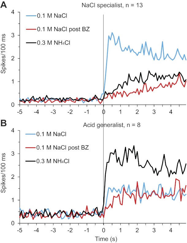
Average response profiles from NaCl specialists (A) and acid generalists (B) to 0.1 M NaCl, 0.1 M NaCl post benzamil treatment, and 0.3 M NH4Cl in 100-ms bins. Vertical line indicates stimulus onset (time 0).
Table 3.
Response latencies and peak-response frequencies of NaCl specialists and acid generalisst to test stimuli as measured by Glass's d
| Stimulus | Response latency (NaCl specialists) | Peak Response (NaCl specialists) | Response latency (acid generalists) | Peak Response (acid generalists) |
|---|---|---|---|---|
| 0.5 M sucrose | ||||
| Range, ms | N/A | 2,000–2,099 | 1,500–1,599 | 4,400–4,499 |
| d | 6.9 ± 4.1 | 1.1 ± 0.6 | 2.5 ± 1.0 | |
| 0.1 M NaCl | ||||
| Range, ms | 100–199 | 500–599 | 200–299 | 2,400–2,499 |
| d | 7.1 ± 5.9 | 23.8 ± 8.8 | 2.1 ± 1.0 | 3.8 ± 0.8 |
| 0.01 M citric acid | ||||
| Range, ms | N/A | 2,800–2,899 | 700–799 | 2,900–2,999 |
| d | 0.8 ± 0.4 | 1.1 ± 0.5 | 5.4 ± 1.3 | |
| 0.02 M QHCl | ||||
| Range, ms | N/A | 1,100–1,199 | 700–799 | 3,600–3,699 |
| d | 1.8 ± 1.1 | 0.7 ± 0.2 | 1.9 ± 0.7 | |
| 0.1 M KCl | ||||
| Range, ms | N/A | 4,300–4,399 | 300–399 | 4,400–4,499 |
| d | 3.0 ± 0.8 | 0.9 ± 0.4 | 2.9 ± 0.7 | |
| 0.3 NH4Cl | ||||
| Range, ms | 700–799 | 2,700–2,799 | 100–199 | 1,800–1,899 |
| d | 1.4 ± 0.6 | 5.8 ± 1.7 | 5.1 ± 3.0 | 9.8 ± 2.8 |
| 0.003 M acetic acid | ||||
| Range, ms | N/A | 4,200–4,299 | 2,000–2,099 | 3,900–3,999 |
| d | 0.3 ± 0.3 | 0.8 ± 0.5 | 1.4 ± 0.5 | |
| 0.01 M acetic acid | ||||
| Range, ms | N/A | 4,400–4,499 | 700–799 | 2,900–2,999 |
| d | 0.4 ± 0.2 | 2.2 ± 1.0 | 3.5 ± 0.6 | |
| 0.03 M acetic acid | ||||
| Range, ms | N/A | 4,400–4,499 | 300–399 | 4,300–4,399 |
| d | 0.2 ± 0.4 | 0.9 ± 0.7 | 5.8 ± 1.5 | |
| 0.1 M acetic acid | ||||
| Range, ms | N/A | 1,000–1,099 | 200–299 | 2,200–2,299 |
| d | 1.4 ± 0.6 | 2.6 ± 1.4 | 7.5 ± 1.3 | |
| 0.003 M acetic acid +0.1 M NaCl | ||||
| Range, ms | 100–199 | 800–899 | 200–299 | 4,400–4,499 |
| d | 3.8 ± 2.6 | 22.7 ± 11.1 | 1.0 ± 1.1 | 5.9 ± 1.2 |
| 0.01 M acetic acid +0.1 M NaCl | ||||
| Range, ms | 100–199 | 700–799 | 200–299 | 3,600–3,699 |
| d | 2.1 ± 1.5 | 28.7 ± 14.4 | 1.4 ± 0.5 | 6.1 ± 0.9 |
| 0.03 M acetic acid +0.1 M NaCl | ||||
| Range, ms | 100–199 | 600–699 | 100–199 | 3,900–3,999 |
| d | 3.1 ± 1.9 | 17.9 ± 6.1 | 1.3 ± 0.4 | 8.0 ± 2.2 |
| 0.1 M acetic acid +0.1 M NaCl | ||||
| Range, ms | 100–199 | 700–799 | 100–199 | 900–999 |
| d | 4.7 ± 2.7 | 19.2 ± 7.6 | 1.5 ± 0.9 | 9.8 ± 2.4 |
| 0.1 M NaCl post 1 μM benzamil | ||||
| Range, ms | 1,200–1,299 | 4,500–4,599 | 400–499 | 4,300–4,399 |
| d | 4.2 ± 3.6 | 5.7 ± 1.6 | 1.4 ± 0.6 | 5.3 ± 1.4 |
Values are mean ± SE.
Acid generalists.
Acid generalists responded abruptly to 0.3 M NH4Cl 100 ms after stimulus contact. The mean response to 0.1 M NaCl was less than twice the frequency and twice the latency. There was a significant stimulus × time interaction effect [F(8, 56) = 2.55, P < 0.05] when comparing the spike rate responses to NaCl with and without benzamil pretreatment. Benzamil pretreatment reduced the NaCl spike rate just for the first second of stimulation (P < 0.01) and increased spike latency twofold (see Table 3).
Response Profiles to Acetic Acid and Acetic Acid-NaCl Mixtures
Shown in Fig. 7 are the average responses to 0.1 M NaCl, 0.003–0.1 M acetic acid, and acetic acid-NaCl in NaCl-specialist (left) and acid-generalist neurons (right). As shown, acetic acid alone had little impact on spike frequency of NaCl specialists but had a concentration-dependent impact on spike frequency and response latency of acid generalists. In fact, as acetic acid concentration increased, response frequency increased and response latency decreased (see Table 3) in a parallel fashion. NaCl specialists' responses to the acetic acid-NaCl mixtures never exceeded those to NaCl alone but were in fact less than those to NaCl alone during the first second of stimulation [F(8, 96) = 4.89, P < 0.001]. Each concentration of acetic acid mixed with NaCl was less than the response to NaCl alone (P values < 0.05) and was less than the sum of the component responses (P values <0.05). In contrast, acid generalists' responses to the mixtures were always greater than those to NaCl alone and were equal to the sum of the component responses.
Fig. 7.
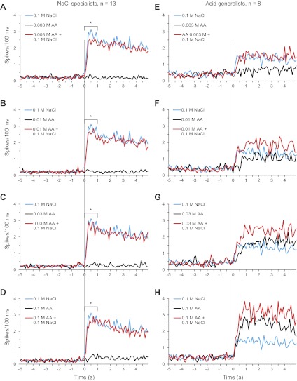
Average response profiles (in 100-ms bins) of NaCl specialists (A–D) and acid generalists (E–H) to 0.1 M NaCl, 0.003–0.1 M acetic acid, and 0.003–0.1 M acetic acid mixed with 0.1 M NaCl. Vertical line indicates stimulus onset (time 0). *Significant differences in 0.1 M NaCl responses from acetic acid-NaCl mixtures during the first second.
DISCUSSION
The taste system is adroit in discriminating mixtures of different taste qualities, yet we understand little about the underlying physiological mechanisms at the whole nerve or single-cell levels. For example, we know little about sour-salty taste interactions even though acids and salts serving as preservatives and food stabilizers are common in processed foods and avidly consumed in industrialized countries. Accordingly, we recorded the summated responses from the whole nerve and the spike discharges from sodium-selective and acid/sodium-responsive neurons in the rat geniculate ganglion to mixtures of acetic acid and NaCl under controlled conditions that approximate the physiological parameters of the oral cavity. It was imperative that the stimulus protocols for the whole nerve and single-cell experiments be similar and that the analyses be anchored temporally to a meaningful stimulus onset marker served reliably by the stimulus-evoked EGG waveform from the anterior tongue.
We hypothesized that CT nerve responses to acetic acid-NaCl mixtures would be less than additive (Lyall et al. 2002). We further predicted that mixture suppression observed at the whole nerve level would be driven largely by responses of NaCl-specialist neurons. That is, the mixture responses of NaCl-specialist neurons would be less than the sum of the component responses (suppression), whereas those of acid generalists would be additive. This latter hypothesis was derived from a prior study (Lyall et al. 2002) showing that only the benzamil-sensitive portion of the whole nerve response was significantly attenuated by acetic acid-NaCl mixtures. Importantly, the hypothesis had not been tested previously at the single-cell level. The results from the present experiments are in general agreement with our hypotheses and the findings from Lyall et al. (2002) and Formaker et al. (2012).
Chorda Tympani: Basic Tastes, Acetic Acid, Acetic Acid-NaCl Mixtures, and Benzamil
We found that the CT nerve responded to a broad range of acetic acid concentrations, spanning over a range of 1.5 log units. The response magnitude to 0.003 M acetic acid observed in the present study corresponds well with the threshold-level responses reported in wild-type mice for comparable concentrations of HCl, citric acid, and acetic acid (Horio et al. 2011). The responses to these three acids began at 0.003 M and increased progressively to a maximum response to 0.02 M HCl, 0.03 M citric acid, and 0.05 M acetic acid. As far as we know, similar findings are not available in rats except for HCl, where the threshold response was reported to be 0.0003 M HCl (Contreras and Frank 1979), 1 log unit lower than that found in the present study for acetic acid. Although we did not test the threshold response to acetic acid, it is conceivable that we could have found responses to concentrations lower that 0.003 M acetic acid, since 0.003 M acetic acid evoked moderate responses.
In the present study, we found that the CT responded with the same magnitude to 0.01 M citric acid (pH = 2.6) and 0.01 M acetic acid (pH = 3.3) even though citric acid had a lower pH. Additionally, the CT responded with more than twice the magnitude to 0.1 M acetic acid (pH = 2.82) than to 0.01 M citric acid even though citric acid had a lower pH. These results indicate that CT nerve response magnitude to acetic acid is not entirely related to extracellular pH.
The present results showed the response of the CT nerve to the acetic acid-NaCl mixtures was less than the sum of the component responses. This suggests that either acetic acid inhibited the CT response to NaCl or that NaCl inhibited the response to acetic acid. Lyall et al. (2002) showed that responses to NaCl through the benzamil-sensitive pathway of the rat were inhibited by acetic acid, whereas responses of the benzamil-insensitive pathway were additive. A similar result has also been shown in the hamster CT with amiloride (Formaker et al. 2012). The suggestion is that a decrease in intracellular pH decreases the sodium current through apical ENaCs. Evidence for a similar mechanism has been shown in Xenopus oocytes (Chalfant et al. 1999). It was concluded from the Chalfant et al. (1999) study that the α-subunit of ENaC was directly regulated by changes in intracellular pH.
Recently, we showed that 1 μM benzamil decreased 0.1 M NaCl responses by 44% if the solution mixture (0.1 M NaCl + 1 μM benzamil) was left on the tongue for 60 s (Breza and Contreras 2012). The effect of a fivefold higher concentration of benzamil was partially recovered after a 60-s wash with 0.1 M NaCl and fully recovered after an additional 60 s with artificial saliva. It was clear from this experiment that benzamil had lasting effects, but the effects were completely reversible. Because benzamil successfully blocked the channel for an extended period of time, we chose to pretreat the tongue with 1 μM benzamil for 60 s before 0.1 M NaCl application to ensure enough time for the drug to bind to its site of action. By delivering the drug in the rinse, NaCl responses from the same stimulus channel (channel 2; see Fig. 2) could be compared before and after treatment to ensure that any difference in response magnitude was not confounded by subtle changes in flow rate between stimulus channels. Pretreatment of 1 μM benzamil for 60 s decreased 5-s NaCl responses in the CT by 40%, consistent with 60-s coapplications of 0.1 M NaCl + 1 μM benzamil (Breza and Contreras 2012); NaCl responses were fully recovered after the third presentation of 0.1 M NaCl.
Single Units: Basic Tastes, Acetic Acid, Acetic Acid-NaCl Mixtures, and Benzamil
We studied the responses of the two salt-sensing neuron groups: NaCl specialists and acid generalists. For comparison sake, the solution delivery procedure was similar to that used in the whole nerve study. We examined the responses to 0.3 M NH4Cl because it is a highly effective stimulus for acid-generalist neurons (Lundy and Contreras 1999) and has been used as a standard/control stimulus in whole nerve recordings for decades. We used 1 μM benzamil to identify NaCl responses mediated via apical ENaC.
The present study further clarified the difference in response characteristics between the two salt-sensing groups. We found the insensitivity of NaCl specialists to acid stimuli applied to acetic acid over a broad concentration range that extended 1 log unit higher than the 0.01 M concentration of citric acid (Breza and Contreras 2012; Breza et al. 2007; 2006; Breza et al. 2010; Lawhern et al. 2011) and HCl (Lundy and Contreras 1999) used previously. In contrast, acid generalists responded in a concentration-dependent manner to all acetic acid concentrations from 0.003 to 0.1 M. As acetic acid concentration increased, the acid generalist's spike rate response increased and its response latency decreased.
The present study further elucidated the selectivity of NaCl specialists to sodium stimulation and relative insensitivity to KCl and NH4Cl. Consistent with our prior studies, NaCl specialists were weakly responsive at best to these two non-sodium salts. In the present study, NaCl specialists responded to the strong 0.3 M NH4Cl with less than 50% the spike rate and 7 times longer response latency than to 0.1 M NaCl. The effect size was 4 times greater for 0.1 M NaCl than for 0.3 M NH4Cl (Table 3) and 17 times greater for 0.1 M NaCl than for the other basic taste stimuli and 0.1 M KCl.
In contrast, acid generalists responded well to all three salts, but best to NH4Cl, second best to NaCl, and least to KCl (Boudreau 1983; Lundy and Contreras 1999). In fact, we found that acid generalists responded more to 0.3 M NH4Cl than to 0.01 M citric acid and 0.01 M acetic acid but responded equally to 0.3 M NH4Cl and 0.1 M acetic acid. Furthermore, we found that acid generalists' response latency to 0.3 M NH4Cl was the quickest among all stimuli and both fiber types. In fact, the response latency to NH4Cl was seven times shorter in acid generalists than in NaCl specialists (see Table 3). This indicates that acid-generalist neurons are well tuned to communicate information about NH4Cl to the brain.
Pretreatment with 1 μM benzamil had a profound impact on NaCl-specialist neurons, virtually eliminating their 5-s response to 0.1 M NaCl. A similar effect has been seen with 10 μM amiloride in NaCl-specific fibers of the hamster CT (Hettinger and Frank 1990). We also found that benzamil pretreatment reduced the spike rate of acid-generalist neurons only during the first second of NaCl stimulation. We suspect that this indicates that benzamil may have been applied a little too long, causing unwanted nonspecific effects across the receptor cell membrane.
As might be expected, NaCl-specialists and acid-generalists responded differently to the acetic acid-NaCl mixtures. NaCl specialists responded to the acetic acid-NaCl mixtures at a spike rate that was lower than the sum of the component spike rate responses. In fact, the spike rate response to the mixture was never greater than the spike rate response to NaCl alone as shown in Fig. 7. Moreover, we found that during first second of stimulation, the spike rate responses to acetic acid-NaCl mixtures were significantly lower than the responses to NaCl alone. In contrast, acid generalists' spike rate responses to the acetic acid-NaCl mixtures were equal to the sum of the component responses; this was coupled with a decrease in response latency to the two highest acetic acid-NaCl mixtures compared with the latency to NaCl or acetic acid alone. Thus the acetic acid-NaCl mixture more quickly depolarized taste receptor cells innervated by acid generalists.
Together, these above findings are in general agreement with results from whole nerve experiments in rats, where only the benzamil-sensitive portion of the response was attenuated (Lyall et al. 2002). Although the inhibitory responses to acetic acid-NaCl mixtures in NaCl specialists were not as robust as those predicted by whole nerve responses (9% reduction in NaCl specialists vs. 21% reduction in CT), the whole nerve is indeed a summated response and many factors need to be considered. For example, it is possible that there are more NaCl specialists than acid generalists in the rat CT. Over the past 12 years, we have sampled from more NaCl specialists than acid generalists (Breza and Contreras 2012; Breza et al. 2006, 2007, 2010; Lawhern et al. 2011; Lundy and Contreras 1999). Together, these samples estimate that there are greater than one-third more NaCl specialists than acid generalist in the rat CT nerve, which may contribute to a larger portion of the summated signal. It is also possible that the fiber diameter of NaCl specialists is significantly larger than that of other neuron types. A recent study in the geniculate ganglion (Nakamura and Bradley 2011) showed that CT neurons in the geniculate ganglion were not equal in diameter, suggesting that fiber or soma diameter may be linked to neuron type. Over the last decade we have noticed that extracellular spike amplitudes from NaCl specialists are larger than those of any other CT neuron type, such as those shown in Breza et al. (2010). Although this would need to be confirmed with intracellular recording and cell labeling, it is possible that NaCl specialists have larger soma and fiber diameters. Aside from these factors, if the percent differences between actual responses to acetic acid-NaCl mixtures and predicted responses are calculated for each neuron, regardless of type, the percent decrease is more similar to the percent decrease of the whole nerve. In fact, NaCl specialists and acid generalists together responded 17% less to stimulus mixtures than to predicted responses: the percent decrease of the CT nerve was 21%. Together, all these factors would certainly impact the amplitude of the whole nerve, since the whole nerve is indeed a summated potential and therefore needs to be considered.
NaCl Specialists and Acid Generalists: Functional Connections with Fungiform Taste Cells
The fungiform taste bud contains a heterogeneous population of taste receptor cells. Type I cells of the fungiform papillae are the only known cell type that is sensitive to amiloride (Vandenbeuch et al. 2008). Type III cells have traditional synapses with afferent fibers that respond well to citric acid, acetic acid, and KCl (Huang et al. 2008; Tomchik et al. 2007). It is unknown how type I cells communicate with afferent fibers, but it is possible that communication to afferent fibers may be accomplished without a synapse, by way of paracrine signaling. Indeed, knocking out the ATP receptor abolishes NaCl taste responses from the mouse CT (Finger et al. 2005). Thus ATP is crucial for communication with afferent fibers but also has been shown to play a role in cell-to-cell communication within the bud (Huang et al. 2007). Type III cells have been implemented in communicating information from neighboring taste receptor cells (Tomchik et al. 2007). Type III cells express PKD2L1 channels on their plasma membrane that may be involved in the transduction of acids (Chang et al. 2010; Horio et al. 2011; Huang et al. 2006). Weak acids are membrane soluble, decrease intracellular pH (Lyall et al. 2001), and activate type III cells exclusively (Huang et al. 2008). Genetic ablation of taste cells expressing the PKD2L1 channel severely compromises whole nerve responses to acids (Huang et al. 2006), and mutants lacking the PKD2L1 receptor have decreased, but not abolished, nerve responses to acids (Horio et al. 2011). Although there are a number of mechanisms that have been proposed for acid transduction, PKD2L1 has consistently shown to be involved in acid transduction in neurons with fungiform receptive fields (Horio et al. 2011; Huang et al. 2006).
With these factors in mind, it is tempting to speculate that NaCl specialists are in contact with type I taste bud cells, whereas acid generalists are in contact with type III cells, and perhaps another cell type that is highly responsive to sodium and ammonium salts. NaCl specialists were unresponsive to a broad concentration range of acetic acid, suggesting that they lack PKD2L1 channel expression. In contrast, acid generalists were responsive to a broad concentration of acetic acid, suggesting that they express PKD2L1 channels. Not only were NaCl specialists virtually unresponsive to acetic acid, but acetic acid decreased their responses to NaCl, suggesting that a single transduction mechanism is involved. Therefore, it is reasonable to assume that intracellular acidification via acetic acid decreases intracellular pH (Chalfant et al. 1999; Lyall et al. 2001, 2002), thereby decreasing ENaC activity in type I cells as shown in Xenopus oocytes (Chalfant et al. 1999). Acid generalists responded to acetic acid and NaCl, but their responses to acetic acid-NaCl mixtures were additive, suggesting that acetic acid and NaCl were transduced through different mechanisms, perhaps through different receptor cell types. Type III cells express an α-ENaC subunit, and ablation of this subunit had no consequences on sodium taste in the mouse CT (Chandrashekar et al. 2010), but we saw a small reduction in NaCl spike rate in acid generalists during the first second after the tongue was pretreated with benzamil. It is quite possible that the Zuker group could not see this modest effect at the whole nerve level, and clearly acid generalists are using an additional mechanism to detect salts other than the α-ENaC subunit. At this juncture, it is unclear whether additive responses in acid generalists are accomplished through cell-to-cell communication or whether the type III cell expresses receptor mechanisms for transduction of both sour and salt taste qualities.
Conclusions
Acetic acid was an effective stimulus in the whole CT nerve and differentially affected NaCl-specialist and acid-generalist neuron types. NaCl specialists were unresponsive to a broad concentration range (0.003–0.1 M) of acetic acid, whereas acid generalists responded to acetic acid in a concentration-dependent manner. Increasing acetic acid concentration decreased response latency and increased responses frequency of acid generalists. Acetic acid and citric acid responses were equal at the same concentration, but acetic acid was more than twice as effective at a similar pH. The response by acid-generalist neurons to an acetic acid-NaCl mixture was equal to the sum of the component responses, whereas that by NaCl-specialist neurons was less than the sum of the component responses. The additive response by acid generalists is coupled with a reduction in response latency for the two highest acetic acid concentrations. Together, these data suggest that acetic acid reduces ENaC activity via intracellular acidification in type I fungiform taste bud cells that are in communication with NaCl-specialist neurons, whereas acetic acid-NaCl mixtures activate type III fungiform taste bud cell via separate receptor/cellular mechanisms (possibly through cell-to-cell communication) and communicate an additive signal to the central nervous system through acid-generalist neurons.
GRANTS
National Institute on Deafness and Communication Disorders Grants R01 DC-004785 (to R. J. Contreras) and 1F31 DC-009920 (to J. M. Breza) supported this research.
DISCLOSURES
No conflicts of interest, financial or otherwise, are declared by the authors.
AUTHOR CONTRIBUTIONS
J.M.B. and R.J.C. conception and design of research; J.M.B. performed experiments; J.M.B. analyzed data; J.M.B. and R.J.C. interpreted results of experiments; J.M.B. prepared figures; J.M.B. and R.J.C. drafted manuscript; J.M.B. and R.J.C. edited and revised manuscript; J.M.B. and R.J.C. approved final version of manuscript.
ACKNOWLEDGMENTS
We thank Paul Hendrick, Fred Fletcher, and Dr. Te Tang for excellent technical support and Dr. Chris Schatschneider for advice on statistical analyses. Data presented in this article were part of J. M. Breza's dissertation.
REFERENCES
- Boudreau JC. Rat neurophysiological taste responses to salt solutions. Chem Senses 8: 131–150, 1983 [Google Scholar]
- Breza JM, Contreras RJ. Anion size modulates salt taste in rats. J Neurophysiol 107: 1632–1648, 2012 [DOI] [PMC free article] [PubMed] [Google Scholar]
- Breza JM, Curtis KS, Contreras RJ. Monosodium glutamate but not linoleic acid differentially activates gustatory neurons in the rat geniculate ganglion. Chem Senses 32: 833–846, 2007 [DOI] [PubMed] [Google Scholar]
- Breza JM, Curtis KS, Contreras RJ. Temperature modulates taste responsiveness and stimulates gustatory neurons in the rat geniculate ganglion. J Neurophysiol 95: 674–685, 2006 [DOI] [PubMed] [Google Scholar]
- Breza JM, Nikonov AA, Contreras RJ. Response latency to lingual taste stimulation distinguishes neuron types within the geniculate ganglion. J Neurophysiol 103: 1771–1784, 2010 [DOI] [PMC free article] [PubMed] [Google Scholar]
- Chalfant ML, Denton JS, Berdiev BK, Ismailov II, Benos DJ, Stanton BA. Intracellular H+ regulates the α-subunit of ENaC, the epithelial Na+ channel. Am J Physiol Cell Physiol 276: C477–C486, 1999 [DOI] [PubMed] [Google Scholar]
- Chandrashekar J, Kuhn C, Oka Y, Yarmolinsky DA, Hummler E, Ryba NJ, Zuker CS. The cells and peripheral representation of sodium taste in mice. Nature 464: 297–301, 2010 [DOI] [PMC free article] [PubMed] [Google Scholar]
- Chang RB, Waters H, Liman ER. A proton current drives action potentials in genetically identified sour taste cells. Proc Natl Acad Sci USA 107: 22320–22325, 2010 [DOI] [PMC free article] [PubMed] [Google Scholar]
- Cohen J. A power primer. Psychol Bull 112: 155–159, 1992 [DOI] [PubMed] [Google Scholar]
- Contreras RJ, Frank M. Sodium deprivation alters neural responses to gustatory stimuli. J Gen Physiol 73: 569–594, 1979 [DOI] [PMC free article] [PubMed] [Google Scholar]
- Finger TE, Danilova V, Barrows J, Bartel DL, Vigers AJ, Stone L, Hellekant G, Kinnamon SC. ATP signaling is crucial for communication from taste buds to gustatory nerves. Science 310: 1495–1499, 2005 [DOI] [PubMed] [Google Scholar]
- Formaker BK, Hettinger TP, Savoy LD, Frank ME. Amiloride-sensitive and amiloride-insensitive responses to NaCl + acid mixtures in hamster chorda tympani nerve. Chem Senses 37: 603–612, 2012 [DOI] [PMC free article] [PubMed] [Google Scholar]
- Heck GL, Mierson S, DeSimone JA. Salt taste transduction occurs through an amiloride-sensitive sodium transport pathway. Science 223: 403–405, 1984 [DOI] [PubMed] [Google Scholar]
- Hettinger TP, Frank ME. Specificity of amiloride inhibition of hamster taste responses. Brain Res 513: 24–34, 1990 [DOI] [PubMed] [Google Scholar]
- Horio N, Yoshida R, Yasumatsu K, Yanagawa Y, Ishimaru Y, Matsunami H, Ninomiya Y. Sour taste responses in mice lacking PKD channels. PLoS One 6: e20007, 2011 [DOI] [PMC free article] [PubMed] [Google Scholar]
- Huang AL, Chen X, Hoon MA, Chandrashekar J, Guo W, Trankner D, Ryba NJ, Zuker CS. The cells and logic for mammalian sour taste detection. Nature 442: 934–938, 2006 [DOI] [PMC free article] [PubMed] [Google Scholar]
- Huang YA, Maruyama Y, Stimac R, Roper SD. Presynaptic (type III) cells in mouse taste buds sense sour (acid) taste. J Physiol 586: 2903–2912, 2008 [DOI] [PMC free article] [PubMed] [Google Scholar]
- Huang YJ, Maruyama Y, Dvoryanchikov G, Pereira E, Chaudhari N, Roper SD. The role of pannexin 1 hemichannels in ATP release and cell-cell communication in mouse taste buds. Proc Natl Acad Sci USA 104: 6436–6441, 2007 [DOI] [PMC free article] [PubMed] [Google Scholar]
- Ishimaru Y, Inada H, Kubota M, Zhuang H, Tominaga M, Matsunami H. Transient receptor potential family members PKD1L3 and PKD2L1 form a candidate sour taste receptor. Proc Natl Acad Sci USA 103: 12569–12574, 2006 [DOI] [PMC free article] [PubMed] [Google Scholar]
- Lawhern V, Nikonov AA, Wu W, Contreras RJ. Spike rate and spike timing contributions to coding taste quality information in rat periphery. Front Integr Neurosci 5: 18, 2011 [DOI] [PMC free article] [PubMed] [Google Scholar]
- Lundy RF, Jr, Contreras RJ. Gustatory neuron types in rat geniculate ganglion. J Neurophysiol 82: 2970–2988, 1999 [DOI] [PubMed] [Google Scholar]
- Lundy RF, Jr, Contreras RJ. Temperature and amiloride alter taste nerve responses to Na+, K+, and NH4+ salts in rats. Brain Res 744: 309–317, 1997 [DOI] [PubMed] [Google Scholar]
- Lyall V, Alam RI, Phan DQ, Ereso GL, Phan TH, Malik SA, Montrose MH, Chu S, Heck GL, Feldman GM, DeSimone JA. Decrease in rat taste receptor cell intracellular pH is the proximate stimulus in sour taste transduction. Am J Physiol Cell Physiol 281: C1005–C1013, 2001 [DOI] [PubMed] [Google Scholar]
- Lyall V, Alam RI, Phan TH, Russell OF, Malik SA, Heck GL, DeSimone JA. Modulation of rat chorda tympani NaCl responses and intracellular Na+ activity in polarized taste receptor cells by pH. J Gen Physiol 120: 793–815, 2002 [DOI] [PMC free article] [PubMed] [Google Scholar]
- Lyall V, Heck GL, Vinnikova AK, Ghosh S, Phan TH, Alam RI, Russell OF, Malik SA, Bigbee JW, DeSimone JA. The mammalian amiloride-insensitive non-specific salt taste receptor is a vanilloid receptor-1 variant. J Physiol 558: 147–159, 2004 [DOI] [PMC free article] [PubMed] [Google Scholar]
- Matsuo R, Yamamoto T. Effects of inorganic constituents of saliva on taste responses of the rat chorda tympani nerve. Brain Res 583: 71–80, 1992 [DOI] [PubMed] [Google Scholar]
- Nakamura S, Bradley RM. Characteristics of calcium currents in rat geniculate ganglion neurons. J Neurophysiol 105: 224–234, 2011 [DOI] [PMC free article] [PubMed] [Google Scholar]
- Ninomiya Y, Funakoshi M. Amiloride inhibition of responses of rat single chorda tympani fibers to chemical and electrical tongue stimulations. Brain Res 451: 319–325, 1988 [DOI] [PubMed] [Google Scholar]
- Rehnberg BG, MacKinnon BI, Hettinger TP, Frank ME. Anion modulation of taste responses in sodium-sensitive neurons of the hamster chorda tympani nerve. J Gen Physiol 101: 453–465, 1993 [DOI] [PMC free article] [PubMed] [Google Scholar]
- Smith DV, Travers JB. A metric for the breadth of tuning of gustatory neurons. Chem Senses Flavor 4: 215–229, 1979 [Google Scholar]
- Spector AC, Travers SP. The representation of taste quality in the mammalian nervous system. Behav Cogn Neurosci Rev 4: 143–191, 2005 [DOI] [PubMed] [Google Scholar]
- Tomchik SM, Berg S, Kim JW, Chaudhari N, Roper SD. Breadth of tuning and taste coding in mammalian taste buds. J Neurosci 27: 10840–10848, 2007 [DOI] [PMC free article] [PubMed] [Google Scholar]
- Travers JB, Smith DV. Gustatory sensitivities in neurons of the hamster nucleus tractus solitarius. Sens Processes 3: 1–26, 1979 [PubMed] [Google Scholar]
- Vandenbeuch A, Clapp TR, Kinnamon SC. Amiloride-sensitive channels in type I fungiform taste cells in mouse. BMC Neurosci 9: 1, 2008 [DOI] [PMC free article] [PubMed] [Google Scholar]


