Abstract
Retroperitoneal fibrosis is a rare disease characterized by the development of inflammation and fibrosis in the soft tissues of the retroperitoneum and other abdominal organs. Retroperitoneal fibrosis can be of 2 types: idiopathic and secondary. The recently advocated concept and diagnostic criteria of immunoglobulin G4 (IgG4)-related disease, derived from research on autoimmune pancreatitis (AIP), has led to widespread recognition of retroperitoneal fibrosis as a condition caused by IgG4-related disease. We now know that previously diagnosed idiopathic retroperitoneal fibrosis includes IgG4-related disease; however, the actual prevalence is unclear. Conversely, some reports on AIP suggest that retroperitoneal fibrosis is concurrently found in about 10% of IgG4-related disease. Because retroperitoneal fibrosis has no specific symptoms, diagnosis is primarily based on diagnostic imaging (computed tomography and magnetic resonance imaging), which is also useful in evaluating the effect of therapy. Idiopathic retroperitoneal fibrosis can occur at different times with other lesions of IgG4-related disease including AIP. Thus, the IgG4 assay is recommended to diagnose idiopathic retroperitoneal fibrosis. High serum IgG4 levels should be treated and monitored as a symptom of IgG4-related disease. The first line of treatment for retroperitoneal fibrosis is steroid therapy regardless of its cause. For patients with concurrent AIP, i.e., IgG4-related retroperitoneal fibrosis, the starting dose of steroid is usually 30-40 mg/d. The response to steroid therapy is generally favorable. In most cases, the pancreatic lesion and retroperitoneal fibrosis improve after the initial treatment. However, the epidemiology, treatment for recurring retroperitoneal fibrosis, and long-term prognosis are still largely unknown. Further analysis of such cases and research are necessary.
Keywords: Autoimmune pancreatitis, Extrapancreatic lesion, Immunoglobulin G4, Immunoglobulin G4-related disease, Retroperitoneal fibrosis
INTRODUCTION
Retroperitoneal fibrosis is a rare disease characterized by development of inflammation and fibrosis in the soft tissue of the retroperitoneum and other abdominal organs. The fibro-inflammatory tissue is most frequently found peripheral to the abdominal aorta, and the next most frequent location is peripheral to the iliac arteries, urinary duct, and renal arteries. In some cases, fibro-inflammatory tissue is most frequently found surrounding the pancreas. In a recently advocated concept, retroperitoneal fibrosis is regarded as part of chronic periaortitis[1]. Retroperitoneal fibrosis is generally divided into 2 types: idiopathic retroperitoneal fibrosis for which no clear cause is found, and secondary retroperitoneal fibrosis which occurs secondary to, for example, drug therapy and malignant tumors[1]. Idiopathic retroperitoneal fibrosis was first reported as retroperitoneal fibrosis causing ureteral obstruction by Albarran, a French urologist, in 1905. In 1948, 2 similar cases were reported by Ormond. Since then, idiopathic retroperitoneal fibrosis is also known as Ormond’s disease[1,2]. About 30% of retroperitoneal fibrosis occurs secondary to drug therapy and malignant tumors[1,3].
The recently advocated concept of immunoglobulin G4 (IgG4)-related disease has led to widespread recognition of retroperitoneal fibrosis as one of the conditions caused by IgG4-related disease. The aforementioned concept of IgG4-related disease was derived from clinical experience and research on autoimmune pancreatitis (AIP). AIP was first reported as pancreatitis caused by autoimmunity in Japan[4,5]; subsequently, AIP was found to be often associated with increased serum IgG4 levels[6]. Hamano et al[7] reported in 2002 that abundant infiltration of IgG4-positive plasma cells was found in both pancreatic and retroperitoneal lesions from patients with AIP having concurrent retroperitoneal fibrosis. AIP is concurrently found not only with retroperitoneal fibrosis, but also with sclerosing cholangitis, sclerosing sialadenitis, and other various extrapancreatic lesions. Histopathologically, these lesions are characterized by the infiltration of IgG4-positive plasma cells as seen in pancreatic lesions. Based on these observations, the concept of IgG4-related sclerosing disease was proposed by Kamisawa et al[8,9]. Since the same disease condition can be found all over the body, the concept and diagnostic criteria for IgG4-related disease have been published not only in association with AIP, but also with rheumatological and nephrological diseases, and have attracted considerable attention in recent times[10,11]. In a consensus meeting, Japanese investigators recommended the adoption of the term “IgG4-related disease” from among many suggested names, and proposed comprehensive diagnostic criteria for this condition[12,13]. The concept of IgG4-related disease has been more or less established, and AIP is now regarded as a pancreatic lesion of IgG4-related disease. The number of reports on retroperitoneal fibrosis as an extrapancreatic lesion of AIP has increased, and retroperitoneal fibrosis is now regarded as a typical lesion of IgG4-related disease[12].
Because retroperitoneal fibrosis is essentially a rare disease, and the concept of IgG4-related disease is relatively new, not many studies have been published on IgG4-related retroperitoneal fibrosis. Moreover, the percentage of retroperitoneal fibrosis that is IgG4-related and the clinical differences, if any, between IgG4- and non-IgG4-related retroperitoneal fibrosis are unknown. In this report, based on our observation of patients with AIP, we describe the relationship between retroperitoneal fibrosis and IgG4-related disease, their clinical features, and the effect of therapies.
EPIDEMIOLOGY
Idiopathic retroperitoneal fibrosis is a rare disease, and epidemiological data are seldom found. However, epidemiological data from Finland indicate that idiopathic retroperitoneal fibrosis affects 0.1 in every 100 000 people, and the prevalence is 1.38 in every 100 000 people[14]. Onset of idiopathic retroperitoneal fibrosis most frequently occurs at and above the age of 50 years. Idiopathic retroperitoneal fibrosis is 2-3 times more common in men than in women[1,14]. About 30% of retroperitoneal fibrosis occurs secondary to a particular cause such as drug therapy, malignant tumors, infection, radiation therapy, or surgery[1,3] (Table 1).
Table 1.
Etiological factors of retroperitoneal fibrosis
| Etiological factors | |
| Idiopathic RF | |
| IgG4-related lesions | Frequency unknown |
| Secondary RF | |
| Drugs | Analgesics, β-blockers, etc. |
| Malignant diseases | Malignant lymphoma, etc. |
| Infections | Tuberculosis, etc. |
| Radiotherapy | Colon cancer, pancreatic cancer, etc. |
| Surgery | Lymphadenectomy, colectomy, etc. |
| Others | Trauma, etc. |
RF: Retroperitoneal fibrosis.
Owing to the establishment of the concepts of AIP and IgG4-related disease, we now know that many previously diagnosed cases of idiopathic retroperitoneal fibrosis are IgG4-related (extrapancreatic lesions of AIP)[3,15]. However, the concept of IgG4-related disease is relatively new, and no large-scale epidemiological data are available to estimate what percentage of retroperitoneal fibrosis is actually IgG4-related. Thus, the exact frequency of IgG4-related retroperitoneal fibrosis is unknown. Corradi et al[3] reported that IgG4-positive cells were found in most cases of idiopathic retroperitoneal fibrosis. Neild et al[16] studied 12 patients with idiopathic retroperitoneal fibrosis and reported that the number of IgG4-positive plasma cells was > 20 per high power field in 7 of 9 patients whose retroperitoneal tissues were examined. Zen et al[15] reported that among 17 patients who had a histological diagnosis of retroperitoneal fibrosis, 10 patients (59%) had increased serum IgG4 levels and extensive IgG4-positive plasma cell infiltration, all of whom were men 50 years or older. Yamashita et al[17] reported that among 29 patients studied, the ratio of IgG4-positive plasma cells to IgG-positive plasma cells was > 20% in 9 patients (31%), and these 9 patients were considered to have IgG4-related retroperitoneal fibrosis. As described above, the proportion of IgG4-related retroperitoneal fibrosis among all cases of retroperitoneal fibrosis varies between the studies (30%-60%), and the scale of the studies is invariably small. Further studies at a larger scale need to be conducted.
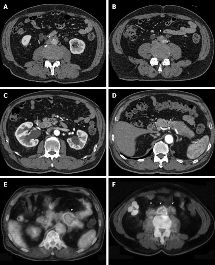
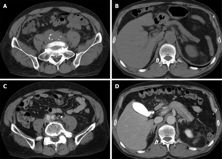
On the other hand, most of the studies to assess the frequency of concurrent retroperitoneal fibrosis in IgG4-related disease were conducted in patients with AIP[18-27]. In many of the studies, concurrent retroperitoneal fibrosis, represented by an extrapancreatic lesion, was reported in about 10% of patients with AIP. Takuma et al[18] reported that concurrent retroperitoneal fibrosis was found in 4 out of 56 patients with AIP (7%); of these, the onset of retroperitoneal fibrosis coincided with the onset of AIP in 2 patients, preceded the onset of AIP in 1 patient, and occurred 1 year after bypass surgery performed for AIP in 1 patient. Hamano et al[22] reported that concurrent retroperitoneal fibrosis was found in 8 out of 64 patients with AIP (12.5%). The onset of retroperitoneal fibrosis was heterochronic to the onset of AIP in 6 patients; the onset of retroperitoneal fibrosis preceded the onset of AIP in 3 patients and followed the onset of AIP in the other 3 patients[22]. Hirano et al[20] reported that concurrent retroperitoneal fibrosis was found in 8 out of 42 patients with AIP (19%). Ohara et al[21] reported that retroperitoneal fibrosis, as represented by an extrapancreatic lesion, was found in 8 out of 132 Japanese patients with AIP (6.1%). Moreover, the results of a Japanese multicenter study indicate that concurrent retroperitoneal fibrosis was found in 50 out of 459 patients with AIP who received steroid therapy (11%)[19]. The frequencies of concurrent retroperitoneal fibrosis reported in other countries are as follows: about 3%-4% in Asian countries including South Korea and China[23,24], about 8%-10% in the United States[25,26], and 3.6% in Europe[27]. In our experience, retroperitoneal fibrosis, as represented by an extrapancreatic lesion, was found in 8 of 51 patients (15.7%) diagnosed with AIP and treated at Kyushu University Hospital (Table 2). Of these, the onset of retroperitoneal fibrosis was synchronized with the onset of AIP in 5 patients (case presented in Figure 1), and was heterochronic to the onset of AIP in 3 patients. Regarding heterochronic onset of retroperitoneal fibrosis, the onset of retroperitoneal fibrosis preceded the onset of AIP in 2 patients (cases presented in Figure 2) and occurred 6.5 years after pancreatectomy in 1 patient with suspected pancreatic cancer.
Table 2.
Retroperitoneal fibrosis as extrapancreatic lesions of autoimmune pancreatitis
| Cases studied: 51 patients with AIP (2002–2011) | |
| Concurrent RF | 8 patients (15.7) |
| Synchronized onset | 5 patients |
| Heterochronic onset | 3 patients |
| Preceding the diagnosis of AIP | 2 patients |
| Following the diagnosis of AIP | 1 patient |
| Treatment: Steroid therapy at a dose of 30–40 mg per day | |
| Improved | 7 patients (87.5) |
| Unchanged | 1 patient (12.5) |
RF: Retroperitoneal fibrosis; AIP: Autoimmune pancreatitis.
Figure 1.

Typical retroperitoneal fibrosis associated with autoimmune pancreatitis. A, B: Soft tissue (arrow) surrounds the abdominal aorta and common iliac artery; C: Bilateral hydronephrosis (arrows); D: Swelling and capsule-like rim are seen in the body and tail of the pancreas. This case was diagnosed as autoimmune pancreatitis; E, F: Fluorodeoxyglucose positron emission tomography images from the same patient; high fluorodeoxyglucose uptake is seen in the body and tail of the pancreas (arrowhead) and in the retroperitoneal fibrosis lesions (arrowhead) surrounding the common iliac artery (arrows indicate dilated urinary ducts).
Figure 2.

Retroperitoneal fibrosis occurring before the onset of autoimmune pancreatitis. A 65-year-old man developed hydronephrosis and renal failure. A, B: Soft tissue was observed surrounding the abdominal aorta and common iliac artery. No abnormality was found in the pancreas. The patient was diagnosed with idiopathic retroperitoneal fibrosis, and started receiving prednisolone (PSL) at the dose of 40 mg/d. The dose was gradually reduced over approximately 6 mo and discontinued. The hydronephrosis improved, and the patient continued to be followed up; C, D: Swelling of the pancreas observed on computed tomography images taken about 1 yr and 6 mo after the discontinuation of PSL. Compared with the initial observation, retroperitoneal fibrosis improved, but persisted. The serum immunoglobulin G4 level was 366 mg/dL. At that point, the patient was diagnosed with autoimmune pancreatitis, and started receiving PSL again at the dose of 40 mg/d. The swelling of the pancreas and retroperitoneal fibrosis were improved.
There are reports on IgG4-related retroperitoneal fibrosis in diseases other than AIP. Ohta et al[28] reported concurrent retroperitoneal fibrosis in 1 out of 10 patients (10%) with IgG4-related sclerosing sialadenitis. Zen et al[29] reported concurrent retroperitoneal fibrosis in 13 out of 114 patients (11.4%) with histologically diagnosed IgG4-related sclerosing disease. Although further accumulation of case data is necessary to determine the accurate frequency of concurrent retroperitoneal fibrosis, the aforementioned studies suggest that concurrent retroperitoneal fibrosis occurs in about 10% of IgG4-related disease (Table 3). The concept of IgG4-related disease encompasses various disease conditions occurring in various organs; however, the primary cause of IgG4-related disease has not been elucidated. Moreover, it is unclear what clinical features determine the involvement of various organs, including the retroperitoneum. This needs to be elucidated in the future.
Table 3.
Frequency of concurrent retroperitoneal fibrosis in immunoglobulin G4-related disease
| Ref. | Disease | Total number of patients | Patients with RF n | Frequency of concurrent RF |
| Takuma et al[18] | AIP | 56 | 4 | 7.1 |
| Kamisawa et al[19] | AIP | 459 | 50 | 10.9 |
| Hirano et al[20] | AIP | 42 | 8 | 19.0 |
| Ohara et al[21] | AIP | 132 | 8 | 6.1 |
| Hamano et al[22] | AIP | 64 | 8 | 12.5 |
| Ryu et al[23] | AIP | 67 | 2 | 3.0 |
| Song et al[24] | AIP | 25 | 1 | 4.0 |
| Chari et al[25] | AIP | 29 | 3 | 10.3 |
| Raina et al[26] | AIP | 26 | 2 | 7.7 |
| Maire et al[27] | AIP | 28 | 1 | 3.6 |
| Ohta et al[28] | IgG4-related sclerosing sialadenitis | 10 | 1 | 10.0 |
| Zen et al[29] | IgG4-related sclerosing disease | 114 | 13 | 11.4 |
| Our cases | AIP | 51 | 8 | 15.7 |
| Total | IgG4-related disease | 1103 | 109 | 9.9 |
RF: Retroperitoneal fibrosis; AIP: Autoimmune pancreatitis; IgG4: Immunoglobulin G4.
DIAGNOSIS
Retroperitoneal fibrosis has no specific symptoms. However, dull abdominal (flank) pain, back pain, general malaise, fever, and weight loss have been known to occur. Retroperitoneal fibrosis often surrounds the urinary duct or inferior vena cava, and is often found in association with hydronephrosis or swelling of the lower extremities[1]. In recent times, retroperitoneal fibrosis is often detected during diagnostic imaging, such as a computed tomography (CT) scan, in asymptomatic patients undergoing a work-up for AIP (Figure 1). However, it should be noted that the onset of retroperitoneal fibrosis may or may not be synchronized with the onset of AIP. Furthermore, the onset of retroperitoneal fibrosis may either follow or precede the onset of AIP as described earlier. The histological features of retroperitoneal fibrosis associated with AIP (i.e., IgG4-related retroperitoneal fibrosis) include diffused fibrosis, infiltration of IgG4-positive plasma cells, obliterative phlebitis, eosinophilic infiltration, and the formation of lymphoid follicles; these are similar to other lesions of IgG4-related disease[15,16]. Zen et al[15] reported that obliterative phlebitis is significantly more frequent in IgG4-related retroperitoneal fibrosis than in non-IgG4-related retroperitoneal fibrosis. However, because surgery for retroperitoneal fibrosis is rarely performed and because a biopsy of the retroperitoneum cannot be easily carried out, it is often difficult to obtain the tissue specimens needed for diagnosis. Clinically, retroperitoneal fibrosis is diagnosed based on the results of diagnostic imaging such as CT scan and magnetic resonance imaging (MRI)[30]. Involvement of IgG4 is usually determined based on serum IgG4 levels and the presence/absence of other lesions such as AIP. Moreover, the effect of therapies is usually assessed using diagnostic imaging techniques.
There is no hematological parameter specifically associated with retroperitoneal fibrosis; however, an increased white blood cell count and increased C-reactive protein levels can be seen as a reflection of acute inflammation. Moreover, renal dysfunction may be observed due to ureteral obstruction. Diagnostic imaging techniques, such as ultrasound (US), CT, and MRI, are used to diagnose retroperitoneal fibrosis. US is usually performed first, because it is a simple and minimally invasive technique. Typically, retroperitoneal fibrosis is depicted as a hypo- to iso-echoic mass encasing an aorta; however, the sensitivity is low, and US is rather important in determining the presence/absence of concurrent hydronephrosis. The most useful diagnostic imaging techniques are CT and MRI. On CT images, retroperitoneal fibrosis is depicted as a soft tissue mass encasing an aorta, which often spreads laterally to involve the inferior vena cava and urinary duct[31] (Figure 1A-C). On MRI images, retroperitoneal fibrosis shows low signal intensity on T1-weighted images, and high signal intensity on T2-weighted images[1,31]. Moreover, although CT and MRI excel in their specificity, fluorodeoxyglucose positron emission tomography is also a useful tool for determination of the presence/absence of retroperitoneal fibrosis as well as other lesions of IgG4-related disease[1,32,33] (Figure 1D-F).
The retroperitoneum is also a site where tumor lesions, such as malignant lymphoma, desmoid fibromatosis, and liposarcoma, often develop. Thus, these diseases need to be differentiated from retroperitoneal fibrosis[1,3]. Although the onset of retroperitoneal fibrosis surrounding the pancreas is rare, this condition is sometimes difficult to distinguish from the invasion of pancreatic cancer to the area surrounding the superior mesenteric artery. In patients with previously diagnosed AIP in whom the onset of retroperitoneal fibrosis has occurred simultaneously with the onset (Figure 1) or relapse of AIP, retroperitoneal fibrosis can be relatively easily diagnosed based on the aforementioned characteristic findings on US/CT/MRI images and the presence of increased serum IgG4 levels. On the other hand, in some patients, the onset of retroperitoneal fibrosis precedes the onset of AIP (Figure 2); thus, the measurement of serum IgG4 levels is recommended for idiopathic retroperitoneal fibrosis[34]. When the IgG4 level is high, it should be treated and monitored as an IgG4-related disease.
TREATMENT AND PROGNOSIS
The first line of treatment for retroperitoneal fibrosis is steroid therapy, regardless of its cause. Although a few asymptomatic patients might need only monitoring, steroid therapy is usually necessary for patients with clinical symptoms (e.g., fatigue, weight loss, and abdominal or back pain) or those with hydronephrosis and acute renal failure caused by ureteral obstruction[35]. The efficacy of immunosuppressants (e.g., azathioprine) and tamoxifen has been reported mainly in Europe and the United States[1,36,37]. However, a randomized controlled trial recently conducted in patients with idiopathic retroperitoneal fibrosis demonstrated that the efficacy of steroids in preventing the relapse of retroperitoneal fibrosis was significantly better than that of tamoxifen[35]. Moreover, Zen et al[15] reported that steroid therapy was effective regardless of the presence/absence of IgG4-related disease or serum IgG4 levels[15].
Steroid therapy is also recognized as a standard therapy for AIP[38]. The indications of steroid are symptoms such as obstructive jaundice due to sclerosing cholangitis, abdominal pain, and hydronephrosis due to associated retroperitoneal fibrosis[19]. Therefore, steroid therapy is strongly recommended for patients with concurrent AIP, that is, IgG4-related retroperitoneal fibrosis. The starting dose of steroid is usually 30-40 mg/d (0.6 mg/kg per day)[38]. The response to steroid therapy is generally favorable. In most cases, the pancreatic lesion and retroperitoneal fibrosis improve after the initial treatment. Among the 8 patients we studied, clinical improvement was achieved in 7 patients. In 1 patient, the disease condition did not change (Table 2). As mentioned earlier, the response to steroid therapy is generally good; however, some patients are resistant to steroid. Further, the mechanism of steroid resistance is unclear, and it is difficult to predict the efficacy of steroid therapy before treatment. In our patient who was refractory to steroid, soft tissue mass surrounding the abdominal aorta and hydronephrosis did not improve; however, his pancreatic lesion was improved and renal function was stabilized. Therefore, we continued with the maintenance steroid treatment. Zen et al[15] reported that 8 of 10 patients with IgG4-related retroperitoneal fibrosis received steroid therapy. One of these steroid-treated patients experienced relapse twice; however, remission of retroperitoneal fibrosis was achieved by re-administration of steroid. In some cases, relapse occurs after withdrawal from steroid therapy. In other cases, the ureteral stent cannot be removed because of inadequate response to steroid therapy. Thus, further study should be conducted to assess the long-term prognoses of retroperitoneal fibrosis. Although the use of immunosuppressants has been reported in patients with IgG4-related disease that is recurrent or non-responsive to steroid therapy, no consensus has been reached yet[26,27,39].
CONCLUSION
Owing to the newly emerged recognition of retroperitoneal fibrosis as an extrapancreatic lesion of AIP and the establishment of the concept of IgG4-related disease, diagnostic methods and therapies for retroperitoneal fibrosis have made great progress. Retroperitoneal fibrosis, as well as other lesions of IgG4-related disease, responds to steroid therapy in most cases. However, data from more cases need to be collected in order to assess the long-term prognoses of retroperitoneal fibrosis.
Footnotes
Supported by The Research Program of Intractable Disease and the Research Committee of Intractable Pancreatic Diseases of the Ministry of Health, Labor and Welfare of Japan
P- Reviewer Masamun A S- Editor Gou SX L- Editor A E- Editor Xiong L
References
- 1.Vaglio A, Salvarani C, Buzio C. Retroperitoneal fibrosis. Lancet. 2006;367:241–251. doi: 10.1016/S0140-6736(06)68035-5. [DOI] [PubMed] [Google Scholar]
- 2.Ormond JK. Bilateral ureteral obstruction due to envelopment and compression by an inflammatory retroperitoneal process. J Urol. 1948;59:1072–1079. doi: 10.1016/S0022-5347(17)69482-5. [DOI] [PubMed] [Google Scholar]
- 3.Corradi D, Maestri R, Palmisano A, Bosio S, Greco P, Manenti L, Ferretti S, Cobelli R, Moroni G, Dei Tos AP, et al. Idiopathic retroperitoneal fibrosis: clinicopathologic features and differential diagnosis. Kidney Int. 2007;72:742–753. doi: 10.1038/sj.ki.5002427. [DOI] [PubMed] [Google Scholar]
- 4.Yoshida K, Toki F, Takeuchi T, Watanabe S, Shiratori K, Hayashi N. Chronic pancreatitis caused by an autoimmune abnormality. Proposal of the concept of autoimmune pancreatitis. Dig Dis Sci. 1995;40:1561–1568. doi: 10.1007/BF02285209. [DOI] [PubMed] [Google Scholar]
- 5.Ito T, Nakano I, Koyanagi S, Miyahara T, Migita Y, Ogoshi K, Sakai H, Matsunaga S, Yasuda O, Sumii T, et al. Autoimmune pancreatitis as a new clinical entity. Three cases of autoimmune pancreatitis with effective steroid therapy. Dig Dis Sci. 1997;42:1458–1468. doi: 10.1023/a:1018862626221. [DOI] [PubMed] [Google Scholar]
- 6.Hamano H, Kawa S, Horiuchi A, Unno H, Furuya N, Akamatsu T, Fukushima M, Nikaido T, Nakayama K, Usuda N, et al. High serum IgG4 concentrations in patients with sclerosing pancreatitis. N Engl J Med. 2001;344:732–738. doi: 10.1056/NEJM200103083441005. [DOI] [PubMed] [Google Scholar]
- 7.Hamano H, Kawa S, Ochi Y, Unno H, Shiba N, Wajiki M, Nakazawa K, Shimojo H, Kiyosawa K. Hydronephrosis associated with retroperitoneal fibrosis and sclerosing pancreatitis. Lancet. 2002;359:1403–1404. doi: 10.1016/s0140-6736(02)08359-9. [DOI] [PubMed] [Google Scholar]
- 8.Kamisawa T, Funata N, Hayashi Y, Eishi Y, Koike M, Tsuruta K, Okamoto A, Egawa N, Nakajima H. A new clinicopathological entity of IgG4-related autoimmune disease. J Gastroenterol. 2003;38:982–984. doi: 10.1007/s00535-003-1175-y. [DOI] [PubMed] [Google Scholar]
- 9.Kamisawa T, Okamoto A. Autoimmune pancreatitis: proposal of IgG4-related sclerosing disease. J Gastroenterol. 2006;41:613–625. doi: 10.1007/s00535-006-1862-6. [DOI] [PMC free article] [PubMed] [Google Scholar]
- 10.Masaki Y, Dong L, Kurose N, Kitagawa K, Morikawa Y, Yamamoto M, Takahashi H, Shinomura Y, Imai K, Saeki T, et al. Proposal for a new clinical entity, IgG4-positive multiorgan lymphoproliferative syndrome: analysis of 64 cases of IgG4-related disorders. Ann Rheum Dis. 2009;68:1310–1315. doi: 10.1136/ard.2008.089169. [DOI] [PubMed] [Google Scholar]
- 11.Kawano M, Saeki T, Nakashima H, Nishi S, Yamaguchi Y, Hisano S, Yamanaka N, Inoue D, Yamamoto M, Takahashi H, et al. Proposal for diagnostic criteria for IgG4-related kidney disease. Clin Exp Nephrol. 2011;15:615–626. doi: 10.1007/s10157-011-0521-2. [DOI] [PubMed] [Google Scholar]
- 12.Stone JH, Zen Y, Deshpande V. IgG4-related disease. N Engl J Med. 2012;366:539–551. doi: 10.1056/NEJMra1104650. [DOI] [PubMed] [Google Scholar]
- 13.Umehara H, Okazaki K, Masaki Y, Kawano M, Yamamoto M, Saeki T, Matsui S, Yoshino T, Nakamura S, Kawa S, et al. Comprehensive diagnostic criteria for IgG4-related disease (IgG4-RD), 2011. Mod Rheumatol. 2012;22:21–30. doi: 10.1007/s10165-011-0571-z. [DOI] [PubMed] [Google Scholar]
- 14.Uibu T, Oksa P, Auvinen A, Honkanen E, Metsärinne K, Saha H, Uitti J, Roto P. Asbestos exposure as a risk factor for retroperitoneal fibrosis. Lancet. 2004;363:1422–1426. doi: 10.1016/S0140-6736(04)16100-X. [DOI] [PubMed] [Google Scholar]
- 15.Zen Y, Onodera M, Inoue D, Kitao A, Matsui O, Nohara T, Namiki M, Kasashima S, Kawashima A, Matsumoto Y, et al. Retroperitoneal fibrosis: a clinicopathologic study with respect to immunoglobulin G4. Am J Surg Pathol. 2009;33:1833–1839. doi: 10.1097/pas.0b013e3181b72882. [DOI] [PubMed] [Google Scholar]
- 16.Neild GH, Rodriguez-Justo M, Wall C, Connolly JO. Hyper-IgG4 disease: report and characterisation of a new disease. BMC Med. 2006;4:23. doi: 10.1186/1741-7015-4-23. [DOI] [PMC free article] [PubMed] [Google Scholar]
- 17.Yamashita K, Haga H, Mikami Y, Kanematsu A, Nakashima Y, Kotani H, Ogawa O, Manabe T. Degree of IgG4+ plasma cell infiltration in retroperitoneal fibrosis with or without multifocal fibrosclerosis. Histopathology. 2008;52:404–409. doi: 10.1111/j.1365-2559.2007.02959.x. [DOI] [PubMed] [Google Scholar]
- 18.Takuma K, Kamisawa T, Anjiki H, Egawa N, Igarashi Y. Metachronous extrapancreatic lesions in autoimmune pancreatitis. Intern Med. 2010;49:529–533. doi: 10.2169/internalmedicine.49.3038. [DOI] [PubMed] [Google Scholar]
- 19.Kamisawa T, Shimosegawa T, Okazaki K, Nishino T, Watanabe H, Kanno A, Okumura F, Nishikawa T, Kobayashi K, Ichiya T, et al. Standard steroid treatment for autoimmune pancreatitis. Gut. 2009;58:1504–1507. doi: 10.1136/gut.2008.172908. [DOI] [PubMed] [Google Scholar]
- 20.Hirano K, Tada M, Isayama H, Yagioka H, Sasaki T, Kogure H, Nakai Y, Sasahira N, Tsujino T, Yoshida H, et al. Long-term prognosis of autoimmune pancreatitis with and without corticosteroid treatment. Gut. 2007;56:1719–1724. doi: 10.1136/gut.2006.115246. [DOI] [PMC free article] [PubMed] [Google Scholar]
- 21.Ohara H, Nakazawa T, Ando T, Joh T. Systemic extrapancreatic lesions associated with autoimmune pancreatitis. J Gastroenterol. 2007;42 Suppl 18:15–21. doi: 10.1007/s00535-007-2045-9. [DOI] [PubMed] [Google Scholar]
- 22.Hamano H, Arakura N, Muraki T, Ozaki Y, Kiyosawa K, Kawa S. Prevalence and distribution of extrapancreatic lesions complicating autoimmune pancreatitis. J Gastroenterol. 2006;41:1197–1205. doi: 10.1007/s00535-006-1908-9. [DOI] [PubMed] [Google Scholar]
- 23.Ryu JK, Chung JB, Park SW, Lee JK, Lee KT, Lee WJ, Moon JH, Cho KB, Kang DW, Hwang JH, et al. Review of 67 patients with autoimmune pancreatitis in Korea: a multicenter nationwide study. Pancreas. 2008;37:377–385. doi: 10.1097/MPA.0b013e31817a0914. [DOI] [PubMed] [Google Scholar]
- 24.Song Y, Liu QD, Zhou NX, Zhang WZ, Wang DJ. Diagnosis and management of autoimmune pancreatitis: experience from China. World J Gastroenterol. 2008;14:601–606. doi: 10.3748/wjg.14.601. [DOI] [PMC free article] [PubMed] [Google Scholar]
- 25.Chari ST, Smyrk TC, Levy MJ, Topazian MD, Takahashi N, Zhang L, Clain JE, Pearson RK, Petersen BT, Vege SS, et al. Diagnosis of autoimmune pancreatitis: the Mayo Clinic experience. Clin Gastroenterol Hepatol. 2006;4:1010–1016; quiz 934. doi: 10.1016/j.cgh.2006.05.017. [DOI] [PubMed] [Google Scholar]
- 26.Raina A, Yadav D, Krasinskas AM, McGrath KM, Khalid A, Sanders M, Whitcomb DC, Slivka A. Evaluation and management of autoimmune pancreatitis: experience at a large US center. Am J Gastroenterol. 2009;104:2295–2306. doi: 10.1038/ajg.2009.325. [DOI] [PMC free article] [PubMed] [Google Scholar]
- 27.Maire F, Le Baleur Y, Rebours V, Vullierme MP, Couvelard A, Voitot H, Sauvanet A, Hentic O, Lévy P, Ruszniewski P, et al. Outcome of patients with type 1 or 2 autoimmune pancreatitis. Am J Gastroenterol. 2011;106:151–156. doi: 10.1038/ajg.2010.314. [DOI] [PubMed] [Google Scholar]
- 28.Ohta N, Kurakami K, Ishida A, Furukawa T, Saito F, Kakehata S, Izuhara K. Clinical and pathological characteristics of IgG4-related sclerosing sialadenitis. Laryngoscope. 2012;122:572–577. doi: 10.1002/lary.22449. [DOI] [PubMed] [Google Scholar]
- 29.Zen Y, Nakanuma Y. IgG4-related disease: a cross-sectional study of 114 cases. Am J Surg Pathol. 2010;34:1812–1819. doi: 10.1097/PAS.0b013e3181f7266b. [DOI] [PubMed] [Google Scholar]
- 30.Kermani TA, Crowson CS, Achenbach SJ, Luthra HS. Idiopathic retroperitoneal fibrosis: a retrospective review of clinical presentation, treatment, and outcomes. Mayo Clin Proc. 2011;86:297–303. doi: 10.4065/mcp.2010.0663. [DOI] [PMC free article] [PubMed] [Google Scholar]
- 31.Vivas I, Nicolás AI, Velázquez P, Elduayen B, Fernández-Villa T, Martínez-Cuesta A. Retroperitoneal fibrosis: typical and atypical manifestations. Br J Radiol. 2000;73:214–222. doi: 10.1259/bjr.73.866.10884739. [DOI] [PubMed] [Google Scholar]
- 32.Ozaki Y, Oguchi K, Hamano H, Arakura N, Muraki T, Kiyosawa K, Momose M, Kadoya M, Miyata K, Aizawa T, et al. Differentiation of autoimmune pancreatitis from suspected pancreatic cancer by fluorine-18 fluorodeoxyglucose positron emission tomography. J Gastroenterol. 2008;43:144–151. doi: 10.1007/s00535-007-2132-y. [DOI] [PubMed] [Google Scholar]
- 33.Shimosegawa T, Kanno A. Autoimmune pancreatitis in Japan: overview and perspective. J Gastroenterol. 2009;44:503–517. doi: 10.1007/s00535-009-0054-6. [DOI] [PubMed] [Google Scholar]
- 34.Miura H, Miyachi Y. IgG4-related retroperitoneal fibrosis and sclerosing cholangitis independent of autoimmune pancreatitis. A recurrent case after a 5-year history of spontaneous remission. JOP. 2009;10:432–437. [PubMed] [Google Scholar]
- 35.Vaglio A, Palmisano A, Alberici F, Maggiore U, Ferretti S, Cobelli R, Ferrozzi F, Corradi D, Salvarani C, Buzio C. Prednisone versus tamoxifen in patients with idiopathic retroperitoneal fibrosis: an open-label randomised controlled trial. Lancet. 2011;378:338–346. doi: 10.1016/S0140-6736(11)60934-3. [DOI] [PubMed] [Google Scholar]
- 36.Marcolongo R, Tavolini IM, Laveder F, Busa M, Noventa F, Bassi P, Semenzato G. Immunosuppressive therapy for idiopathic retroperitoneal fibrosis: a retrospective analysis of 26 cases. Am J Med. 2004;116:194–197. doi: 10.1016/j.amjmed.2003.08.033. [DOI] [PubMed] [Google Scholar]
- 37.van Bommel EF, Hendriksz TR, Huiskes AW, Zeegers AG. Brief communication: tamoxifen therapy for nonmalignant retroperitoneal fibrosis. Ann Intern Med. 2006;144:101–106. doi: 10.7326/0003-4819-144-2-200601170-00007. [DOI] [PubMed] [Google Scholar]
- 38.Ito T, Nishimori I, Inoue N, Kawabe K, Gibo J, Arita Y, Okazaki K, Takayanagi R, Otsuki M. Treatment for autoimmune pancreatitis: consensus on the treatment for patients with autoimmune pancreatitis in Japan. J Gastroenterol. 2007;42 Suppl 18:50–58. doi: 10.1007/s00535-007-2051-y. [DOI] [PubMed] [Google Scholar]
- 39.Ghazale A, Chari ST, Zhang L, Smyrk TC, Takahashi N, Levy MJ, Topazian MD, Clain JE, Pearson RK, Petersen BT, et al. Immunoglobulin G4-associated cholangitis: clinical profile and response to therapy. Gastroenterology. 2008;134:706–715. doi: 10.1053/j.gastro.2007.12.009. [DOI] [PubMed] [Google Scholar]


