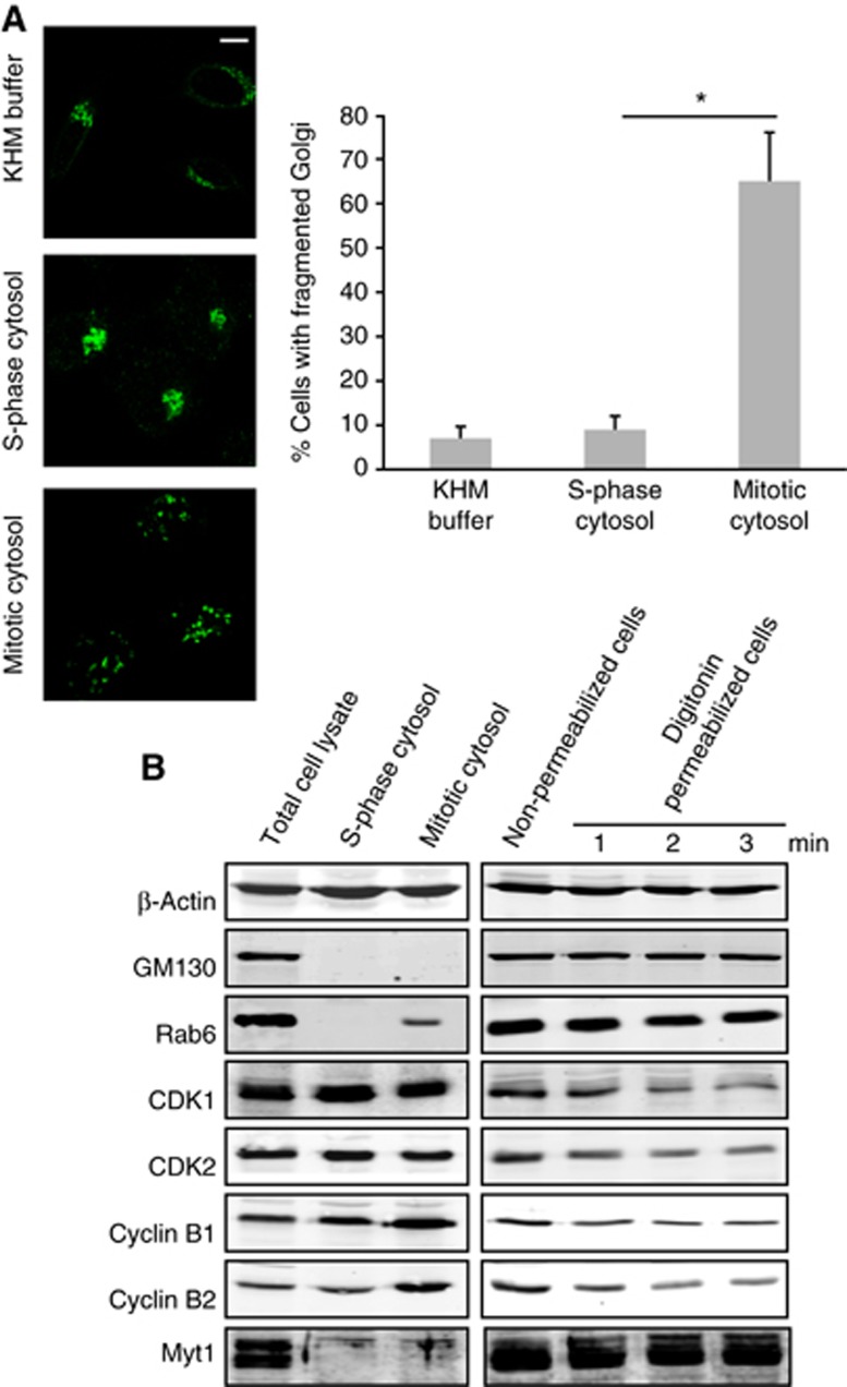
Figure 3.
Fragmentation of the Golgi complex with mitotic cytosol in permeabilized cells. (A) Left panel. HeLa cells stably expressing ManII-GFP were grown on coverslips and incubated with thymidine for 12 h before permeabilization. Permeabilized cells were incubated with an ATP-regenerating system and either KHM buffer (top panel), S-phase cytosol (centre panel), or mitotic cytosol (bottom panel), at 32°C for 1 h. Cells were fixed and visualized by fluorescence microscopy. Scale bar is 10 μm. Right panel. Quantitation of the experimental data. 200 cells on 2 different coverslips were counted (mean±s.d., n=3, *P<0.05) for each experimental condition to obtain the percentage of cells with fragmented Golgi complex. (B) Left panel. Total cell lysate, S-phase, and mitotic cytosol were western blotted with the antibodies shown. Right panel. The level of the same (indicated) proteins was analysed by western blotting the total cell lysate of non-permeabilized cells and cells permeabilized for 1, 2 and 3 min with digitonin and washed with 1M KCl in KHM buffer.
Source data for this figure is available on the online supplementary information page.