Abstract
B-cell lymphoma 2 (Bcl-2) is an important regulator of cellular plasticity and resilience. In bipolar disorder (BD), studies have shown a key role for a Bcl-2 gene single-nucleotide polymorphism (SNP) rs956572 in the regulation of intracellular calcium (Ca2+) dynamics, Bcl-2 expression/levels, and vulnerability to cellular apoptosis. At the same time, Bcl-2 decreases glutamate (Glu) toxicity in neural cells. Abnormalities in Glu function have been implicated in BD. In magnetic resonance spectroscopy (MRS) studies, anterior cingulated cortex (ACC) Glu levels have been reported to be increased in bipolar depression and mania, but no study specifically evaluated ACC Glu levels in BD-euthymia. Here, we compared ACC Glu levels in BD-euthymia compared with healthy subjects using 1H-MRS and also evaluated the selective role of the rs956572 Bcl-2 SNP in modulating ACC Glu and Glx (sum of Glu and glutamine) in euthymic-BD. Forty euthymic subjects with BD type I and forty healthy controls aged 18–40 were evaluated. All participants were genotyped for Bcl-2 rs956572 and underwent a 3-Tesla brain magnetic resonance imaging examination including the acquisition of an in vivo PRESS single voxel (2 cm3) 1H-MRS sequence to obtain metabolite levels from the ACC. Euthymic-BD subjects had higher Glu/Cre (creatine) and Glx/Cre compared with healthy controls. The Bcl-2 SNP AA genotype was associated with elevated ACC Glu/Cre and Glx/Cre ratio in the BD group but not in controls. The present study reports for the first time an increase in ACC Glu/Cre and Glx/Cre ratios in BD-euthymia. Also, Bcl-2 AA genotype, previously associated with lower Bcl-2 expression and increase intracellular Ca2+, showed to be associated with increased ACC Glu and Glx levels in euthymic-BD subjects. The present findings reinforce a key role for glutamatergic system dysfunction in the pathophysiology of BD, potentially involving modulatory effects by Bcl-2 in the ACC.
Keywords: Bcl-2, bipolar disorder, depression, calcium, glutamate, spectroscopy
INTRODUCTION
Bipolar disorder (BD) has been consistently associated with glutamatergic system abnormalities (Machado-Vieira et al, 2009a; Manji 2008). Glutamate (Glu) is released into the synaptic cleft, taken up by glial cells, converted to glutamine (Gln), and cycled back to neurons. Gln/Glu ratios may represent a marker of glutamatergic synapse integrity and neuronal–glial coupling in the tripartite glutamatergic synapse (Machado-Vieira 2009b). Glu excess is known to lead to excitotoxicity and apoptosis (Hashimoto et al, 2002), associated with increased intracellular calcium (Ca+2) levels and generation of mitochondrial reactive species (Kumar et al, 2010).
The evidence about Glu abnormalities in patients with BD comes from several different angles, including peripheral biomarkers, postmortem, genetic, treatment, as well as magnetic resonance spectroscopy (MRS) imaging studies (reviewed in Zarate et al, 2010). For example, glutamatergic abnormalities have been reported in plasma (Kim et al, 1982; Altamura et al, 1993; Mauri et al, 1998), serum (Mitani et al, 2006), cerebral spinal fluid (CSF) (Levine et al, 2000; Frye et al, 2007), and brain tissue (Francis et al, 1989; Eastwood and Harrison, 2010) of individuals afflicted with BD, as well as unipolar depression. A recent postmortem study specifically controlling for the effects of postmortem interval found increased levels of Glu in the frontal cortex of patients with BD and unipolar depression (Hashimoto et al, 2007; Lan et al, 2009). Postmortem studies have also reported decreased NR1 and NR2A transcripts, and suggest a decrease in the total number of NMDA receptors, as well as a decrease in the number of open ion channels in BD (Scarr et al, 2003; Beneyto et al, 2007; Beneyto and Meador-Woodruff, 2008). Pharmacological studies reinforce the association between BD and the glutamatergic system by reporting that first line agents to treat BD, such as lithium, valproate, carbamazepine, and lamotrigine, also modulate the glutamatergic system (Yatham et al, 2009). In view of the evidence that excessive synaptic Glu may contribute to neuronal atrophy and loss, it is notable that chronic treatment with lithium has been shown to upregulate synaptosomal uptake of Glu in mice (Hokin et al, 1996). Chronic administration of VPA led to a dose-dependent increase in hippocampal Glu uptake capacity, as measured by uptake of [3H] Glu into proteoliposomes, by increasing the levels of the Glu transporters EAAT1 and EAAT2 in hippocampus. Overall, chronic treatment with VPA or lithium is likely to decrease intrasynaptic Glu levels through a variety of mechanisms. Of all the medications currently used in the treatment of BD, lamotrigine probably has the most direct effects on the Glu system. There is evidence that it inhibits the release of Glu in the hippocampus of rats (Ahmad et al, 2005) and that it increases AMPA subunit receptor expression (Du et al, 2007). Moreover, glutamatergic abnormalities in BD have been also detected through the use of MRS; MRS studies report higher Glu and Glx (Glu+Gln) levels in different brain regions across depression and mania in patients with BD (Yüksel and Ongur, 2010). Preliminary studies done in patients in euthymia show elevation of Glx and Glu in hippocampus, orbitofrontal cortex, and occipital cortex (Bhagwagar et al, 2007; Colla et al, 2009; Senaratne et al, 2009) but no study has specifically evaluated anterior cingulate cortex (ACC) Glu-related metabolites. The ACC is a key region implicated in mood regulation (Drevets, 2000; Phillips et al, 2003) and it shows extensive functional and structural abnormalities in patients with BDs, which are also present during periods of euthymia, suggesting that ACC dysfunction might represent a possible trait marker of BD (Liu et al, 2012; Foland-Ross et al, 2011). Therefore, we hypothesized that ACC glutamatergic abnormalities would also be present in euthymic-BD patients compared with healthy control subjects.
Recent evidence suggests that antiapoptotic protein B-cell lymphoma 2 (Bcl-2) might be an important modulator of glutamatergic functioning (Machado-Vieira et al, 2011); Bcl-2 regulates diverse neurobiological processes such as neurogenesis, morphogenesis, and synaptic plasticity (Machado-Vieira et al, 2011) by conferring increased cell survival by blocking proapoptotic family members like Bax and BH3 proteins(Chao and Korsmeyer, 1998). Bcl-2 protein has two main protective functions; first it regulates Ca2+ release from the endoplasmatic reticulum by directly interacting with the inositol 1,4,5-triphosphate receptor (Chen et al, 2004). Second, it acts as a sensor for the redox status of mitochondria by detecting and regulating the mitochondrial glutathione pool (Zimmermann et al, 2007).
Evidences have shown that Bcl-2 protects neural cells from glutamatergic insults, which are thought to lead to cell death, neuropil reshaping, and subsequent abnormalities in brain volume. It has been reported that Bcl-2 expression could be an important factor promoting neuronal survival after increased exposure to Glu. An increase in Bcl-2 expression has been also reported to protect cells against the deleterious effect of Glu toxicity in neural cells (Zhong et al, 1993). On the other hand, Bcl-2 gene expression was shown to significantly decrease after Glu exposure, associated with an increase in intracellular neuronal Ca2+ and oxidative stress levels (Machado-Vieira et al, 2009a; Kumar et al, 2010; Manji, 2008). It was described a significant decrease in Bcl-2 protein and mRNA expression in the frontal cortex of subjects with BD (Machado-Vieira et al 2009b; Kim et al, 2010). Recently, the Bcl-2 single-nucleotide polymorphism (SNP) (rs956572 variant AA) was reported to increase basal and stimulated cytosolic Ca2+ levels in lymphoblasts from BD subjects, associated with a significant reduction in Bcl-2 levels and mRNA expression in this variant (Hashimoto et al, 2002; Machado-Vieira et al, 2011). However, the interplay between the glutamatergic system and Bcl-2 function in BD is not fully understood.
The present study investigates the potential role for ACC glutamatergic-related metabolites (ie, Glu and Glx) in BD-euthymia as compared with healthy control subjects. We hypothesize that glutamatergic metabolites (Glu and Glx) are increased during BD-euthymia. We also investigated whether the Bcl-2 rs956572 SNP, known to modulate Bcl-2 levels and which has been implicated in BD, modulates ACC glutamatergic metabolites. We hypothesize that BD-euthymic subjects AA carriers would display higher Glu levels in the ACC compared with other genotypes.
MATERIALS AND METHODS
Forty euthymic-BD I subjects (26F) aged 18–40 years old were included in the study. Diagnoses were determined by trained psychiatrists based on the Structured Clinical Interview (SCID-I/P) (Frye et al, 2007; First et al, 1996; Ongur et al, 2008) for DSM-IV TR (Bhagwagar et al, 2007; DSM-IV, 2000; Colla et al, 2009; Senaratne et al, 2009). Subjects had been on stable medication regimen for at least 2 months before the scanning session. Subjects with neurological disorders or medical disorders, head trauma, current substance abuse, or treated with electroconvulsive therapy in the last 6 months, were excluded. The Young Mania Rating Scale (YMRS) (Machado-Vieira et al, 2011; Young et al, 1978) and the Hamilton Depression Rating Scale (HDRS-21) (Chao and Korsmeyer, 1998; Hamilton, 1960) were used to assess residual subthreshold depressive and manic symptoms. Euthymia was defined as <12 YMRS and <7 HDRS (Chen et al, 2004; Nierenberg and DeCecco, 2001; Tohen et al, 2000). Patients also fulfilled DSM-IV criteria for remission.
Forty healthy individuals (19F, aged 18–40 years old) were also recruited in the study. All healthy controls had no current or past history of psychiatric disorder according to the evaluation conducted by trained psychiatrists using the Mini International Neuropsychiatric Interview (MINI) (Zimmermann et al, 2007; Sheehan et al, 1998). In addition, healthy control subjects had no family history in first-degree relatives with mood or psychotic disorders and were not taking any psychotropic medicine for at least 3 months before enrollment, based on a semi-structured interview. Subjects with a history of substance abuse within the previous 3 months before enrollment were excluded from the study. The research ethics board of the Hospital das Clinicas—University of Sao Paulo approved the study. Written informed consent was obtained from all study participants.
Image Acquisition
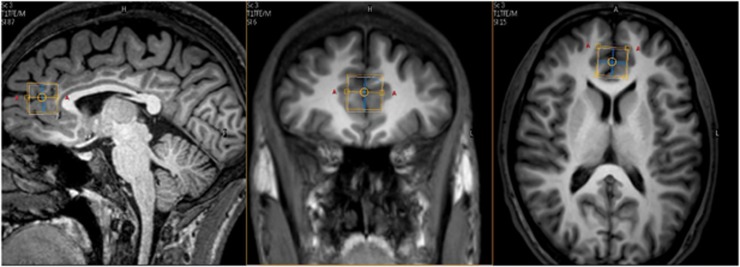
MRS imaging sequences were performed using a 3.0T magnet (Intera Achieva, Philips, Best, the Netherlands) and an eight-channel head coil. Metabolite levels of Glu; myo-inositol; N-acetylaspartate, creatine (Cre); choline (Cho); and Glx were obtained using the PRESS sequence (TE/TR=80/1500 ms) with 160 scan averages. The size of the MRS voxel was 2 cm3 and it was placed in the ACC as shown in Figure 1. The MRS voxel was placed, on midsagittal T1-weighted images, anterior to genu of the corpus callosum, with the ventral edge aligned with the dorsal corner of the genu, and centered on the midline of axial images. Spectra were quantified with LCModel software (Zhong et al, 1993; Provencher, 1993), using internal water as a reference, and metabolite levels with CRLBs>20% were excluded from the analysis. We herein report the metabolite concentrations as ratios over Cre, because absolute values are usually considered less reliable as they are more susceptible to relaxation and partial volume effects than ratios over Cre (Provencher, 2012; http://s-provencher.com/pages/lcm-manual.shtml).
Figure 1.
T1-weighted magnetic resonance imaging (MRI) location for anterior cingulate prefrontal cortex 1H-MRS single-voxel acquisition (2 cm3).
To investigate brain tissue composition of the voxel of interest, three-dimensional volumetric images were obtained using the 3D-T1FFE technique (FA=8° TE/TR/TI=3.2/7/900 ms) with an isotropic voxels of 1 mm3. Briefly, the brain tissue was extracted using the brain extraction tool (BET) and segmentation into white matter (WM), gray matter (GM), and CSF was achieved using the automated brain segmentation tool FAST, both apart of the FSL suite (http://www.fmrib.ox.ac.uk/fsl).
Genotyping
DNA was extracted from peripheral blood according to the salting-out protocol (Laitinen et al, 1994) and then genotyped for Bcl-2 rs956572 using real-time PCR allelic discrimination. PCR amplification for rs956572 was performed in 5 μl reactions with 5 ng of template DNA, 1 × TaqMan Universal Master Mix (Applied Biosystems, Foster City, CA), 1 × each primer and probe assay, and H2O. Thermal cycling consisted of initial denaturation for10 min at 95 °C, followed by 40 cycles of denaturation at 95 °C for 15 s and annealing at 60 °C for 1 min. Fluorescence detection occurred in the annealing step. The amplification and allelic discrimination were performed in a 7500 Real-Time System (Applied Biosystems). Quality control of Real time PCR results was done by direct sequencing using the ABI PRISM 3100 Genetic Analyzer (Applied Biosystems).
Statistical Analysis
χ2 test was used for comparison of categorical data, and the ANOVA for continuous data. To analyze the influence of Bcl-2 genotype on MRS results, we used a multivariate analysis of covariance (MANOVA) entering MRS metabolites as dependent variables, and age, gender, group, and Bcl-2 genotype as covariates. To assess the effect of medication exposure on the metabolites of interest, we repeated the MANOVA model using treatment divided into four main subclasses (ie, lithium, anticonvulsants, antipsychotics, and benzodiazepines) as a categorical factor (yes/no). Results shown were corrected by Bonferroni post-hoc test for multiple variables. A significance level of P<0.05 was adopted and all analysis were performed using the IBM PASW 20 (Supplementary Figure S1)
RESULTS
The Bcl-2 genotype distribution in the experimental sample was in accordance with the Hardy–Weinberg equilibrium (χ2=0.06 p=0.80), and genotype (p=0.6) and allele A (p=0.24) frequency did not differ between BD and control group. The three genotypes groups did not differ on age or gender (Table 1). Sociodemographic and illness characteristics, as well as medications, use did not differ between the three Bcl-2 genotype groups (Table 1).
Table 1. Sociodemographic and Clinical Variables by Bcl-2 Genotype in Each Group.
| Variable differences | AA (n=10) | AG (n=38) | GG (n=32) | Between-group differences | ||||
|---|---|---|---|---|---|---|---|---|
| |
Mean |
SD |
Mean |
SD |
Mean |
SD |
F |
Sig. (2-tailed) |
| Healthy controls (n=40) | ||||||||
| Age (years) | 25.2 | 6.7 | 27 | 4.5 | 26.5 | 6.7 | 0.14 | 0.90a |
| Gender (men/women) | 2/2 | 7/11 | 12/6 | 0.24b | ||||
| Bipolar disorder (n=40) | ||||||||
| Age (years) | 25.8 | 5.8 | 28.5 | 5.8 | 30.7 | 6.8 | 1.4 | 0.24a |
| Gender (men/women) | 2/4 | 9/11 | 3/11 | 0.40b | ||||
| YMRS | 1.6 | 0.5 | 2.5 | 1.7 | 3.10 | 2.3 | 0.6 | 0.5a |
| HDRS | 5.0 | 1.7 | 4.0 | 1.9 | 5.0 | 1.7 | 1.1 | 0.34a |
| Length of illness (years) | 7 | 7.8 | 8.8 | 5.1 | 7.8 | 5.8 | 0.1 | 0.82a |
| Age at first episode (years) | 17.6 | 1.5 | 19.0 | 5 | 21.3 | 3.4 | 1.1 | 0.34a |
| Number of manic episodes | 2.6 | 2.8 | 3.7 | 2.7 | 3.1 | 1.6 | 0.3 | 0.7a |
| Medication use in bipolar disorder | ||||||||
| Lithium | 4/6 | 18/20 | 10/14 | 0.27b | ||||
| Anticonvulsants | 0/6 | 8/20 | 9/14 | 0.03b | ||||
| Antipsychotics | 0/6 | 3/20 | 2/14 | 0.60b | ||||
| Antidepressants | 2/6 | 4/20 | 2/14 | 0.62b | ||||
| Benzodiazepines | 0/6 | 1/20 | 1/14 | 0.79b | ||||
ANOVA.
χ2 test.
Significance level p<0.05.
There were no significant differences between BD patients and healthy subjects in the voxel of interest (Figure 1) regarding volumes of GM (F=1.84, df=78, p=0.12), WM (F=2.15, df=78, p=0.19), or CSF (F=2.75, df=78, p=0.63). Also, no differences on GM, WM, and CSF in voxel content were observed among the three Bcl-2 genotypes. We here report the metabolites ratios by Cre, although the analysis of absolute values (without Cre) revealed similar results (Supplementary Tables S1–3). Furthermore, metabolites levels correction by voxel content (GM, WM, CSF) also yielded similar results.
Increased ACC Glu/Cre and Glx/Cre in Subjects with BD Compared with Healthy Controls Using 1H-MRS
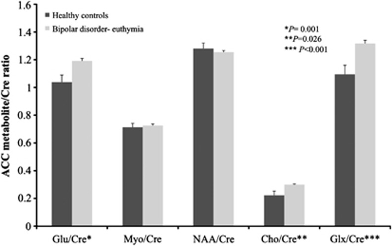
Significant main effects of metabolites levels were observed by diagnostic group. BD group demonstrated higher ratios of glutamatergic metabolites Glu/Cre (F=11.0, df=1, p=0.001), and Glx/Cre (F=14.0, df=1, p<0.001) in the ACC voxel compared with healthy controls.
Cho/Cre levels (F=5.1, df=1, p=0.026) were also higher in the ACC voxel compared with the control group. No difference in other metabolites was observed. (Figure 2)
Figure 2.
Mean metabolite/Cre ratios between bipolar disorder and healthy controls.
A Selective Increase in ACC Glu/Cre and Glx/Cre Ratios in Euthymic-BD Subjects Carrying the Bcl2 SNP rs956572AA
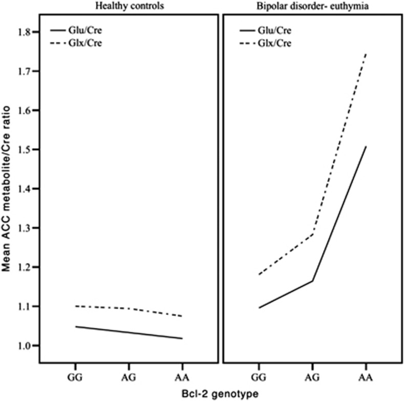
Euthymic-BD subjects with the AA genotype had higher ACC Glu/Cre ratio in AA compared with AG (B=−0.35, p=0.03) and GG (B=−0.46, p=0.008) (Figure 3). Also, BD subjects carrying the AA variant had Glx/Cre compared with AG (B=−0.48, p=0.02) and GG (B=−0.61, p=0.05). (Table 2; Supplementary Figure S2) No evidence of Bcl-2 genotype impact on MRS metabolite ratios was found in healthy controls (Figure 3).
Figure 3.
Interaction between mean anterior cingulated cortex (ACC) Glu/Cre and Glx/Cre levels, and B-cell lymphoma 2 (Bcl-2) genotype.
Table 2. MANCOVA Model: Metabolites/Cre Ratio as Dependent Variables and Age, Gender, Group, and Bcl-2 Genotype as Covariates.
| Dependent variable | Covariate | B | SE | t | Sig. | Partial eta squared (%) | Observed power (%) |
|---|---|---|---|---|---|---|---|
| Glu/Cre | |||||||
| Age | 0.006 | 0.005 | 1314 | 0.193 | 2.3 | 25.4 | |
| Gender | 0.013 | 0.056 | 0.233 | 0.816 | 0.1 | 5.6 | |
| Group | 0.484 | 0.148 | 3261 | 0.002 | 12.9 | 89.6 | |
| Bcl-2 genotype (AG) | 0.001 | 0.127 | 0.007 | 0.995 | 0.0 | 5.0 | |
| Bcl-2 genotype (GG) | 0.024 | 0.127 | 0.192 | 0.848 | 0.1 | 5.4 | |
| Bcl-2 genotype (AA) | 0a | – | – | – | – | – | |
| Group*Bcl-2 genotype (AG) | −0.359 | 0.167 | −2154 | 0.035 | 6.1 | 56.6 | |
| Group*Bcl-2 genotype (GG) | −0.468 | 0.171 | −2745 | 0.008 | 9.5 | 77.3 | |
| Group*Bcl-2 genotype (AA) | 0a | – | – | – | – | – | |
| Glx/Cre | |||||||
| Age | 0.009 | 0.006 | 1496 | 0.139 | 3.0 | 31.5 | |
| Gender | −0.002 | 0.071 | −0.034 | 0.973 | 0.0 | 5.0 | |
| Group | 0.667 | 0.187 | 3557 | 0.001 | 14.9 | 93.9 | |
| Bcl-2 genotype (AG) | 0.002 | 0.161 | 0.011 | 0.991 | 0.0 | 5.0 | |
| Bcl-2 genotype (GG) | 0.014 | 0.161 | 0.085 | 0.933 | 0.0 | 5.1 | |
| Bcl-2 genotype (AA) | 0a | – | – | – | – | – | |
| Group*Bcl-2 genotype (AG) | −0.488 | 0.211 | −2.32 | 0.023 | 7.0 | 62.9 | |
| Group*Bcl-2 genotype (GG) | −0.621 | 0.215 | −2887 | 0.005 | 10.4 | 81.3 | |
| Group*Bcl-2 genotype (AA) | 0a | – | – | – | – | – | |
Abbreviations: Cre, creatine phosphocreatine; Glu, glutamate; Glx, glutamate+glutamine.
This parameter is set to zero because it is redundant.
Significance level p<0.05.
When lithium, anticonvulsant, antipsychotic, and benzodiazepines were used as factors in the analysis, results for Glu/Cre and Glx/Cre remained similar. AA genotype had higher Glu/Cre ratio compared with GG (B=−0.50, p=0.006) and AG (B=−0.47, p=0.004). Glx/Cre ratio was also higher in AA compared with GG (B=0.70, p=0.001) and AG (B=−0.67, p=0.001).
Medication Effects
Glu/Cre and Glx/Cre ratios and metabolite ratio were no different comparing BD-euthymic subjects who were taking lithium ((Glu/Cre F=1.80, p=0.16), (Glx/Cre F=1.40 p=0.24)), benzodiazepines ((Glu/Cre F=1.28, p=0.26), (Glx/Cre F=1.45 Pp=0.23)), or antipsychotics ((Glu/Cre F=2.01, p=0.16), (Glx/Cre F=1.31 p=0.26)) to subjects who were not taking those medications at the time of scanning. Patients treated with anticonvulsants (valproate or carbamazepine) (n=16) had lower Glu/Cre and Glx/Cre ratio ((Glu/Cre F=7.90, p=0.01), (Glx/Cre F=10.78 p=0.003)) compared with the patients not taking anticonvulsants. To further investigate the effects of concomitant treatments with anticonvulsants on Glu-related metabolites, we excluded patients taking anticonvulsants from the MANCOVA model, and the analysis of the data demonstrated the same result of higher levels of Glu/Cre (F=10.53, p=0.02) and Glx/Cre (F=12.99, p=0.001) as before the exclusion of these subjects.
DISCUSSION
To the best of our knowledge, this is the first MRS study to specifically evaluate Glu ACC levels by Bcl-2 genotype in euthymic-BD compared with healthy controls. We found that BD subjects during euthymia have higher ACC Glu/Cre and Glx/Cre compared with healthy controls. Overall, our findings of elevated Glu/Cre and Glx/Cre support a potential role for glutamatergic overactivity in the pathophysiology of BD type I, which might represent a trait marker in BD considering that this finding has also been reported during mood episodes (Yüksel and Ongur, 2010). Furthermore, the present study also describes for the first time a selective modulatory effect of the Bcl-2 SNP rs956572 on ACC Glu/Cre and Glx/Cre ratios in euthymic-BD subjects but not in healthy controls. Specifically, AA genotype had higher ACC Glu/Cre and Glx/Cre ratios compared with the AG and GG variants in euthymic-BD (Figure 3).
Altered glutamatergic system has been one of the most consistent findings in the MRS studies in BD (Yüksel and Ongur, 2010). ACC glutamatergic abnormalities have been consistently described during episodes of illness in BD. For example, Frye et al (2007) described increased ACC Glu levels in BD (n=23) compared with healthy controls (n=12) (Frye et al, 2007). Ongur et al (2008) reported higher Gln/Glu in the ACC in subjects with BD in acute mania (n=15) compared with healthy controls (n=21), possibly suggesting an impairment in Glu turn-over between neurons and astroglial cells. In that study, medication status did not impact levels of Gln/Glu (Ongur et al, 2008), which is similar to which was also observed in the present study for lithium, antipsychotics, and benzodiazepines. Friedman et al (2004) reported instead that treatment with lithium but not with valproate might reduce Glx concentrations, a finding which was not confirmed by our results. In the present study, we found that patients treated with anticonvulsants had lower levels of glutamatergic metabolites but it is unlikely that medication status affected the main findings of the study, which remain essentially unchanged after removing patients who were talking anticonvulsants at the time of the scanning. Further longitudinal studies are needed to investigate the effects of anticonvulsants and other treatment classes on Glu-related metabolites.
Previous MRS studies with euthymic-BD subjects reported increased Glu (Colla et al, 2009) and Glx in hippocampus (Senaratne et al, 2009), occipital (Bhagwagar et al, 2007; Senaratne et al, 2009), and the orbitofrontal cortices (Senaratne et al, 2009), but no study has specifically evaluated ACC as the region of interest. Importantly, the ACC has been directly implicated in the pathophysiology and therapeutics of BD. ACC modulates autonomic/neuroendocrine responses and neurotransmitters activity during neural processing associated with control of emotional behavior and stress (Drevets et al, 2008). Furthermore, the ACC shares extensive anatomical connections with the amygdala; subiculum; hypothalamus; accumbens; ventral tegmental area; substantia nigra; raphe; locus ceruleus; periaqueductal gray; and brainstem autonomic nuclei, and other areas of the orbitomedial PFC (Ongur et al, 2003; Drevets et al, 1998). These structures are implicated in the modulation of emotional behavior, raising the possibility that abnormal synaptic interactions between these areas and the ACC may contribute to disturbances in emotional processing or regulation (Ongür et al, 1998).The ACC seems particularly vulnerable to potential disease associated changes such as oxidative stress (Wang et al, 2009), which might be modulated by Bcl-2 levels (Hockenbery et al, 1993).
Overall, our findings suggest that elevated Glu and Glx support a potential role for glutamatergic overactivity in the pathophysiology of BD during mood episodes and euthymia, which might represent a trait marker in BD.
The Bcl-2 AA genotype has been previously associated with reduced Bcl-2 levels/expression and increased cytosolic Ca2+ in human lymphoblasts (Machado-Vieira et al, 2011). AA variant is also associated with lower GM volume in healthy individuals (Salvadore et al, 2009). Furthermore, lower Bcl-2 protein and mRNA levels were described in the frontal cortex of BD subjects (Kim et al, 2010). Further supporting the neuroprotective profile of Bcl-2, preclinical models have also demonstrated that 4 weeks of lithium treatment upregulates Bcl-2 in the dentate gyrus and hippocampus (Hammonds and Shim, 2009). Also, behavioral studies suggest a potential role for Bcl-2 in BD phenotype (Lien et al, 2008), and as a key target for mood stabilizers therapeutic actions (Manji et al, 2000). Considering that Bcl-2 AA genotype has been associated to Ca2+ homeostasis, which accumulation mediates Glu cellular excitotoxicity (Lazarewicz and Salińska, 1993), Bcl-2 AA genotype would be a risk factor for neuronal apoptosis and oxidative stress, potentially displaying less adaptive mechanisms to glutamatergic insult associated with lower Bcl-2 expression and higher ACC Glu levels.
Bcl-2 is a key protein involved in neurons survival after Glu exposure. Deleterious cellular effects of Glu seem to involve Bcl-2 at early stages. The AA genotype in BD subjects would have increased levels of glutamatergic metabolites, which might lead to greater deleterious cellular effects and excitotoxicity. Bcl-2 gene expression decreases after Glu exposure (Kumar et al, 2010), thus potentially altering key processes mediated by Bcl-2, such as neurogenesis, morphogenesis, Ca2+ dynamics, and synaptic plasticity (Machado-Vieira et al, 2011). We might also speculate that higher Glu/Cre ratio observed in patients with the rs956572 SNP AA genotype might be reflecting cytosolic Ca2+ dysregulation previously associated with this Bcl-2 SNP (Machado-Vieira et al, 2011; Uemura et al, 2011).
This study has several strengths. First, this study has the largest sample size evaluating Glu metabolites with MRS in BD research. Previous MRS studies have had small sample sizes; only 5 out of 11 previous studies of individuals with BD had more than 20 patients (Yüksel and Ongur, 2010). Also, we choose to analyze the TE/TR=80 ms to better evaluate Glu levels. One possible limitation of this study is the small sample size in the Bcl-2 AA genotype group, which is a relatively uncommon genotype with a prevalence ranging of 5–20% in the general population (National Center for Biotechnology Information, 2012). Moreover, subjects in the Bcl-2 AA genotype were under pharmacotherapy with lithium or antidepressants (but not anticonvulsants), which could be a limitation. The evidence provided herein about the Bcl-2 rs956572 SNP as a regulator of glutamatergic function in patients with BD adds to the evidence from previous in vitro studies in patients with BD, but it needs to be replicated in a larger cohort of patients. Finally, given that BD is a chronic illness with dramatic psychosocial consequences, all patients were taking medications at the time of the evaluation and this limits the interpretation of our results, although we included treatment as a covariate in our analysis and we found no significant effect on our results.
In sum, the present findings highlight a role for altered ACC glutamatergic activity in BD. The modulatory effects of the rs956572 Bcl-2 SNP on ACC glutamatergic metabolites in euthymic-BD (but not in controls) support an association between Bcl-2 activity and glutamatergic system dysregulation in the ACC, which might be relevant for the pathophysiology and therapeutics of this devastating illness. Further studies are warranted to confirm the association between Bcl-2 and ACC glutamatergic metabolism.
Acknowledgments
We would like to thank the members of Mood Disorders Unit (GRUDA) and Laboratory of Neuroscience (LIM27), Institute of Psychiatry, University of Sao Paulo for their hard work, as well as the volunteers for their collaboration. We also thank Sao Paulo Research Foundation (Fapesp) and Associação Beneficente Alzira Denise Hertzog da Silva (ABADHS). This study was financed by Sao Paulo Research Foundation (Fapesp). Dr Salvadore is a full time employee of Janssen pharmaceutical company. Dr Moreno has acted as a consultant to and conducted research sponsored by companies with developments in the area of bipolar and depressive disorders (Servier, BMS, Eli Lilly, Abbott, Astra Zêneca, GSK) and received research grants from Research Foundation Support Agency of the State of Sao Paulo, Brazil (FAPESP). Dr Zarate is listed as a co-inventor on a patent application for the use of ketamine and its metabolites in major depression. Dr Zarate has assigned his rights in the patent to the US government but will share a percentage of any royalties that may be received by the government. Dr Zarate is also the recipient of the Brain & Behavior Research Foundation Award. Dr Machado-Vieira has received research grants from Research Foundation Support Agency of the State of Sao Paulo, Brazil (FAPESP). We declare that, except for income received from our primary employer, no financial support or compensation has been received from any individual or corporate entity over the past 3 years for research or professional service.
The authors declare no conflict of interest.
Footnotes
Supplementary Information accompanies the paper on the Neuropsychopharmacology website (http://www.nature.com/npp)
Supplementary Material
References
- Ahmad S, Fowler LJ, Whitton PS. Effects of combined lamotrigine and valproate on basal and stimulated extracellular amino acids and monoamines in the hippocampus of freely moving rats. Naunyn-Schmiedeberg's Arch Pharmacol. 2005;371:1–8. doi: 10.1007/s00210-004-1008-4. [DOI] [PubMed] [Google Scholar]
- Altamura CA, Mauri MC, Ferrara A, Moro AR, D'Andrea G, Zamberlan F. Plasma and platelet excitatory amino acids in psychiatric disorders. Am J Psychiatry. 1993;150:1731–1733. doi: 10.1176/ajp.150.11.1731. [DOI] [PubMed] [Google Scholar]
- Beneyto M, Kristiansen LV, Oni-Orisan A, McCullumsmith RE, Meador-Woodruff JH. Abnormal glutamate receptor expression in the medial temporal lobe in schizophrenia and mood disorders. Neuropsychopharmacology. 2007;32:1888–1902. doi: 10.1038/sj.npp.1301312. [DOI] [PubMed] [Google Scholar]
- Beneyto M, Meador-Woodruff JH. Lamina-specific abnormalities of NMDA receptor-associated postsynaptic protein transcripts in the prefrontal cortex in schizophrenia and bipolar disorder. Neuropsychopharmacology. 2008;33:2175–2186. doi: 10.1038/sj.npp.1301604. [DOI] [PubMed] [Google Scholar]
- Bhagwagar Z, Wylezinska M, Jezzard P, Evans J, Ashworth F, Sule A, et al. Reduction in occipital cortex gamma-aminobutyric acid concentrations in medication-free recovered unipolar depressed and bipolar subjects. Biol Psychiatry. 2007;61:806–812. doi: 10.1016/j.biopsych.2006.08.048. [DOI] [PubMed] [Google Scholar]
- Chao DT, Korsmeyer SJ. BCL-2 family: regulators of cell death. Annu Rev Immunol. 1998;16:395–419. doi: 10.1146/annurev.immunol.16.1.395. [DOI] [PubMed] [Google Scholar]
- Chen R, Valencia I, Zhong F, McColl KS, Roderick HL, Bootman MD, et al. Bcl-2 functionally interacts with inositol 1,4,5-trisphosphate receptors to regulate calcium release from the ER in response to inositol 1,4,5-trisphosphate. J Cell Biol. 2004;166:193–203. doi: 10.1083/jcb.200309146. [DOI] [PMC free article] [PubMed] [Google Scholar]
- Colla M, Schubert F, Bubner M, Heidenreich JO, Bajbouj M, Seifert F, et al. 2009Glutamate as a spectroscopic marker of hippocampal structural plasticity is elevated in long-term euthymic bipolar patients on chronic lithium therapy and correlates inversely with diurnal cortisol Mol Psychiatry 14696–704,.647. [DOI] [PubMed] [Google Scholar]
- Drevets WC. Functional anatomical abnormalities in limbic and prefrontal cortical structures in major depression. Prog Brain Res. 2000;126:413–431. doi: 10.1016/S0079-6123(00)26027-5. [DOI] [PubMed] [Google Scholar]
- Drevets WC, Ongür D, Price JL.1998Neuroimaging abnormalities in the subgenual prefrontal cortex: implications for the pathophysiology of familial mood disorders Mol Psychiatry 3220–226,190–191. [DOI] [PubMed] [Google Scholar]
- Drevets WC, Savitz J, Trimble M. The subgenual anterior cingulate cortex in mood disorders. CNS Spectrums. 2008;13:663–681. doi: 10.1017/s1092852900013754. [DOI] [PMC free article] [PubMed] [Google Scholar]
- DSM-IV, PATFO . Diagnostic and Statistical Manual of Mental Disorders: DSM-IV-TR. American Psychiatric Publishing; 2000. [Google Scholar]
- Du J, Suzuki K, Wei Y, Wang Y, Blumenthal R, Chen Z, et al. The anticonvulsants lamotrigine, riluzole, and valproate differentially regulate AMPA receptor membrane localization: relationship to clinical effects in mood disorders. Neuropsychopharmacology. 2007;32:793–802. doi: 10.1038/sj.npp.1301178. [DOI] [PubMed] [Google Scholar]
- Eastwood SL, Harrison PJ. Markers of glutamate synaptic transmission and plasticity are increased in the anterior cingulate cortex in bipolar disorder. Biol Psychiatry. 2010;67:1010–1016. doi: 10.1016/j.biopsych.2009.12.004. [DOI] [PMC free article] [PubMed] [Google Scholar]
- First MB, Spitzer RL, Williams JB. Structured Clinical Interview for DSM-IV Axis I Disorders SCID-I. American Psychiatric Press: Washington, DC; 1996. [Google Scholar]
- Foland-Ross LC, Thompson PM, Sugar CA, Madsen SK, Shen JK, Penfold C, et al. Investigation of cortical thickness abnormalities in lithium-free adults with bipolar I disorder using cortical pattern matching. Am J Psychiatry. 2011;168:530–539. doi: 10.1176/appi.ajp.2010.10060896. [DOI] [PMC free article] [PubMed] [Google Scholar]
- Francis PT, Poynton A, Lowe SL, Najlerahim A, Bridges PK, Bartlett JR, et al. Brain amino acid concentrations and Ca2+-dependent release in intractable depression assessed antemortem. Brain Res. 1989;494:315–324. doi: 10.1016/0006-8993(89)90600-8. [DOI] [PubMed] [Google Scholar]
- Friedman SD, Dager SR, Parow A, Hirashima F, Demopulos C, Stoll AL, et al. Lithium and valproic acid treatment effects on brain chemistry in bipolar disorder. Biol Psychiatry. 2004;56:340–348. doi: 10.1016/j.biopsych.2004.06.012. [DOI] [PubMed] [Google Scholar]
- Frye MA, Watzl J, Banakar S, O'Neill J, Mintz J, Davanzo P, et al. Increased anterior cingulate/medial prefrontal cortical glutamate and creatine in bipolar depression. Neuropsychopharmacology. 2007;32:2490–2499. doi: 10.1038/sj.npp.1301387. [DOI] [PubMed] [Google Scholar]
- Hamilton M. A rating scale for depression. J Neurol Neurosurgery Psychiatry. 1960;23:56–62. doi: 10.1136/jnnp.23.1.56. [DOI] [PMC free article] [PubMed] [Google Scholar]
- Hammonds MD, Shim SS. Effects of 4-week treatment with lithium and olanzapine on levels of brain-derived neurotrophic factor, B-cell CLL/lymphoma 2 and phosphorylated cyclic adenosine monophosphate response element-binding protein in the sub-regions of the hippocampus. Basic Clin Pharmacol Toxicol. 2009;105:113–119. doi: 10.1111/j.1742-7843.2009.00416.x. [DOI] [PubMed] [Google Scholar]
- Hashimoto K, Sawa A, Iyo M. Increased levels of glutamate in brains from patients with mood disorders. Biol Psychiatry. 2007;62:1310–1316. doi: 10.1016/j.biopsych.2007.03.017. [DOI] [PubMed] [Google Scholar]
- Hashimoto R, Hough C, Nakazawa T, Yamamoto T, Chuang DM. Lithium protection against glutamate excitotoxicity in rat cerebral cortical neurons: involvement of NMDA receptor inhibition possibly by decreasing NR2B tyrosine phosphorylation. J Neurochemistry. 2002;80:589–597. doi: 10.1046/j.0022-3042.2001.00728.x. [DOI] [PubMed] [Google Scholar]
- Hockenbery DM, Oltvai ZN, Yin XM, Milliman CL, Korsmeyer SJ. Bcl-2 functions in an antioxidant pathway to prevent apoptosis. Cell. 1993;75:241–251. doi: 10.1016/0092-8674(93)80066-n. [DOI] [PubMed] [Google Scholar]
- Hokin LE, Dixon JF, Los GV. A novel action of lithium: stimulation of glutamate release and inositol 1,4,5 trisphosphate accumulation via activation of the N-methyl D-aspartate receptor in monkey and mouse cerebral cortex slices. Adv Enzyme Regul. 1996;36:229–244. doi: 10.1016/0065-2571(95)00021-6. [DOI] [PubMed] [Google Scholar]
- Kim H-W, Rapoport SI, Rao JS. Altered expression of apoptotic factors and synaptic markers in postmortem brain from bipolar disorder patients. Neurobiol Disease. 2010;37:596–603. doi: 10.1016/j.nbd.2009.11.010. [DOI] [PMC free article] [PubMed] [Google Scholar]
- Kim JS, Schmid-Burgk W, Claus D, Kornhuber HH. Increased serum glutamate in depressed patients. Archiv für Psychiatrie und Nervenkrankheiten. 1982;232:299–304. doi: 10.1007/BF00345492. [DOI] [PubMed] [Google Scholar]
- Kumar A, Singh RL, Babu GN. Cell death mechanisms in the early stages of acute glutamate neurotoxicity. Neurosci Res. 2010;66:271–278. doi: 10.1016/j.neures.2009.11.009. [DOI] [PubMed] [Google Scholar]
- Laitinen J, Samarut J, Hölttä E. A nontoxic and versatile protein salting-out method for isolation of DNA. Biotechniques. 1994;17:316–322. [PubMed] [Google Scholar]
- Lan MJ, McLoughlin GA, Griffin JL, Tsang TM, Huang JT, Yuan P. Metabonomic analysis identifies molecular changes associated with the pathophysiology and drug treatment of bipolar disorder. Mol Psychiatry. 2009;14:269–279. doi: 10.1038/sj.mp.4002130. [DOI] [PubMed] [Google Scholar]
- Lazarewicz JW, Salińska E. Role of calcium in glutamate-mediated toxicity: mechanisms of calcium fluxes in rabbit hippocampus in vivo investigated with microdialysis. Acta Neurobiologiae Experimentalis. 1993;53:3–13. [PubMed] [Google Scholar]
- Levine J, Panchalingam K, Rapoport A, Gershon S, McClure RJ, Pettegrew JW. Increased cerebrospinal fluid glutamine levels in depressed patients. Biol Psychiatry. 2000;47:586–593. doi: 10.1016/s0006-3223(99)00284-x. [DOI] [PubMed] [Google Scholar]
- Lien R, Flaisher-Grinberg S, Cleary C, Hejny M, Einat H. Behavioral effects of Bcl-2 deficiency: implications for affective disorders. Pharmacol Rep. 2008;60:490–498. [PubMed] [Google Scholar]
- Liu J, Blond BN, van Dyck LI, Spencer L, Wang F, Blumberg HP, et al. Trait and state corticostriatal dysfunction in bipolar disorder during emotional face processing. Bipolar Disorders. 2012;14:432–441. doi: 10.1111/j.1399-5618.2012.01018.x. [DOI] [PMC free article] [PubMed] [Google Scholar]
- Machado-Vieira R, Manji HK, Zarate CA. The role of the tripartite glutamatergic synapse in the pathophysiology and therapeutics of mood disorders. Neuroscientist. 2009b;15:525–539. doi: 10.1177/1073858409336093. [DOI] [PMC free article] [PubMed] [Google Scholar]
- Machado-Vieira R, Pivovarova NB, Stanika RI, Yuan P, Wang Y, Zhou R, et al. The Bcl-2 gene polymorphism rs956572AA increases inositol 1,4,5-trisphosphate receptor-mediated endoplasmic reticulum calcium release in subjects with bipolar disorder. Biol Psychiatry. 2011;69:344–352. doi: 10.1016/j.biopsych.2010.10.019. [DOI] [PMC free article] [PubMed] [Google Scholar]
- Machado-Vieira R, Salvadore G, Ibrahim LA, Diaz-Granados N, et al. Targeting glutamatergic signaling for the development of novel therapeutics for mood disorders. Curr Pharmaceut Design. 2009a;15:1595–1611. doi: 10.2174/138161209788168010. [DOI] [PMC free article] [PubMed] [Google Scholar]
- Manji H. Bcl-2: A key regulator of affective resilience in the pathophysiology and treatment of severe mood disorders. Biol Psychiatry. 2008;63 (Suppl 1:243S. [Google Scholar]
- Manji HK, Moore GJ, Chen G. Lithium up-regulates the cytoprotective protein Bcl-2 in the CNS in vivo: a role for neurotrophic and neuroprotective effects in manic depressive illness. J Clin Psychiatry. 2000;61 (Suppl 9:82–96. [PubMed] [Google Scholar]
- Mauri MC, Ferrara A, Boscati L, Bravin S, Zamberlan F, Alecci M, et al. Plasma and platelet amino acid concentrations in patients affected by major depression and under fluvoxamine treatment. Neuropsychobiology. 1998;37:124–129. doi: 10.1159/000026491. [DOI] [PubMed] [Google Scholar]
- Mitani H, Shirayama Y, Yamada T, Maeda K, Ashby CR, Kawahara R, et al. Correlation between plasma levels of glutamate, alanine and serine with severity of depression. Prog Neuro-Psychopharmacol Biol Psychiatry. 2006;30:1155–1158. doi: 10.1016/j.pnpbp.2006.03.036. [DOI] [PubMed] [Google Scholar]
- National Center for Biotechnology Information 2012dbSNP2012th edn,Available at: http://www.ncbi.nlm.nih.gov/projects/SNP/snp_ref.cgi?rs=956572 (accessed 18 July 2012).
- Nierenberg AA, DeCecco LM. Definitions of antidepressant treatment response, remission, nonresponse, partial response, and other relevant outcomes: a focus on treatment-resistant depression. J Clin Psychiatry. 2001;62 (Suppl 16:5–9. [PubMed] [Google Scholar]
- Ongur D, Ferry AT, Price JL. Architectonic subdivision of the human orbital and medial prefrontal cortex. J Comparative Neurol. 2003;460:425–449. doi: 10.1002/cne.10609. [DOI] [PubMed] [Google Scholar]
- Ongur D, Jensen JE, Prescot AP, Stork C, Lundy M, Cohen BM, et al. Abnormal glutamatergic neurotransmission and neuronal-glial interactions in acute mania. Biol Psychiatry. 2008;64:718–726. doi: 10.1016/j.biopsych.2008.05.014. [DOI] [PMC free article] [PubMed] [Google Scholar]
- Ongür D, Drevets WC, Price JL. Glial reduction in the subgenual prefrontal cortex in mood disorders. Proc Natl Acad Sci USA. 1998;95:13290–13295. doi: 10.1073/pnas.95.22.13290. [DOI] [PMC free article] [PubMed] [Google Scholar]
- Phillips ML, Drevets WC, Rauch SL, Lane R. Neurobiology of emotion perception I: The neural basis of normal emotion perception. Biol Psychiatry. 2003;54:504–514. doi: 10.1016/s0006-3223(03)00168-9. [DOI] [PubMed] [Google Scholar]
- Provencher SWS. Estimation of metabolite concentrations from localized in vivo proton NMR spectra. Magnet Resonance Med. 1993;30:672–679. doi: 10.1002/mrm.1910300604. [DOI] [PubMed] [Google Scholar]
- Provencher2012. LCModel and LCM gui User's Manual. Available at http://s-provencher.com/pages/lcm-manual.shtml .
- Salvadore G, Nugent AC, Chen G, Akula N, Yuan P, Cannon DM, et al. Bcl-2 polymorphism influences gray matter volume in the ventral striatum in healthy humans. Biol Psychiatry. 2009;66:804–807. doi: 10.1016/j.biopsych.2009.05.025. [DOI] [PMC free article] [PubMed] [Google Scholar]
- Scarr E, Pavey G, Sundram S, MacKinnon A, Dean B. Decreased hippocampal NMDA, but not kainate or AMPA receptors in bipolar disorder. Bipolar Disorders. 2003;5:257–264. doi: 10.1034/j.1399-5618.2003.00024.x. [DOI] [PubMed] [Google Scholar]
- Senaratne R, Milne AM, MacQueen GM, Hall GB. Increased choline-containing compounds in the orbitofrontal cortex and hippocampus in euthymic patients with bipolar disorder: a proton magnetic resonance spectroscopy study. Psychiatry Res. 2009;172:205–209. doi: 10.1016/j.pscychresns.2008.07.007. [DOI] [PubMed] [Google Scholar]
- Sheehan DV, Lecrubier Y, Sheehan KH, Amorim P, Janavs J, Weiller E, et al. 1998The Mini-International Neuropsychiatric Interview (M.I.N.I.): the development and validation of a structured diagnostic psychiatric interview for DSM-IV and ICD-10 J Clin Psychiatry 59(Suppl 20)22–33;.quiz 34–57. [PubMed] [Google Scholar]
- Tohen M, Jacobs TG, Grundy SL, McElroy SL, Banov MC, Janicak PG, et al. Efficacy of olanzapine in acute bipolar mania: a double-blind, placebo-controlled study. The Olanzipine HGGW Study Group. Arch General Psychiatry. 2000;57:841–849. doi: 10.1001/archpsyc.57.9.841. [DOI] [PubMed] [Google Scholar]
- Uemura T, Green M, Corson TW, Perova T, Li PP, Warsh JJ. Bcl-2 SNP rs956572 associates with disrupted intracellular calcium homeostasis in bipolar I disorder. Bipolar Disorders. 2011;13:41–51. doi: 10.1111/j.1399-5618.2011.00897.x. [DOI] [PubMed] [Google Scholar]
- Wang J-F, Shao L, Sun X, Young LT. Increased oxidative stress in the anterior cingulate cortex of subjects with bipolar disorder and schizophrenia. Bipolar Disorders. 2009;11:523–529. doi: 10.1111/j.1399-5618.2009.00717.x. [DOI] [PubMed] [Google Scholar]
- Yatham LN, Kennedy SH, Schaffer A, Parikh SV, Beaulieu S, O'Donovan C, et al. Canadian Network for Mood and Anxiety Treatments (CANMAT) and International Society for Bipolar Disorders (ISBD) collaborative update of CANMAT guidelines for the management of patients with bipolar disorder: update 2009. Bipolar Disorders. 2009;11:225–255. doi: 10.1111/j.1399-5618.2009.00672.x. [DOI] [PubMed] [Google Scholar]
- Young RC, Biggs JT, Ziegler VE, Meyer DA. A rating scale for mania: reliability, validity and sensitivity. British J Psychiatry. 1978;133:429–435. doi: 10.1192/bjp.133.5.429. [DOI] [PubMed] [Google Scholar]
- Yüksel C, Ongur D. Magnetic resonance spectroscopy studies of glutamate-related abnormalities in mood disorders. Biol Psychiatry. 2010;68:785–794. doi: 10.1016/j.biopsych.2010.06.016. [DOI] [PMC free article] [PubMed] [Google Scholar]
- Zarate C, Machado-Vieira R, Henter I, Ibrahim L, Diazgranados N, Salvadore G. Glutamatergic modulators: the future of treating mood disorders. Harv Rev Psychiatry. 2010;18:293–303. doi: 10.3109/10673229.2010.511059. [DOI] [PMC free article] [PubMed] [Google Scholar]
- Zhong LT, Kane DJ, Bredesen DE. BCL-2 blocks glutamate toxicity in neural cell lines. Brain research. Molecular Brain Res. 1993;19:353–355. doi: 10.1016/0169-328x(93)90139-g. [DOI] [PubMed] [Google Scholar]
- Zimmermann AK, Loucks FA, Schroeder EK, Bouchard RJ, Tyler KL, et al. Glutathione binding to the Bcl-2 homology-3 domain groove: a molecular basis for Bcl-2 antioxidant function at mitochondria. J Biol Chemistry. 2007;282:29296–29304. doi: 10.1074/jbc.M702853200. [DOI] [PMC free article] [PubMed] [Google Scholar]
Associated Data
This section collects any data citations, data availability statements, or supplementary materials included in this article.


