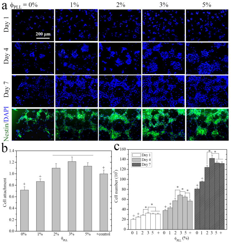
Figure 5.

E14 mouse NPC attachment and proliferation on the neutral and PLL-grafted PEGDA hydrogels. (a) Fluorescence images of neurospheres stained with DAPI (blue) at days 1, 4, and 7, and immunostained with anti-nestin (green, undifferentiated NPCs) in the same field at day 7 post-seeding. Scale bar of 200 μm is applicable to all. (b) Cell attachment at 12 h, and (c) cell numbers at days 1, 4, and 7, compared with cell-seeded TCPS as positive (+) control. *, p < 0.05. +, p < 0.05 relative to other hydrogels.
