Abstract
Background
Previous studies have found fewer clinical infections in wounds closed with monofilament suture compared with braided suture. Recently, barbed monofilament sutures have shown improved strength and increased timesavings over interrupted braided sutures. However, the adherence of bacteria to barbed monofilament sutures and other commonly used suture materials is unclear.
Questions/Purposes
We therefore determined: (1) the adherence of bacteria to five suture types including a barbed monofilament suture; (2) the ability to culture bacteria after gentle washing of each suture type; and (3) the pattern of bacterial adherence.
Methods
We created an experimental contaminated wound model using planktonic methicillin-resistant Staphylococcus aureus (MRSA). Five types of commonly used suture material were used: Vicryl™, Vicryl™ Plus, PDS™, PDS™ Plus, and Quill™. To determine adherence, we determined the number of bacteria removed from the suture by sequential washes. Sutures were plated to determine bacterial growth. Sutures were examined under confocal microscopy to determine adherence patterns.
Results
The barbed monofilament suture showed the least bacterial adherence of any suture material tested. Inoculated monofilament and barbed monofilament sutures placed on agar plates had less bacterial growth than braided suture, whereas antibacterial monofilament and braided sutures showed no growth. Confocal microscopy showed more adherence to braided suture than to the barbed monofilament or monofilament sutures.
Conclusions
Barbed monofilament suture showed similar bacterial adherence properties to standard monofilament suture.
Clinical Relevance
Our findings suggest barbed monofilament suture can be substituted for monofilament suture, at the surgeon’s discretion, without fear of increased risk of infection.
Introduction
Although the closure of clean surgical wounds can be accomplished successfully using a wide variety of suture materials, closure of contaminated wounds such as with an infected total joint arthroplasty magnifies the properties of the individual suture types and their association with bacterial infection. Previous studies showed there were less infections in contaminated wounds closed with monofilament suture compared with braided suture [1, 10, 20]. Several studies have shown bacteria adhere more tightly to braided suture [2, 10, 12, 13, 25, 27] and that braided suture leads to more infections as compared with monofilament suture [1, 9, 10, 14, 28].
Recently, a barbed bidirectional monofilament suture, Quill™ (Angiotech Pharmaceuticals, Vancouver, BC, Canada), has allowed for knotless closure of wounds [18]. The barbs allow the suture to distribute tension across the length of a wound without the need for knots, whereas the bidirectionality of barbed monofilament suture with needles on both ends makes it possible for two surgeons to work simultaneously to close the wound [5, 18, 21, 26]. Studies using barbed monofilament suture have shown decreased operative time, decreased costs from operating room charges and anesthesia fees [26], and wound strength and tissue reaction scores comparable to those of monofilament suture tied with knots [17, 19]. Some have suggested that knotting the suture exaggerates the physical characteristics of the monofilament and multifilament sutures causing inflammatory reactions [3]. Therefore, a barbed suture that does not require knots may be less likely to provoke inflammatory reactions. However, the bacterial properties of barbed monofilament suture, such as bacterial adherence and correlation with postoperative wound infection, have not been defined in comparison to standard monofilament and braided sutures.
Using an in vitro model of contaminated soft tissues we determined (1) the adherence of bacteria to five suture types including a barbed monofilament suture; (2) the ability to culture bacteria after gentle washing of each suture type; and (3) the pattern of bacterial adherence.
Materials and Methods
An experimental model was created to simulate the passage of suture through contaminated soft tissues while closing a wound. A bacterial broth consisting of brain heart infusion broth (BHI) inoculated with planktonic methicillin-resistant Staphylococcus aureus (MRSA) was created and two concentrations were developed: 5 × 10¹⁰ CFU/mL (high concentration broth) and 1 × 10⁶ CFU/mL (physiologic concentration broth). The bacterial load required for infection in healthy hosts is reportedly 1 × 10⁵ to 1 × 10⁶ CFU/mL [6, 11]. Sutures were taken from sterile, unexpired packages and cut into 2-cm strands. The sutures then were incubated in either the high concentration broth or physiologic concentration broth for 5 minutes, then removed and placed in a normal saline vortex wash for 15 seconds. The sutures were placed in the normal saline vortex zero, one, two, three, or four times and the number of vortex washes recorded (Fig. 1). The normal saline from the vortex washes then was examined and the CFU/mL recorded. After the vortex washes, the sutures were placed on agar plates and growth observed after 24 hours. A suture of each type underwent no vortex washes and then was examined under confocal microscopy after Syto® 9 (Invitrogen, Grand Island, NY, USA) staining. Five types of suture material were compared in this study: (1) braided polyglactin 910 (Vicryl™; Ethicon Inc, Somerville, NJ, USA); (2) monofilament polydioxanone (PDS™ II; Ethicon Inc); (3) braided polyglactin 910 with Irgacare MP™ (Vicryl™ Plus Antibacterial; Ethicon Inc); (4) monofilament polydioxanone with Irgacare MP (PDS Plus Antibacterial; Ethicon Inc); and (5) barbed monofilament knotless tissue-closure device (Quill). The antibacterial coating on Vicryl Plus and PDS Plus is triclosan, an antimicrobial agent that achieves its effects by blocking an essential enzyme in bacterial fatty acid synthesis [8].
Fig. 1.
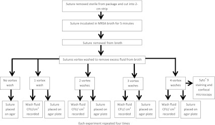
A diagram of the experimental design is shown. Each suture was placed in MRSA broth for 5 minutes. The sutures were removed and underwent a vortex wash in normal saline for 15 seconds. The wash solution then was examined and CFU/mL recorded. The suture was placed on an agar plate and cultured overnight. Sutures from the four vortex wash groups underwent Syto® 9 DNA staining and were examined under confocal microscopy to determine adherence patterns (CFU = colony forming units).
To avoid carryover of bacteria on the forceps between washes, the forceps were flamed with ethanol between each wash. The vortex washes of the high bacteria concentration (5 × 10¹⁰ CFU/mL) consisted of each suture strand being placed in 2 mL of sterile 0.9% saline in a 15-mL Falcon™ tube (BD Biosciences, San Jose, CA, USA) and vortex washed for 10 seconds to remove bacteria that did not tightly adhere to the suture. For the physiologic bacteria concentration experiments (1 × 10⁶ CFU/mL), each suture strand was placed in 1 mL of sterile 0.9% saline in a 15-mL Falcon™ tube and vortex washed for 10 seconds.
To determine the number of CFU/mL in the vortex wash solutions, 500-μL spread plates of the saline solution were incubated overnight. These plates were examined for bacterial growth. The data are reported as CFU/mL recovered at each wash and total CFU/mL recovered. To detect bacteria remaining on the suture after the vortex washes, sutures were placed on mannitol agar plates and incubated overnight at 37°C. Bacterial growth was detected in two ways. Formation of opaque colonies around the suture and the formation of a yellow halo produced by the color change in a pH indicator on production of acidic end products of mannitol fermentation by the bacteria. Laser scanning confocal microscopy and Syto 9 DNA staining were used to observe the adherence. Stained sutures were imaged using a Leica Sp5 scanning confocal microscope (Leica Microsystems Inc, Buffalo Grove, IL, USA). Syto 9 was excited at 488 nm and emission was detected from 496 to 534 nm. After a 5-minute incubation in the MRSA solution (approximately 5 × 10¹⁰ CFU/mL) and four washes in 3 mL of sterile 0.9% saline, suture strands were stained with fluorescent DNA stain Syto® 9. With this fluorescent green stain, bacteria could be seen on the suture using a laser scanning confocal microscope. The culture of bacteria from the suture and use of confocal microscopy were performed to determine if tight adherence to the suture resulted in less bacteria washed off the suture. If this were the case, then we would expect there to be increased growth from these sutures and more bacterial adherence on confocal microscopy.
An a priori power calculation was not performed for this pilot model. We determined the differences in the number of CFU/mL between the different suture types. The number of CFU/mL was recorded for each trial and the average number of CFU/mL calculated for each suture type. An independent weighted ANOVA was performed on the data from each trial. Tukey’s test was used to determine if a difference existed between the individual suture types. The amount of bacteria cultured from each suture was viewed qualitatively, observing the relative amount of growth on the agar plates, rather than quantitatively. Statistical analysis was performed using SPSS 20.0 Software (IBM, Armonk, NY, USA).
Results
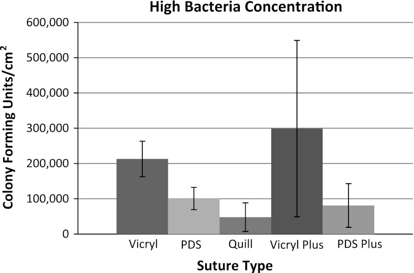
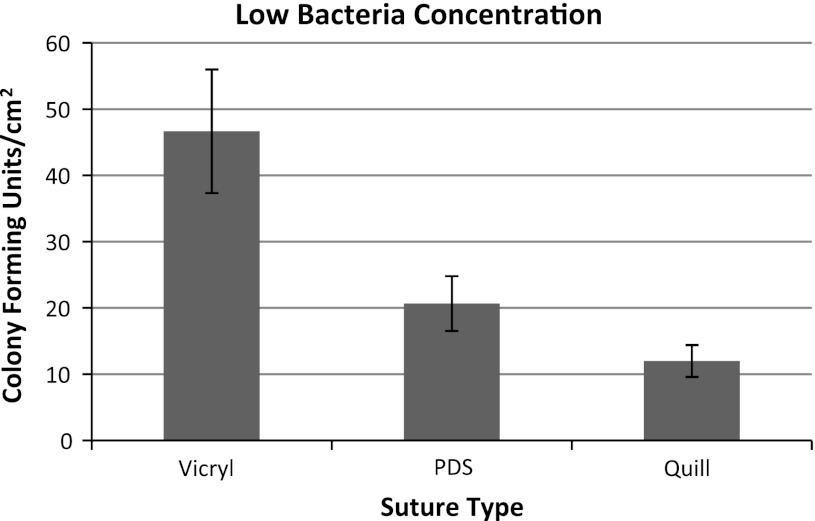
Quill™ showed the least amount of bacterial adherence (48,000 CFU/cm2) of the five sutures tested (Table 1). Vicryl (213,000 CFU/cm2) and Vicryl Plus (299,000 CFU/cm2) had the most bacterial adherence (Fig. 2). At high bacteria concentrations (5 × 10¹⁰ CFU/mL), Quill showed less bacterial adherence than Vicryl and Vicryl Plus (p = 0.05 and 0.04, respectively). The bacterial adherence to PDS plus (81,000 CFU/mL/suture) was less than Vicryl* Plus (Table 2), p = 0.02. There were no statistical differences between other suture types regarding bacterial adherence at high bacterial concentrations. At physiologic bacteria concentrations (5 × 10⁶ CFU/mL), Quill (12 CFU/cm2) again showed the lowest amount of bacterial adherence (Fig. 3) when compared with Vicryl (47 CFU/cm2) and PDS (21 CFU/cm2), p = 0.5 and p = 0.9, respectively.
Table 1.
Number of CFU/cm2 recovered from sutures after washing by suture type and initial bacterial concentration
| Type of suture | Supraphysiologic bacterial concentration (CFU/cm2) | Physiologic bacterial concentration (CFU/cm2) |
|---|---|---|
| Vicryl™ (braided) | 213,000 | 47 |
| PDS™ (monofilament) | 101,000 | 21 |
| Quill™ (barbed) | 48,000 | 12 |
| Vicryl™ Plus (braided) | 299,000 | |
| PDS™ Plus (monofilament) | 81,000 |
CFU = colony-forming units.
Fig. 2.
The number of recovered bacteria from each vortex wash were added together for a total number of bacteria recovered from the suture by four washes. The mean number of bacteria for each suture is displayed. The error bars show two standard deviations (95% confidence intervals).
Table 2.
ANOVA followed by Tukey’s test to determine significance between individual groups
| Sutures | p value |
|---|---|
| Vicryl* versus PDS* | 0.3 |
| Vicryl* versus Quill™ | 0.05 |
| Vicryl* versus Vicryl* Plus | 0.8 |
| Vicryl* versus PDS* Plus | 0.2 |
| PDS* versus Quill™ | 0.8 |
| PDS™ versus Vicryl™ Plus | 0.1 |
| PDS™ versus PDS™ Plus | 0.8 |
| Quill™ versus Vicryl™ Plus | 0.04 |
| Quill™ versus PDS™ Plus | 0.7 |
| Vicryl™ Plus versus PDS™ Plus | 0.02 |
Fig. 3.
The number of colony forming units (CFU) initially associating with the sutures after using physiologic bacterial concentrations is shown. The bacteria decreased below the lower limit of detection (2 CFU/suture) after one wash. The error bars indicate two standard deviations (95% confidence intervals).
Photographs of the suture material on agar plates (Fig. 4) qualitatively showed substantial growth of bacteria around the Vicryl sutures (shown as yellow), even after four washes. The PDS* and Quill sutures showed visibly less growth. The antibacterial braided and monofilament sutures qualitatively showed no growth, even after no washes.
Fig. 4.
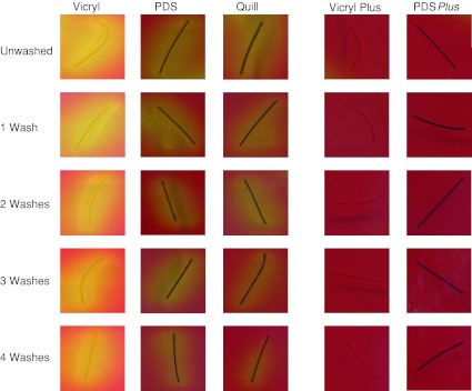
Sutures were incubated for 5 minutes in approximately 5 × 10¹⁰ CFU of MRSA. After a rinse vortex wash to remove broth, sutures were vortex washed zero, one, two, three, or four times. The sutures then were plated on mannitol salt agar and incubated overnight at 37°C. Data shown are representative of three independent experiments. The yellow around the suture indicates bacterial growth.
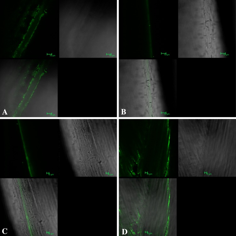
By confocal microscopy a qualitatively greater amount of bacteria adhered to Vicryl* as compared with PDS* (Fig. 5). A brighter green signal was seen in the grooves throughout the length of the Vicryl suture, whereas few bacteria were seen throughout the length of the PDS suture. Confocal microscopy of the Quill suture showed adherence patterns similar to those of the PDS suture.
Fig. 5A–D.
Confocal microscopic images of (A) Vicryl™, (B) PDS™, (C) Quill™, and (D) Vicryl™ Plus sutures after incubation in MRSA solution and four vortex washes in saline are shown. Images of PDS™ Plus are not shown as they are similar to those for PDS™.
Discussion
Previous research has shown the superiority of monofilament sutures in contaminated wounds [1–3, 10, 13, 14, 20, 25]. Barbed monofilament suture is being used more frequently as a result of its efficiency, cost savings, and improved or comparable wound closure outcomes [5, 7, 15–17, 19, 21, 23, 24, 26, 28]. The use of barbed monofilament suture for closure in a TKA has shown superiority in tightness of closure and failure analysis [16, 24]. However, the barbed nature of this monofilament suture has the theoretical concern of the barbs acting as a place for bacteria to hide, resulting in higher infection rates if contaminated wounds are closed with barbed monofilament suture. We therefore determined (1) the adherence of bacteria to each of five suture types including a barbed monofilament suture; (2) the ability to culture bacteria after gentle washing of each suture type; and (3) the pattern of bacterial adherence.
There are several limitations of this study. First, these in vitro data may not translate directly to the in vivo setting. Although we have attempted to simulate incubation of the sutures with bacteria, this scenario would not necessarily be replicated in vivo. Second, this study does not control for numerous factors that would be present in vivo, such as the properties of specific bacteria, presence of gross tissue infection or biofilm formation at the site, an active immune system, and the flow of fluids over the sutures. Third, we chose to use MRSA for our study as it is the most common isolate at our institution (66% of all Staphylococcus cultures). One study suggested coagulase negative Staphylococcus aureus to be the most common bacteria isolated from wounds [22]. Different bacteria could have different adherence properties and could affect our findings. Further studies comparing the adherence patterns of different bacteria are needed. Fourth, we chose to use planktonic bacteria rather than a biofilm. Bacteria in a biofilm, a common finding in infected total joint arthroplasties, likely would show increased adherence and that could alter our findings.
Our current study echoes those of Masini et al. [13]. They used 108 CFU/mL of Staphylococcus aureus engineered to emit photons, allowing them to quantify the amount of bacteria adhered to each suture using imaging software. They found a greater amount of bacteria adhered to Vicryl than monofilament sutures (Monocryl and Prolene). In contrast to our study, Masini et al. observed less bacterial adherence to Vicryl Plus than to Vicryl sutures. It is possible that prolonged exposure to the antibacterial properties of the Vicryl* Plus (24-hour incubation) killed the bacteria before it was able to emit photons to be detected by their imaging software, whereas, in our study, the bacteria were washed off the suture in 5 minutes and the number of bacteria were recorded from the wash. Based on the findings of their study and ours, it is unclear whether the increased adherence was the result of the braided nature of Vicryl or the suture material, given that silk suture did not adhere a different amount of bacteria compared with monofilament sutures. Our study adds to the literature as Masini et al. did not examine the most commonly used absorbable monofilament suture for deep layer closure [13].
Edmiston et al. [4], using scanning electron microscopy, compared bacterial adherence with antibiotic-(triclosan) coated suture (Vicryl Plus) and traditional absorbable braided suture (Vicryl) using three different bacterial broths: MRSA, Staphylococcus epidermidis, and Escherichia coli. They exposed the suture materials to the bacterial broths for either 5 seconds or 2 minutes, gently washed the suture with normal saline, and then incubated the suture for 24 hours. They observed fewer bacteria adhered to the sutures coated with triclosan compared with those not coated. Although fewer bacteria appeared to adhere to the antibiotic suture by scanning electron microscopy after 24 hours of incubation, our findings suggest that this is a function of the bactericidal activity of the antibacterial coating, rather than a function of adherence. We found qualitatively equivalent adherence of bacteria to Vicryl and Vicryl Plus sutures.
Barbed monofilament suture appears to have comparable performance to monofilament suture in a contaminated wound model. The barbed monofilament suture, Quill, showed less bacterial adherence than Vicryl and Vicryl Plus, both absorbable braided sutures. Antibacterial-coated sutures effectively prevented bacterial growth, although adherence to the suture was unchanged compared with nonantibacterial-coated sutures. Although in vivo models will be needed to confirm use of this in vitro model in the clinical setting, our observations suggest barbed monofilament suture might be substituted for monofilament suture, at the surgeon’s discretion, without fear of increased risk of infection.
Footnotes
The institution of one or more of the authors (JRF) has received, during the study period, funding from the John Lachman Orthopaedic Research Foundation, a nonprofit organization funding resident research at Temple University Hospital, Philadelphia, PA, USA.
All ICMJE Conflict of Interest Forms for authors and Clinical Orthopaedics and Related Research editors and board members are on file with the publication and can be viewed on request.
Clinical Orthopaedics and Related Research neither advocates nor endorses the use of any treatment, drug, or device. Readers are encouraged to always seek additional information, including FDA-approval status, of any drug or device prior to clinical use.
Each author certifies that his or her institution approved or waived approval for the reporting of this investigation and that all investigations were conducted in conformity with ethical principles of research.
This work was performed at Temple University Hospital and School of Medicine, Philadelphia, PA, USA.
References
- 1.Alexander JW, Kaplan JZ, Altemeier WA. Role of suture materials in the development of wound infection. Ann Surg. 1967;165:192–199. doi: 10.1097/00000658-196702000-00005. [DOI] [PMC free article] [PubMed] [Google Scholar]
- 2.Ananthakrishnan N, Rao RS, Shivam S. Bacterial adherence to cotton and silk sutures. Natl Med J India. 1992;5:217–218. [PubMed] [Google Scholar]
- 3.Edlich RF, Panek PH, Rodeheaver GT, Turnbull VG, Kurtz LD, Edgerton MT. Physical and chemical configuration of sutures in the development of surgical infection. Ann Surg. 1973;177:679–688. doi: 10.1097/00000658-197306000-00006. [DOI] [PMC free article] [PubMed] [Google Scholar]
- 4.Edmiston CE, Seabrook GR, Goheen MP, Krepel CJ, Johnson CP, Lewis BD, Brown KR, Towne JB. Bacterial adherence to surgical sutures: can antibacterial-coated sutures reduce the risk of microbial contamination? J Am Coll Surg. 2006;203:481–489. doi: 10.1016/j.jamcollsurg.2006.06.026. [DOI] [PubMed] [Google Scholar]
- 5.Einarsson JI, Vellinga TT, Twijnstra AR, Chavan NR, Suzuki Y, Greenberg JA. Bidirectional barbed suture: an evaluation of safety and clinical outcomes. JSLS. 2010;14:381–385. doi: 10.4293/108680810X12924466007566. [DOI] [PMC free article] [PubMed] [Google Scholar]
- 6.Elek SD, Conen PE. The virulence of staphylococcus pyogenes for man: a study of the problems of wound infection. Br J Exp Pathol. 1957;38:573–586. [PMC free article] [PubMed] [Google Scholar]
- 7.Jandali S, Nelson JA, Bergey MR, Sonnad SS, Serletti JM. Evaluating the use of a barbed suture for skin closure during autologous breast reconstruction. J Reconstr Microsurg. 2011;27:277–286. doi: 10.1055/s-0031-1275491. [DOI] [PubMed] [Google Scholar]
- 8.Jones RD, Jampani HB, Newman JL, Lee AS. Triclosan: a review of effectiveness and safety in health care settings. Am J Infect Control. 2000;28:184–196. doi: 10.1016/S0196-6553(00)90027-0. [DOI] [PubMed] [Google Scholar]
- 9.Kathju S, Nistico L, Hall-Stoodley L, Post JC, Ehrlich GD, Stoodley P. Chronic surgical site infection due to suture-associated polymicrobial biofilm. Surg Infect (Larchmt). 2009;10:457–461. doi: 10.1089/sur.2008.062. [DOI] [PMC free article] [PubMed] [Google Scholar]
- 10.Katz S, Izhar M, Mirelman D. Bacterial adherence to surgical sutures: a possible factor in suture induced infection. Ann Surg. 1981;194:35–41. doi: 10.1097/00000658-198107000-00007. [DOI] [PMC free article] [PubMed] [Google Scholar]
- 11.Krizek TJ, Robson MC. Evolution of quantitative bacteriology in wound management. Am J Surg. 1975;130:579–584. doi: 10.1016/0002-9610(75)90516-4. [DOI] [PubMed] [Google Scholar]
- 12.Masini BD, Stinner DJ, Waterman SM, Wenke JC. Bacterial adherence to high-tensile strength sutures. Arthroscopy. 2011;27:834–838. doi: 10.1016/j.arthro.2011.02.003. [DOI] [PubMed] [Google Scholar]
- 13.Masini BD, Stinner DJ, Waterman SM, Wenke JC. Bacterial adherence to suture materials. J Surg Educ. 2011;68:101–104. doi: 10.1016/j.jsurg.2010.09.015. [DOI] [PubMed] [Google Scholar]
- 14.Mehta PH, Dunn KA, Bradfield JF, Austin PE. Contaminated wounds: infection rates with subcutaneous sutures. Ann Emerg Med. 1996;27:43–48. doi: 10.1016/S0196-0644(96)70295-0. [DOI] [PubMed] [Google Scholar]
- 15.Murtha AP, Kaplan AL, Paglia MJ, Mills BB, Feldstein ML, Ruff GL. Evaluation of a novel technique for wound closure using a barbed suture. Plast Reconstr Surg. 2006;117:1769–1780. doi: 10.1097/01.prs.0000209971.08264.b0. [DOI] [PubMed] [Google Scholar]
- 16.Nett M, Avelar R, Sheehan M, Cushner F. Water-tight knee arthrotomy closure: comparison of a novel single bidirectional barbed self-retaining running suture versus conventional interrupted sutures. J Knee Surg. 2011;24:55–59. doi: 10.1055/s-0031-1275400. [DOI] [PubMed] [Google Scholar]
- 17.Parikh PM, Davison SP, Higgins JP. Barbed suture tenorrhaphy: an ex vivo biomechanical analysis. Plast Reconstr Surg. 2009;124:1551–1558. doi: 10.1097/PRS.0b013e3181babb77. [DOI] [PubMed] [Google Scholar]
- 18.Quill™ device. Available at: http://www.angiotech.com/focus-markets/wound-closure/quill/. Accessed June 30, 2012.
- 19.Rashid RM, Sartori M, White LE, Villa MT, Yoo SS, Alam M. Breaking strength of barbed polypropylene sutures: rater-blinded, controlled comparison with nonbarbed sutures of various calibers. Arch Dermatol. 2007;143:869–872. doi: 10.1001/archderm.143.7.869. [DOI] [PubMed] [Google Scholar]
- 20.Shambaugh P, Dunphy J. Postoperative wound infections and the use of silk: an experimental study. Surgery. 1937;1:379–385. [Google Scholar]
- 21.Shermak MA, Mallalieu J, Chang D. Barbed suture impact on wound closure in body contouring surgery. Plast Reconstr Surg. 2010;126:1735–1741. doi: 10.1097/PRS.0b013e3181ef8fa3. [DOI] [PubMed] [Google Scholar]
- 22.Shuman EK, Urquhart A, Malani PN. Management and prevention of prosthetic joint infection. Infect Dis Clin North Am. 2012;26:29–39. doi: 10.1016/j.idc.2011.09.011. [DOI] [PubMed] [Google Scholar]
- 23.Sulamanidze M. Evaluation of a novel technique for wound closure using a barbed suture. Plast Reconstr Surg. 2007;120:349–50; author reply 350. [DOI] [PubMed]
- 24.Vakil JJ, O’Reilly MP, Sutter EG, Mears SC, Belkoff SM, Khanuja HS. Knee arthrotomy repair with a continuous barbed suture: a biomechanical study. J Arthroplasty. 2011;26:710–713. doi: 10.1016/j.arth.2010.07.003. [DOI] [PubMed] [Google Scholar]
- 25.Varma S, Ferguson HL, Breen H, Lumb WV. Comparison of seven suture materials in infected wounds: an experimental study. J Surg Res. 1974;17:165–170. doi: 10.1016/0022-4804(74)90103-6. [DOI] [PubMed] [Google Scholar]
- 26.Villa MT, White LE, Alam M, Yoo SS, Walton RL. Barbed sutures: a review of the literature. Plast Reconstr Surg. 2008;121:102e–108e. doi: 10.1097/01.prs.0000299452.24743.65. [DOI] [PubMed] [Google Scholar]
- 27.Zamora N, Esteban J, Kinnari TJ, Celdran A, Granizo JJ, Zafra C. In-vitro evaluation of the adhesion to polypropylene sutures of non-pigmented, rapidly growing mycobacteria. Clin Microbiol Infect. 2007;13:902–907. doi: 10.1111/j.1469-0691.2007.01769.x. [DOI] [PubMed] [Google Scholar]
- 28.Zaruby J, Gingras K, Taylor J, Maul D. An in vivo comparison of barbed suture devices and conventional monofilament sutures for cosmetic skin closure: biomechanical wound strength and histology. Aesthet Surg J. 2011;31:232–240. doi: 10.1177/1090820X10395010. [DOI] [PubMed] [Google Scholar]


