

Abstract
The historical development of a scientific knowledge on calcium orthophosphates from the 1770s until 1940 is described. Many forgotten and poorly known historical facts and approaches have been extracted from old publications and then they have been analyzed, systematized and reconsidered from the modern point of view. The chosen time scale starts with the earliest available studies of 1770s (to the best of my findings, calcium orthophosphates had been unknown before), passes through the entire 19th century and finishes in 1940, because since then the amount of publications on calcium orthophosphates rapidly increases and the subject becomes too broad. Furthermore, since publications of the second half of the 20th century are easily accessible, a substantial amount of them have already been reviewed by other researchers. The reported historical findings clearly demonstrate that the substantial amount of the scientific facts and experimental approaches have been known for very many decades and, in fact, the considerable quantity of relatively recent investigations on calcium orthophosphates is just either a further development of the earlier studies or a rediscovery of the already forgotten knowledge.
Keywords: apatite, calc phosphate, calcium orthophosphate, history, lime phosphate
Introduction
By virtue of abundance in the nature and presence in the living organisms, calcium apatites[a] and other calcium orthophosphates (Table 1) appear to be the chemical compounds of a special interest in many fields of science, including geology, chemistry, biology and medicine.1,2 As follows from the designation, calcium orthophosphates contain both calcium (Ca, atomic number 20) and phosphorus (P, atomic number 15) as the major constituencies. Concerning the history of both chemical elements, according to Wikipedia, the free encyclopedia, calcium (from Latin calx, genitive calcis, meaning “lime”) compounds were known as early as the first century, when the ancient Romans prepared lime as calcium oxide. However, calcium sulfate (also known as plaster of Paris or lime plaster) had been known much earlier: three statues were discovered in a buried pit at 'Ain Ghazal in Jordan those were sculpted with lime plaster over armatures of reeds and twine. They were made in the pre-pottery Neolithic period, around 7200 BC. However, calcium in a pure state was not isolated until 1808, when the famous British chemist and inventor Sir Humphry Davy (1778–1829) electrolyzed a mixture of lime and mercuric oxide.3,4
Table 1. Existing calcium orthophosphates and their major properties1,2.
| Ca/P molar ratio | Compound | Formula | Solubility at 25°C, -log(Ks) |
Solubility at 25°C, g/L |
pH stability range in aqueous solutions at 25°C |
|---|---|---|---|---|---|
| 0.5 |
Monocalcium phosphate monohydrate (MCPM) |
Ca(H2PO4)2·H2O |
1.14 |
~18 |
0.0 – 2.0 |
| 0.5 |
Monocalcium phosphate anhydrous (MCPA or MCP) |
Ca(H2PO4)2 |
1.14 |
~17 |
[c] |
| 1.0 |
Dicalcium phosphate dihydrate (DCPD), mineral brushite |
CaHPO4·2H2O |
6.59 |
~0.088 |
2.0 – 6.0 |
| 1.0 |
Dicalcium phosphate anhydrous (DCPA or DCP), mineral monetite |
CaHPO4 |
6.90 |
~0.048 |
[c] |
| 1.33 |
Octacalcium phosphate (OCP) |
Ca8(HPO4)2(PO4)4·5H2O |
96.6 |
~0.0081 |
5.5 – 7.0 |
| 1.5 |
α-Tricalcium phosphate (α-TCP) |
α-Ca3(PO4)2 |
25.5 |
~0.0025 |
[a] |
| 1.5 |
β-Tricalcium phosphate (β-TCP) |
β-Ca3(PO4)2 |
28.9 |
~0.0005 |
[a] |
| 1.2 – 2.2 |
Amorphous calcium phosphates (ACP) |
CaxHy(PO4)z·nH2O, n = 3 – 4.5; 15 – 20% H2O |
[b] |
[b] |
~5 – 12 [d] |
| 1.5 – 1.67 |
Calcium-deficient hydroxyapatite (CDHA or Ca-def HA)[e] |
Ca10-x(HPO4)x(PO4)6-x(OH)2-x (0 < x < 1) |
~85 |
~0.0094 |
6.5 – 9.5 |
| 1.67 |
Hydroxyapatite (HA, HAp or OHAp) |
Ca10(PO4)6(OH)2 |
116.8 |
~0.0003 |
9.5 – 12 |
| 1.67 |
Fluorapatite (FA or FAp) |
Ca10(PO4)6F2 |
120.0 |
~0.0002 |
7 – 12 |
| 1.67 |
Oxyapatite (OA, OAp or OXA)[f] |
Ca10(PO4)6O |
~69 |
~0.087 |
[a] |
| 2.0 | Tetracalcium phosphate (TTCP or TetCP), mineral hilgenstockite | Ca4(PO4)2O | 38 – 44 | ~0.0007 | [a] |
[a] These compounds cannot be precipitated from aqueous solutions. [b]Cannot be measured precisely. However, the following values were found: 25.7 ± 0.1 (pH = 7.40), 29.9 ± 0.1 (pH = 6.00), 32.7 ± 0.1 (pH = 5.28). The comparative extent of dissolution in acidic buffer is: ACP > > α-TCP > > β-TCP > CDHA > > HA > FA. [c]Stable at temperatures above 100°C. [d]Always metastable. [e]Occasionally, it is called “precipitated HA (PHA).” [f]Existence of OA remains questionable.
Phosphorus is a bit younger. The discovery of this chemical element in a pure state [its name given from Greek mythology, Φωσφόρος meaning “light-bearer” (Latin: Lucifer), referring to the “Morning Star,” the planet Venus] is credited to a German merchant and alchemist Hennig Brand (ca. 1630–1710) in 1669, although other alchemists might have discovered phosphorus around the same time. Brand experimented with urine, which contains considerable quantities of dissolved phosphates from normal metabolism. However, it was the famous French chemist Antoine Laurent Lavoisier (1743–1794), who recognized phosphorus as a chemical element in 1777. Interestingly, phosphorus appears to be the first element discovered since antiquity. To conclude these introductive exercises, the earliest research paper I have been able to find, containing the word “phosphorus” in the title belongs to the famous British natural philosopher, chemist, physicist and inventor Robert Boyle (1627–1691) and was published in 16935 shortly after his death.
General Problems with an Access to Old Publications
Due to large problems accessing the scientific literature published in the first half of the 19th century and even earlier, a deep investigation into the history of the subject still remains to be both fragmental and incomplete. That time, the scientific concepts were rather different from the modern ones and chemical formulae of the substances had not been introduced yet. Furthermore, scientific journals were rare and just a few of them are currently available in the electronic form. Luckily, very many books published before the 20th century have been scanned by Google as a part of its project to make the world’s books discoverable online. This timely project by Google combined with the power of the modern electronic databases of scientific publications allows reconstructing the major historical milestones on calcium orthophosphates, which was often impossible for earlier review writers. For example, a paper of 1994 by Driskell entitled: “Early history of calcium phosphate materials and coatings”6 starts from the classical publication of 1920 by Albee assisted by Morrison.7 In 1999, Shackelford published a paper: “Bioceramics – an historical perspective,”8 in which the same publication by Albee assisted by Morrison7 was mentioned as the earliest reference. The same is valid for the historical papers by Hulbert, et al.9,10 and Shepperd.11 Thus, it might create a false impression that calcium phosphates were unknown before 1920. Certainly, this is not the case; nevertheless, the precise sequence of the scientific events happened in the 18th century still remain poorly restorable, while the historical time scale of even earlier scientific events remain almost irrecoverable. This is mainly due to a lack of the citation practice existed in the scientific literature published in the 19th century and before. Besides, even nowadays, for the entire 18th century and approximately the first quarter of the 19th century, only scientific books digitalized by Google are widely available. No journal papers devoted to apatites and/or any type of calcium phosphates published before ~1820s have been found. However, in no case this means that they do not exist; presumably, they either have not been scanned yet (especially, this might be valid for discontinued scientific journals) or an access to the scanned versions is restricted to a limited amount of subscribers only.
Investigations and Knowledge on Calcium Orthophosphates
In the 18th century
According to the available literature, the history of calcium orthophosphates started in 1769. For example, in 1881 Roscoe and Schorlemmer12 published the following statement: “Gahn[b], in 1769, discovered the existence of calcium phosphate in bones, but it was not until this fact was published by Scheele[c] in 1771 that phosphorus was obtained from bone-ash, which has from that time invariably served for its preparation.” (ref. 12, p. 458). Simultaneously, the presence of phosphoric compounds was discovered in blood serum, which follows from a publication of 177013 (please, note the old-fashioned using a long, medial or descending letter “ſ,” which is a form of the minuscule letter “s” formerly used where “s” occurred in the middle or at the beginning of a word): “The ſerum conſiſts chemically of a coagulable matter, and water in which common ſal ammoniac and phoſphoric ammoniac, and generally common ſalt, and frequently ſelenites, and, fixed ammoniac, are dissolved; …” (ref. 13, p. 309). Furthermore, let me cite a publication of 177714: “I have only been informed of this diſcovery, by the Gazette, Salutaire de Bouillon, October, 1775. It is there ſaid, that Mr. Henry Gahn, a phyſician at Stockholm, has communicated a proceſs for extracting from bones the ſaline matter in question; and that Mr. Scheele had aſcertained, that the earth of animals was compoſed of a calcareous ſubſtance united with the phoſphoric acid. This diſcovery, continues the author of the article of the Gazette, belongs to Mr. Gahn, and has been confirmed by later experiments.” (ref. 14, p. 383). Presumably, this citation might be considered as one of the earliest mentioning on calcium phosphates in the history.
Further, according to Shepperd,11 the famous German chemist Martin Heinrich Klaproth (1743–1817) and the famous French chemist Joseph-Louis Proust (1754–1826) also studied calcium orthophosphates. Unfortunately, Shepperd has not provided any references to the publications by those great chemists. Nevertheless, a search performed among the Google books has provided some results. For example, in a French book published in 1790,15 one can find a subchapter entitled “Apatite. Phosphate de Chaux.” which is started from the following sentence: “C’est Mr. Klaproth, qui à découvert le premier cette combinaison de la terre calcaire avec l’acide phosphorique.” (ref. 15, p. 363). Further, “Mr. Proust dans une Lettre à Mr. d’Arcet a donné l’Analyse de cette pierre, qui est une combinaison de la terre calcaire avec l’acide phosphorique.” (ref. 15, p. 366). One can see that, by that time, apatite was already recognized as a calcium phosphate, which is also confirmed by a German book published in 1789,16 in which “Apatit” was called “die Phosphorsaure mit Kalkerde” (ref. 16, p. 2).
Furthermore, according to a book written by Lavoisier,17 the production process of orthophosphoric acid by decomposition of calcined bones in sulfuric acid has been known since, at least, 1790 (again, pay attention to the old-fashioned using a letter “ſ” instead of “s”): “The bones of adult animals being calcined to whiteneſs, are pounded, and paſſed through a fine ſilk ſiewe; pour upon the fine powder a quantity of dilute ſulphuric acid, leſs than is ſufficient for diſſolving the whole. This acid unites with the calcareous earth of the bones into a ſulphat of lime, and the phoſphoric acid remains free in the liquor.” (ref. 17, p. 205). Further, the production process of white phosphorus has been described: “The liquid is decanted off, and the reſiduum waſhed with boiling water; this water which has been uſed to waſh out the adhering acid is joined with what was before decanted off, and the whole is gradually evaporated; the diſſolved ſulphat of lime criſtallizes in form of ſilky threads, which are removed, and by continuing the evaporation we procure the phoſphoric acid under the appearance of a white pellucid glaſs. When this is powdered, and mixed with one third its weight of charcoal, we procure very pure phoſphorus by ſublimation.” (ref. 17, p. 206).
In the last decade of the 18th century, further investigations on calcium orthophosphates were performed by two famous French chemists Antoine François, comte de Fourcroy (1755 – 1809) and Nicolas Louis Vauquelin (1763–1829), who discovered an existence of acidic calcium orthophosphates, currently known as MCPM, MCPA, DCPD and DCPA (Table 1). For example, according to a book, published in 181618, “Super-Phosphat of Lime was diſcovered in 1795, by Fourcroy and Vauquelin. It had indeed been often formed before, but chemiſts had neglected to examine it.” (ref. 18, p. 458). In addition, let me cite a book by Fourcroy, 180419: “A few years ago this ſalt, which I here denominate the acid phoſphate of lime, was unknown. Scheele very properly remarked that the ſaline earth of bones was diſſolved by an acid in human urine, but he did not obſerve that this union between phoſphoric acid and the bony phoſphate, makes a kind of permanent and particular ſalt different from the latter. It was in the year 1795, the third year of the Republic, that I diſcovered it, with Citizen Vauquelin, in a connected ſeries of experiments upon bony matters; wherein we proved that the calcareous phoſphate which conſtitutes the ſolid baſe, is only in part decompoſable by acids, and that the portion of phoſphoric acid which is ſeparated, retains in ſolution phoſphate of lime, which it then defends from all ſubſequent alteration by other acids.” (ref. 19, p. 347). Furthermore, in his book Fourcroy mentioned other contributors to early studies on calcium orthophosphates: “Citizens Nicholas of Nancy, Pelletrier, Berniard, Bullion, in France; Weſtrumb, and ſeveral chemiſts in Germany; Bonvioſin, at Turin; Tenant, Pearſon, and ſome others in England, multiplied their inquiries and experiments.” (ref. 19, p. 337). Thus, one can notice that in the end of the 18th century, the subject of calcium orthophosphates was extensively investigated by a number of researchers in several countries of the Western Europe.
To conclude this section, one should notice that the basic knowledge on calcium orthophosphates became available by the end of the 18th century.
In the 19th century
For the 19th century, both scientific books and a small amount of scientific journals are available; however, for the first half of the 19th century, journals are almost unavailable. Nevertheless, according to the available literature, a real explosion of the scientific knowledge on calcium orthophosphates occurred in the 19th century. Namely, Fourcroy19 stated that presence of water in acidic calcium orthophosphates was known by 1804: “The acid phoſphate of lime contains a remarkable quantity of water: the cryſtallization immediately ſoftens and becomes liquefied by the fire.” (ref. 19, p. 349). Unfortunately, based on this citation, it is impossible to differentiate which type of an acidic calcium orthophosphate MCPM or DCPD Fourcroy born in mind. Knowledge on acidic calcium orthophosphates was further developed in 180520: “Acid Phosphate of Lime is formed either by taking away a part of the base by sulphuric, nitric, or muriatic acid, or by superadding phosphoric acid to the last described phosphate.” (ref. 20, p. 195) and in 180621: “In this way were distinguished among the salts two combinations, one neutral, and one with an excess of acid; and these were supposed to be determinate, as in sulphate and super-sulphate of potassa, or the phosphate and super-phosphate of lime.” (ref. 21, p. 38). More precise information on acidic calcium orthophosphates became available by 181922: “XI. Biphosphate of Lime. —This salt may be formed by digesting phosphate of lime in phosphoric acid, dissolved in hot water.” (ref. 22, p. 327). “XII. Quadriphosphate of Lime.—Glassy Phosphoric Acid of the Apothecaries.—This salt may be formed by digesting, for some time, finely powdered phosphate of lime in a quantity of sulphuric acid, sufficient to saturate all the lime of the phosphate, and afterwards diluting the mixture with a sufficient quantity of water, and filtering. Sulphate of lime remains on the filter, and a liquid quadriphosphate passes through.” (ref. 22, pp. 327–8). Presumably, aqueous solutions of DCPD and MCPM, respectively, were prepared; therefore, we can claim that MCPM and DCPD were differentiated by 1819. Furthermore: “XIII. Subphosphate of Lime.—This salt occurs native under the names of apatite or asparagus stone.” (ref. 22, p. 328). Thus, the differences between HA (subphosphate of lime) and TCP (phosphate of lime) were established by 1819.
The preparation process of a pure calcium orthophosphate currently known as CDHA was developed by 181323: “a. Phosphat of lime, proper. As this salt constitutes the basis of bones, it is not necessary to prepare it artificially. It may be obtained in a state of purity by the following process: Calcine the bones to whiteness, reduce them to powder, and wash them repeatedly with water, to separate several soluble salts which are present. Dissolve the whole in muriatic acid, and precipitate by means of ammonia. The precipitate, when well washed and dried, is pure phosphat of lime.” (section PHOSPHAT). One can see that the basics of this process are still used to produce chemically pure calcium orthophosphates from the natural resources (phosphate ore and bones). In 1819, the major properties were described as follows22: “4. Phosphate of lime is a white insoluble powder, destitute of taste, and unaltered by exposure to air. It is soluble in hydrochloric (muriatic) and nitric acids, and may be precipitated from solution in them by means of ammonia. When exposed to a very violent heat, it undergoes a kind of fusion, and is converted into a white semi-transparent porcelain.” (ref. 22, p. 327). However, that knowledge on the thermal properties of phosphate of lime was based on the Fourcroy’s investigations19: “The phoſphate of lime is extremely difficult to fuse; nevertheleſs, by ſtrong fire, ſuch as that of a glaſs-house, it ſoftens and acquires a ſemi-tranſparence like the grain of porcelain.” (ref. 19, p. 341). Thus, calcium phosphate ceramics has been known, since, at least, 1804.
In the 4th edition of Encyclopædia Britannica (1810), applications of calcium orthophosphates were described as follows24: “The phoſphate of lime is of great importance in chemiſtry, for the purpoſe of extracting phoſphoric acid, to be decompoſed to obtain phoſphorus. It is also employed for making cupels, for poliſhing metals and precious ſtones, and for removing ſpots of greaſу from linen, paper, and ſilk. It is uſed in medicine as a remedy for rickets, to correct the ſuppoſed effects of acids in ſoftening the bones.” (ref. 24, p. 585). Although it is slightly beyond the subject, one should mention, that calcium phosphite (Ca3(PO3)2) is also mentioned in that edition of Encyclopædia Britannica, as “phoſphite of lime” (ref. 24, p. 586). A chemical term “tribasic phosphate of lime,” which fully corresponds to α-TCP and β-TCP, has been known since, at least, 1832 (ref. 25, p. 90).
Concerning publications in scientific journals, in the first half of the 19th century, research papers on calcium apatites and other calcium orthophosphates were published by the famous Swedish chemist Jöns Jacob Berzelius (1779 – 1848),26,27 as well as by M. Baruel28 and J.D. Smith.29 For example, in a book by Bache, published in 1819,22 the following statement on the Berzelius’s contribution was published: “5. According to an analysis by Berzelius, calculated in numbers, in which the equivalent number for lime is assumed, phosphate of lime is composed of Phosphoric acid 34·3— Lime 29·0—one atom; so that it appears that the composition of this salt cannot be reconciled with the atomic theory.” (ref. 22, p. 327). However, as early as 1804, Fourcroy19 wrote: “100 parts of phoſphate of lime contain, according to the analyſis of Citizens Fourcroy and Vauquelin, Phoſphoric acid 41 Lime 59” (ref. 19, p. 346), while the chemical composition of acid phosphate of lime he described as follows: “An accurate analyſis of this ſalt affords the following proportions of component parts, Lime 45 Phoſphoric acid 54” (ref. 19, p. 351).
A little bit later, the famous German chemist Eilhard Mitscherlich (1794–1863), who was a learner and a friend of J.J. Berzelius and who is, perhaps, best remembered today for his law of isomorphism (1819), also worked in this area. For example, the earliest chemical formulae of calcium phosphates were published in 1844 on page 69a of Mitscherlich’s book30 as 2.Ca2P and 5.Ca2P. Besides, in 1849, it was written that “M. J. L. Lassaigne, at the meeting of the Academy of Sciences of Paris, of the 15th January, presented a memoir upon this subject, showing by experiments that the phosphate and carbonate of lime are introduced into plants in solution in water containing carbonic acid, which had before been shown as to the phosphate by M. Dumas, and has long been known as to the carbonate.”31 Thus, a higher solubility of both calcium orthophosphates and calcium carbonate in weak acids was already known in 1849.
The second half of the 19th century started with “Chemistry” by Wilson, published in 1850.32 The following parts from that book are worth citing. “797. Phosphates of Lime. —There are many compounds of lime and phosphoric acid, owing to the peculiarity of that acid in relation to the number of equivalents of base it combines with at once. The most interesting phosphate of lime is that which occurs in bones, and is distinguished as the bone-earth phosphate, 3CaO,PO5” (ref. 32, p. 219). Thus, various types of calcium orthophosphates were known by 1850. However, the preparation technique sounds unusual to the modern readers: “The phosphorus combines in part with the oxygen of the lime, CaO, to form phosphoric acid, and this with undecomposed lime, to form phosphate of lime, CaO,PO5. At the same time another portion of the phosphorus combines with the calcium of the lime, forming phosphuret of calcium, CaP.” (ref. 32, p. 164). In “Chemistry” by Brande and Taylor, published in 1863,33 one can find the following statements: “Common Phosphate of Lime; Tribasic Phosphate of Lime; Bone Phosphate; [3(CaO),PO5]. This salt occurs abundantly in bone-ash, and is found as a mineral product.” (ref. 33, p. 331). Furthermore, “Native phosphate of lime (bone phosphate) occurs in apatite, moroxite, phosphorite, and asparagus stone; its primitive form is a six-sided prism: it also occurs in some volcanic products” (ref. 33, p. 332). Thus, a similarity between the inorganic phase of bones and calcium orthophosphate rocks of natural origin (apatite and phosphorites) was known by 1863. “When a solution of bone-earth in hydrochloric or nitric acid is boiled to expel all carbonic acid, and decomposed by caustic ammonia, the bone-phosphate separates in the form of a bulky precipitate, which, when perfectly dried, is white and amorphous” (ref. 33, p. 331). This statement is really astonishing because it might be considered as the first mention on ACP,34 32 years before discovering X-rays in 1895 by the famous German scientist Wilhelm Conrad Röntgen (1845–1923)! Next citation: “The substance known under the name of coprolites, and which appear to be the excrements of fossil reptiles, also abound in phosphate of lime” (ref. 33, p. 332) means that by 1863 researchers were aware on this fact.
Further historical description is based on journal publications only. C. Morfit,35 Robert Warington [there were 2 chemists with this name, presumably, a father (1807 – 1867) and a son (1838 – 1907)], who performed the earliest well documented systematic studies of the outstanding quality,36-39 and R. Fresenius40 also worked with calcium orthophosphates. Furthermore, let me cite a paragraph from reference 37: “Mitscherlich tells us, that when chloride of calcium is added to ordinary disodic phosphate, the latter being maintained in excess, the precipitate formed is tricalcic phosphate, while the solution becomes acid from the production of monosodic phosphate. Berzelius, on the contrary, states, that the precipitate formed under these conditions is not tricalcic phosphate, but the octocalcic triphosphate, which lie has elsewhere described. All experimenters agree, that when the operation is reversed, and disodic phosphate is poured into an excess of chloride of calcium, the precipitate is neither tricalcic nor octocalcic, but dicalcic phosphate” (ref. 37, pp. 296–7). Thus, TCP, DCP and OCP have been known since, at least, 1866, while, in fact, a bit earlier. Furthermore, the researchers were already aware on the fact that the type of a precipitated calcium orthophosphate depended on a sequence of the mixing reagents.
Among the available publications written by two Robert Waringtons,36-39 reference 37 by Robert Warington Jr. deserves both a special attention and extensive citations. For example, to prove that OCP indeed was already known in 1866, let me make another citation from reference 37: “Octocalcic phosphate can only be produced by the simultaneous formation of monosodic phosphate: 8CaCl2 + 5Na4H2P2O8 = Ca8H2P6O24 + 16NaCl + 2Na2H4P2O8” (ref. 37, p. 300).
One can see a balanced chemical equation, fully identical to the modern ones. It is hard to believe that it was published in 1866! It is interesting to note that only 22 years appeared to be enough to perform a transition from the earliest primitive chemical formulae without oxygen, such as 2.Ca2P and 5.Ca2P,30 to the modern chemical equations. More to the point, the chemical formulae of hydrated forms of calcium orthophosphates were known by 1866: “8·73 grs. of the vacuum-dried salt, lost on ignition 12·30 grains, or 26·35 per cent.; the formula Ca2H2P2O8·4H2O, demands 26·16 per cent. of water” (ref. 37, p. 299). Needless to explain, that “Ca2H2P2O8·4H2O” represents two molecules of DCPD (see Table 1). Furthermore, “It is interesting to observe that while disodic phosphate is of an alkaline nature, dicalcic phosphate possesses faint acid properties.” (ref. 37, p. 300). More to the point, the form and shape of DCPD crystals were described as well: “The crystalline form of the dicalcic tetrahydrated phosphate has been examined by Professor Church. He describes the crystals as thin rhomboïdal plates, of which the diagonally opposite acute angles are sometimes truncated, hexagonal forms being thus produced. This truncation seems to be occasionally hemihedral, and then may proceed up to the diagonal between obtuse angles; from this change triangular forms arise. Other modifications are also met with” (ref. 37, pp. 300–1). Another interesting conclusion might be found here: “We may then safely affirm that whenever dicalcic phosphate, octocalcic triphosphate, or any phosphate of intermediate composition, is precipitated from solution by ammonia, the salt obtained will be the octocalcic triphosphate; a tricalcic phosphate cannot be obtained in this manner. The following is probably a type of the reaction: 4Ca2H2P2O8 + 6NH3 = Ca8H2P6O24 + 6NH4.P2O8.” (ref. 37, pp. 301–2).
This seems to be the earliest mentioning on the fact that TCP cannot be precipitated from the aqueous solutions (currently we know that ACP or CDHA are precipitated instead). In addition, the following citation from the same publication: “It is quite possible that precipitated tricalcic phosphate may possess somewhat different solubilities, when prepared by different methods; this difference can, however, scarcely be great.” (ref. 37, p. 304) has two important consequences: (1) by 1866 this fact was not quite clear yet; (2) it indirectly points to variability in the Ca/P ratio for the precipitated ACP or CDHA, which is the major reason of different solubilities.
The latest available publication by Warington39 was devoted to the hydrolysis of a freshly precipitated TCP (i.e., either ACP or CDHA) to the stoichiometric HA under continuous (up to 50 h) boiling in distilled water. From the results of numerous chemical analyses the author concluded that during boiling an aqueous suspension of the TCP was slowly transformed to a suspension of 3Ca3P2O8.CaOH2O (i.e., HA, see Table 1) and soluble acidic calcium orthophosphates. The following conclusion was made: “Since it appears that all phosphates of calcium less basic than apatite are unstable under the continued action of pure water, it seems probable that a more exact examination of natural phosphates would show that many phosphates now regarded as tricalcic are in fact of a more basic nature” (ref. 39, p. 989). Thus, the apatitic nature of the majority of natural calcium orthophosphates has been predicted in 1873. The next available journal article on the TCP hydrolysis was published in 1929 only.41
The first accessible journal paper on detection and preservation techniques of various deposits, including calcium orthophosphates, was published in 1852.42 Presence of calcium orthophosphates in teakwood was established in 1862.43 Various analytical topics on calcium orthophosphate chemistry has been studied since, at least, 186344 and remained to be a subject of active research approx. until 1910 sec.45-54
A popular fertilizer superphosphate, which represents “a mixture of the last-mentioned compound and sulphate of lime”55 has been known since, at least, 180356: “The phoſphoric acid ſeparated from that portion of lime, immediately combines with the reſt of the phoſsphate of lime, and forms ſuper phoſphate of lime, which is not farther decompoſable by ſulphuric acid” (pp. 394–5). More than 20 research papers on superphosphate were published by the beginning of the 20th century, which indicated to the importance of this chemical for the human being. To give the appropriate tribute to those early investigators and restore the historical perspective, it is worth citing them all.57-79 One can see that almost all of these studies were performed and published by single researchers (see individual comments to refs. 58 and 65), while the majority of the investigations were devoted to analytical chemistry.
More to the point, let me make a citation from a publication by Wells of 190680: “The apparent constancy of the proportion of carbonate and phosphate of calcium in bones made an impression on Hoppe-Seyler in 1862, and we find him speculating on the possibility of the components of the two salts being joined together to form one giant molecule: 3 [Ca3 (Po4)2]-CaCo3, which he imagined might be united in some such way” (ref. 80, p. 522), see Figure 1. Further, Wells mentioned: “This formula is interesting chiefly from the historical standpoint, but it serves to emphasize the tendency of these salts to appear in nearly constant proportions in the animal body, a fact possibly of some importance” (ref. 80, p. 523). Obviously, the atomic arrangement shown in Figure 1 represents the earliest structural drawing of a single molecule of a calcium orthophosphate currently known as carbonateapatite. An attentive reader will notice two different types of calcium [currently known as Ca(1) and Ca(2)] in that structure.
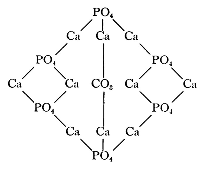
Figure 1. The first available structure of a bone mineral, currently known as carbonateapatite. Reprinted from reference 80.
Although vitrification properties of some forms of calcium orthophosphates at heating have been known since, at least, 1804,19 the first accessible journal paper on this topic was published in 1877.81 The modern chemical formula of perfectly transparent crystals of natural FA as Ca5(PO4)3F has been known since, at least, 1873,82 while the major crystallographic faces of a natural calcium apatite were described in 1883.83 Chemical formulae of DCP (as “Mono-Hydrogen Calcium Orthophosphate, HCaPO4”) and MCP [as “Tetra-Hydrogen Calcium Phosphate, H4Ca(PO4)2”] have been known since, at least, 1879 (ref. 55, pp. 205–6). Interestingly that in a publication of 1871, the chemical formulae of calcium orthophosphates were written in different ways: 3CaO PO5 for apatite and CaO 2HO PO5 for “some acid phosphate of lime”59.
Neutral phosphates of lime were further investigated in 1872.84 Besides, in the 19th century, calcium apatites were considered as combined compounds, which results from this citation: “Calcium phosphate, combined with calcium chloride or calcium fluoride, occurs in the well-known minerals, apatite and osteolite.” (reg. 55, p. 188). One might guess that, in the 19th century, the atomic arrangement of single molecule of carbonateapatite (Fig. 1) could inspire researchers to compose similar drawings for the single molecules of FA, HA and/or chlorapatite; however, I have not succeeded to find anything on this matter.
Chemical equations, describing various interactions between calcium phosphates and other chemicals have been known since at least 1863. For example, the afore-cited production processes of both orthophosphoric acid and white phosphorus from the Lavoisier book,17 in 1863 were written using chemical equations33: “When bone-phosphate is digested in dilute sulphuric acid, it is resolved into sulphate of lime and (if a sufficiency of sulphuric acid be used) phosphoric acid: 3(CaO),PO5 + 3SO3 = 3(CaO,SO3) + PO5.” (ref. 33, p. 331). Furthermore, various types of phosphates (namely, metaphosphate, acid phosphate) and differences in their solubility were known by 188112: “In order to prepare phosphorus, the bone-ash is first mixed with so much dilute sulphuric acid as to form the acid phosphate: Ca3 (PO4)2 + 2H2SO4 = CaH4 (PO4)2 + 2CaSO4.
The solution of this soluble acid phosphate is next poured off from the precipitated gypsum, and evaporated to dryness, after which, the solid residue being heated to redness, water is given off and calcium metaphosphate formed: CaH4 (PO4)2 = Ca (PO3)2 + 2H2O.
This salt is then carefully mixed with charcoal, and heated to bright redness in earthenware retorts shown in Figure 147, when the following change takes place: Ca (PO3)2 + 10C = P4 + Ca3 (PO4)2 + 10CO” (ref. 12, p. 460).
The quantitative analysis of a calcium orthophosphate was performed in 1884,85 followed by remarks by C. Glaser in 1885.86 In 1880 sec, occurrence of a calcium apatite87 and TTCP88-90 in metallurgical slags was discovered. An attempt of a direct estimation of phosphoric acid as the tribasic phosphate of lime was performed in 1888.91 A chemical interaction of TCP with carbon dioxide and iron hydroxide was investigated in 1891.92
To conclude this section, one should notice, that the major discoveries in chemistry of calcium orthophosphates were made in the 19th century.
From 1900 until 1940
As the modern scientific databases comprise scientific papers published since 1900, the history of calcium orthophosphates in the 20th century is based on journal publications only. This approach allows establishing the real chronological order.
The 20th century started with the systematic studies performed by F. K. Cameron with coworkers79,93-99 and H. Bassett.100-103 Namely, in 1908, Bassett composed the first version of the solubility diagram CaO–P2O5–H2O.102 According to the literature,12,55,104 by that time researchers already operated with individual calcium orthophosphates. Binary salts of calcium orthophosphates with orthophosphates of other chemical elements have been known since at least 1911.105 Currently well-known chemical route of CDHA preparation by mixing of aqueous solutions of calcium nitrate and K2HPO4 [now (NH4)2HPO4 is used instead] was investigated conductometrically in 1915,106 while a neutralization reaction of H3PO4 by Ca(OH)2 was investigated in 1923.107 The first paper on in vitro mineralization using aqueous solutions containing ions of calcium and orthophosphate was published in 1926,108 followed by a publication of 1933.109 The latter studies might be considered as predecessors of current mineralization investigations using simulating body fluid (SBF) and other artificial simulating solutions.
The crystal structure of FA was established in 1931,110 followed by a great study on the structural characteristics of apatite-like calcium orthophosphates of various origin in 1933.111 Such terms as hydroxyfluorapatite, Ca10(F,OH)2(PO4)6, OA and carbonateapatite were already known. Besides, currently unknown substance “a hydrate of tricalcium phosphate, Ca9(H2O)2(PO4)6” with the molecular weight of 966.4 and apatite-like diffraction pattern (obviously, it was CDHA with x = 1, see Table 1) was mentioned as well.111 Furthermore, the solubility data of several calcium orthophosphates were updated in 1931.112,113 Additionally, in 1931–1933, Bredig et al.,114 Trömel115 and Schneiderhöhn (a reference was not found) differentiated α- and β-modifications of TCP (see Table 1) and created the initial versions of the high-temperature diagram for the binary system CaO–P2O5. Very popular at the turn of this millennium silicon- (or silica-) contained calcium orthophosphates, in fact, appear to be known since, at least, 1937,116,117 simultaneously with defluorination studies of natural phosphate rocks,117-119 while the earliest research paper on an application of calcium orthophosphates in chromatography was published in 1942.120
In 1940, the available level of knowledge on basic calcium orthophosphates (TCP, TTCP and apatites) was summarized into a big review,121 which might be considered as the first comprehensive review on the subject. Interestingly, but the authors of that review have written about HA that “Existence as a unique stoichiometric compound doubtful” (ref. 121, p. 259, Table), which is a clear indication to the great difficulties to synthesize HA but not CDHA. That time, the existence of OA was uncertain.121 Besides, in 1940, a fundamental study on the equilibrium in the system CaO–P2O5–H2O was published.122
To finalize this chemical section, one should mention that by 1928 it was clearly known that TCP could not be prepared by wet-precipitation. Let me cite: “We have been unable to find any evidence of the existence of a molecular species with the formula Ca3(PO4)2. Precipitates of this “substance” rarely have the correct empirical composition and they cannot be purified by recrystallization. The evidence is such as to lead us to suspect that there may be no such chemical entity as Ca3(PO4)2. No one has succeeded in preparing it by precipitation (12, 35–48), bearing out the theoretical objections to such a reaction on the grounds that fifth order reactions do not occur (49). We do not take the position that there can be no compound with this formula. It may be found possible to synthesize it by other methods, but so far no one has succeeded in preparing it by precipitation.” (ref. 123, p. 128). Similar conclusions were made by other researchers.124 Nevertheless, this knowledge was not common yet, since, in 1935, a report was published that “Tricalcium phosphate monohydrate was prepared by the slow addition of calcium chloride solution to a constantly agitated alkaline solution of disodium phosphate, maintained at 65° to 70°C.”125 This controversy has been explained in reference 121 by a matter of definitions: “The terms ‘tricalcium phosphate’ and ‘hydroxyapatite’ are very widely and very loosely used. For example, some authors use the former for any precipitate more basic than dicalcium phosphate, although such precipitates have been frequently shown to have an apatite lattice or to be mixtures of dicalcium phosphate and an apatite. Others confine the use of the term to those precipitates with P2O5:CaO ratios approaching that of Ca3(PO4)2.” (ref. 121, p. 265).
More recent (after 1940) publications on chemistry of calcium orthophosphates are not considered, since they are well known.
Early Publications on Calcium Orthophosphates of Geological Origin
As written above, apatite was recognized as a calcium phosphate by 1789.15,16 As early as 1794, Kirwan126 wrote a section “Phoſphorite. Apatite of Werner.” (ref. 126, pp. 129–30), in which one can read the following: “In Spain, it is found in extenſive ſtrata forming intire mountains; … Sometimes alſo cryſtallized in Germany, and of greeniſh colour. In Saxony, it is found in low hexahædral priſms with truncated edges and angles, the planes of the ſides longitudinally ſeamed, or in hexangular tables heaped one on the other.” Thus, calcium orthophosphates of geological origin has been known since the end of the 18th century. At the very beginning of the 19th century (in 1805), Nisbet20 wrote: “Phosphated Lime, apatite or phosphorite, has been found in an amorphous state in Hungary, and forming entire mountains in Spain. It is also found in truncated hexahedral, longitudinally striated prisms, laminated in their transverse fracture, and generally with tin and fluor. Klaproth found it to contain acid 45, lime 55” (ref. 20, p. 195).
In 1809, Murray127 wrote on natural phosphorite: “Its colour is yellowish or greyish white, without lustre or transparency; rough to the touch; and having a specific gravity of 2.8. This substance was analysed by Pellerier, who found it to consist of lime 59, phosphoric acid 34, fluoric acid 2.5, silex 2, iron 1, with a trace of carbonic and muriatic acids” (ref. 127, p. 477). Thus, a complex quantitative composition of natural phosphorites was established by 1809. Eleven years afterwards (in 1820), R. Jameson in his fundamental book: “A system of mineralogy”128 published a section entitled “Genus II. APATITE” (ref. 128, pp. 574–87), in which the major information on physical, chemical, crystallographic properties of natural apatites and phosphorites, as well as the geographic locations of major deposits were summarized. Shortly afterwards (in 1822), D. Lowry published a catechism book “Conversations on mineralogy,”129 from which a self-explanatory “discussion” is worth citing:
“The family of phosphates of lime contains but two species, apatite and phosphorite.
Frances: Are they very different from each other?
Mrs. L.: In appearance they are, but not in composition; apatite is generally crystallized, and strongly translucent, or transparent: but phosphorite is always amorphous and opake.” (ref. 129, p. 187). Thus, the key similarities and differences between two major types of calcium orthophosphates of geological origin were established by 1822.
Concerning journal papers on the subject, the earliest publication on calcium orthophosphates of geological origin I have managed to find was published in 1873,82 followed by another one dedicated to deposits of natural calcium orthophosphates was published in 1889.130 Presumably, the latter publication might be considered as the first review paper on calcium orthophosphates, in which the author described various types of natural phosphate deposits, their formation, as well as possible utilization, mainly as fertilizers. Next available scientific publications on natural calcium orthophosphates were published in 1895,131 1901,132,133 1902134-136 and 1904.137 An important study on the presence of orthophosphates of various chemical elements, including calcium, in soil was published in 1906;138 however, this subject is beyond the scope of this review.
Studies on the Presence of Calcium Orthophosphates in Living Organisms
Studies performed in the 18th century
As mentioned above, the presence of calcium orthophosphates in bones has been known since the 1770s.12,13 Although, the presence of phosphorus in urine was established in 1669 by Brand, knowledge on dissolved calcium in urine is available since, at least, 1780s139 (again, pay attention to the old-fashioned using a letter “ſ” instead of “s”): “We have ſeen that urine contains many peculiar ſalts; theſe ſalts are combinations of the phoſphoric acid with volatile alkali, ſoda, and lime, and the baſe of the calculus of the bladder” (ref. 139, p. 353). According to another publication,140 the discovery of calcium orthophosphates in urine is related to the great chemists Scheele and Lavoisier: “The immortal, and ever to be deplored, Lavoiſier ſuppoſed theſe ſubſtances to conſiſt of acidulous phoſphate of lime and animal matter, many of them being partially fuſible; but ſtill it was the unrivalled Scheele who diſcovered that the urine of healthy perſons contains ſuperphoſphate, or acidulous phoſphate, of lime; and who alſo indicated the experiment which verified his opinion, that phoſphate of lime is the baſis of bone” (ref. 140, pp. 39–40).
A special chapter entitled “Concerning the bones of animals” (ref. 139, pp. 400–6) was published in 1788 as a part of the de Fourcroy’s book.139 Several records from that chapter are worth citing: “Theſe hard ſubſtances cannot be conſidered as paſſive in the animal economy, they are true ſecretory organs, which ſeparate from the blood and other humours a peculiar ſaline ſubſtance, of which they are the reſervoir, or place of depoſition” (ref. 139, p. 400). This is the correct description of the role of bones in general metabolism. “The bones of men and of quadrupeds, which alone have been hitherto examined by chemiſts, are not earthy matters, as was formerly ſuppoſed: they contain a certain quantity of gelatinous matter, diſperſed in ſmall cavities, formed by the interval between the ſolid parts which compoſу their texture; and the ſolid parts themſelves, though they ſeem to reſemble earthy ſubſtances by their inſolubility and firmneſs, have been for ſeveral years paſt acknowledged to be a true neutral ſalt, conſiſting of phoſphoric acid and lime” (ref. 139, p. 401). Thus, the bioorganic/inorganic composite nature of bones has been known since, at least, 1788.
Concerning the level of knowledge on the presence of calcium orthophosphates in teeth and other calcified tissues of animals, available by the end of the 18th century, let me cite three self-explanatory sentences from a publication of 1797141: “More recently, Berniard (Journal de Physique, Octobre, 1781,) obtained phosphoric acid not only from fossil bones, from those of the whale and sea-horse, but from the tooth of the manati and the grinder of the elephant. It has also been extracted from ivory. In short, they who have experimentally attended to this subject have agreed, that animal bones are chiefly composed of phosphoric acid and calcareous earth, or are the phosphate of lime: and that their teeth consist in the main, of the like materials.” (ref. 141, p. 62).
Studies performed in the 19th century
Further investigations on calcium orthophosphates in living organisms were performed in the 19th century. For example, the structure, composition, properties and formation mechanisms of bones were described in details in the London medical dictionary, published in 1809.142 The following statements from that dictionary are worth citing: “Bones consist of fibers crossing each other in different directions” (ref. 142, p. 263), “The substance of bones is now known to be a calcareous phosphat, which is deposited by exhalent arteries; and this substance is constantly renovated, while an equal portion is carried off by absorption” (ref. 142, p. 263), “Bones, we have said, owe their solidity to phosphorated lime. The bones of an adult contain the greatest proportion of earth; those of a fœtus, of gelatin. Fish bones, from Mr. Accum’s experiments contain one-sixth part more of phosphoric acid than the bones of quadrupeds. Carbonated alkalis decompose the phosphat of lime in bones. They contain also a small proportion of selenite, carbonate of lime, and, according to Proust, of mineral alkali” (ref. 142, p. 264). Thus, the presence of carbonates and other trace elements in the mineral phase of bones has been known since, at least, 1809. Similar is valid for the self-renovating properties of bones. The following self-explanatory citation from a book published in 1812143 is worth mentioning: “It is remarkable, that though phosphate of lime is always found in the urine of adults, this salt is not evacuated by infants. The rapid formation of the bones, in the first periods of life, requires that there should be no waste of any of the phosphoric salts; and Nature, ever provident, has provided accordingly” (ref. 143, p. 225).
Furthermore, according to the Fourcroy’s book, published in 1804,19 the presence of calcium orthophosphates in milk and blood was already known: “Citizens Berthollet and Fourcroy confimed the diſcovery of Scheele reſpecting the preſence of phoſphate of lime in human urine. They found it alſo in milk, in blood, in the muſcles, in jelly, etc.” (ref. 19, p. 537). Besides, as seen from a catechism book by Sir Humphry Davy, published in 1814,144 the general principles of bone and teeth formation (biomineralization) were already known: “During the earliest period of animal life they consist almost entirely of a gelatinous membrane of the form of the bones, but of a loose spongy texture, the cells or cavities of which are destined to be filled with phosphat of lime; it is the gradual acquisition of this salt which gives to the bones their subsequent hardness and durability. Infants first receive it from their mother’s milk, and afterwards derive it from all animal and from most vegetable food, especially farinaceous substances, such as wheat flour, which contain it in sensible quantities. A portion of the phosphat after the bones of the infant have been sufficiently expanded and solidified, is deposited in the teeth, which consist at first of only a gelatinous membrane or case, fitted for the reception of this salt; and which, after acquiring hardness within the gum, gradually protrude from it.” (ref. 144, p. 302). What is more, such disease as rickets (rachitis) was already known to be caused by deficiency of calcium phosphates: “Pray, Mrs. B. does not the disease of the bones of children, called the rickets, proceed from a deficiency of phosphat of lime?” and response: “… The other cause of this disorder is an imperfect digestion and assimilation of the food, attended with an excess of acid, which counteracts the formation of phosphat of lime” (ref. 144, p. 303). Furthermore, according to the available literature,145 one can conclude, that the correct basic knowledge on the structure, chemical composition and properties of bones was obtained by 1820.
The earliest journal publication, I have been able to find, devoted to the importance of calcium orthophosphates to bone health was published in 1842,146 followed by a study published in 1895,147 while those on calcium phosphates of bones were published by Aeby in 1872148 and 1873,149,150 followed by papers written by other researchers.151,152
In spite of the aforementioned achievements, basic research on bones kept going. Namely, compositional differences between bones belonged to young and old individuals were extensively investigated in the middle of the 19th century. For example, let me cite a summary on this topic, published in 1928153: “In 1855 Frémy (4) concluded that in humans there is, with increasing age, a relative increase of CaCO3 with respect to calcium phosphate. Von Recklinghausen (5) in 1858 concluded from his own analyses and from the work of Bibra (1844), of Stark (1845), of Heintz (1849), and of Frémy, that there was no observable difference in composition between the bones of young and those of old individuals. In 1860 Milne Edwards (6) reported that his own analyses agreed with those of Bibra and of Frémy in showing that the proportion of carbonate in the bones of young animals was smaller than in adult bone. Zalesky (7) in 1866 reviewed the literature and concluded that the bones of children of various ages have the same composition as those of adults, not only as a whole, but also as regards the proportion of individual constituents. He pointed out that the methods of determining CO2 employed by previous investigators were defective. In 1872 Wildt (8) analyzed the ashed bones of rabbits of different ages and found an increase in the proportion of CO2 with increasing age. Weiske (10) in 1889 analyzed the ashed bones of birds and found an increase in CaO and CO2 with advancing age, while the P2O5 remained constant. Graffenberger (11) in 1891 analyzed the ashed bones of rabbits and also found an increase of the ratio calcium carbonate: calcium phosphate with increasing age.” (ref. 153, pp. 155–6). This citation clearly shows that the chemical composition of the inorganic phases of bones was a hot topic of research in the 19th century.
In 1861, the presence of calcium orthophosphates in urine was investigated in further details,154,155 while the presence of calcium orthophosphates in the auditory system of mammals has been known since, at least, 1879.156 The earliest journal paper, I have been able to find, on a behavior of an undisclosed calcium orthophosphate in organisms of carnivores was published in 1883.157
Concerning undesirable in vivo formed calcifications of biologically formed calcium orthophosphates, they have been known since, at least, 1809158: “IV. The calculi found in the urethra consist of ammoniaco-magnesian phosphate, and phosphate of lime, with a small portion of uric acid; …” and “Mr. Brande, in the next section, has given the result of analysis of the calculi found in the horse, ox, sheep, rhinoceros, dog, hog, and rabbit. These were found mostly to consist of phosphate of lime and carbonate of lime in different proportions.” (ref. 158, p. 477). Furthermore, let me cite a publication of 1814159: “2d. Fusible Calculi. These are composed chiefly of phosphate of lime, and the triple phosphate of Magnesia and Ammonia.” and “4th. Bone-earth Calculi. These are composed chiefly of phosphate of lime.” (ref. 159, p. 94).
However, the earliest research paper, I have been able to find, on the undesirable calcifications was published in 1856,160 followed by a book161 written by the famous German doctor, anthropologist, pathologist, prehistorian, biologist and politician Rudolf Ludwig Karl Virchow (1821 – 1902), in which he introduced a term “metastatic calcification.” To the best of my findings, next journal publications on undesirable in vivo formed calcifications containing calcium orthophosphates were in print in 1882162 and 1883.163
Studies performed within 1900–1940
Research papers on the importance of calcium orthophosphates to bone health were kept publishing in 1906164 and 1910.165 Papers on the importance of calcium orthophosphates for lactation were published in 1914,166 1919167 and 1922,168 while the fact that the optimum Ca:P ratio in a diet is the same as that found in human bones or in mother’s milk has been known since at least 1930.169
Research papers on undesirable in vivo formed calcifications were kept publishing in 1901,170 190680 and 1913.171 An important conclusion was made in 190680: “The composition of the inorganic salts in calcified areas in the body seems to be practically the same, if not identical, whether the salts are laid down under normal conditions (ossification) or under pathological conditions” (ref. 80, p. 520). Thus, the similarity between the calcium orthophosphates of bones and those found in metastatic calcification has been known since at least 1906.
The next available studies on a behavior of an undisclosed calcium orthophosphate in organisms were in print in 1912172 and 1921.173 A paper on the importance of calcium and phosphorus for growing children was published in 1913,174 while that on the necessity of normal calcium and phosphorus metabolism was published in 1930.175 Determination of the amounts of dissolved calcium orthophosphates in spinal fluid176 and serum177-179 were performed in the 1920 sec, while that in saliva180 and in serum of pregnant women181,182 and diseased people182,183 in the beginning of the 1930s.
To conclude this topic, one should mention a huge review published in 1935184 and another one on bone composition published in 1937185 summarizing the available by that time knowledge on calcium orthophosphates in living organisms.
Early Attempts to Treat Various Diseases by Calcium Orthophosphates
Attempts to treat various diseases by calcium orthophosphates have been known since, at least, 1804 and, initially, they were devoted to treat rickets19: “In medicine it has begun to be preſcribed in the Rachitis, to diminiſh the effects of acids which ſoften the bones, &c.” (ref. 19, p. 346). Further details became available by 181922: “4. Phosphate of lime has been employed as a remedy in rickets, a disease in which there appears to be a deficiency of this salt deposited in the bones. Its usefulness in this disease may be considered rather doubtful, unless the digestive powers of the system should be improved at the same time” (ref. 22, p. 327).
However, the earliest journal articles, I have been able to find, were in print as early as 1851.186,187 The author performed a thorough study on the biomedical influence of phosphate of lime that was “taken with the breakfast, dinner, and supper, in order to have it mixed as well as possible with the food taken” on treatment of various diseases. According to the author186: “… the phosphate of lime proved most beneficial in children who suffered from scrofula, diarrhoea, ulcerations and excoriations of the skin and the bowels, general waste of the cellular tissue, loss of power, &c. In these cases, the mere use of six to ten grains per diem was often quite sufficient to effect the cure, and I have met with such striking and satisfactory results, as to leave no doubt that the want of the phosphate of lime was the real cause of the symptoms alluded to.” (ref. 186, p. 434, left column). Furthermore, the author states: “As to other affections, in which I have tried the phosphate of lime, I have to mention rickets, caries, inflammations, and consecutive abundant suppurations of the cellular tissue, and also fractures of the bones. In all these cases, the administration proved most beneficial, and I would strongly advocate its further experimental use. With respect to fractures of the bones, I have to state in particular, that the consolidation of the callus took place in a much shorter period than is generally the case; however, too large doses of phosphate of lime must be shunned in these cases, as I have observed an abundant callus, causing a deformity of the bones, produced by the daily administration of twenty-four grains of the phosphate for a fortnight” (ref. 186, p. 434, right column). In the second paper, the same author mentioned an interesting hypothesis that “phosphate of lime contained in a certain quantity of blood” (thus, the presence of calcium orthophosphates in blood was already known in 1851) might be partly “originated from the wear and tear of the bones and the muscular tissue.” Let me cite187: “Supposing now the wear and tear of bones to be abnormally increased, an abnormal quantity of phosphates will consequently re-enter into the composition of the blood, and by analysing the latter we shall find an increased quantity of phosphates in it. The general waste of bones is therefore leading to an increase of the phosphates in the blood, and nothing really would be more erroneous, than to suppose the general quantity of phosphates in the system to be increased, whenever we find their quantity increased in the blood” (ref. 187, p. 668, right column). The readers are strongly advised to get through the both papers to rediscover the medical ideas and approaches published 160 years ago!
The next available medical study involving phosphate of lime was published in 1874.188 An interesting idea to cure cancer “by embedding a piece of bone material or calcium phosphate in a cancer and exposing it to a pencil of X rays …” to create a secondary radiation in calcium orthophosphates (either natural or implanted ones) was announced in 1913.189 Strange enough, but seeking in the scientific databases on farther development of this idea has resulted in zero publications (researchers introduced radioactive isotopes of other chemical elements instead190-193). Nevertheless, this might be a worth reinvestigating approach.
The value of phosphorus and calcium in asthma, hay fever and allied diseases was investigated in 1928,194 while the earliest paper, I have been able to find, on addition of calcium orthophosphates to forage of animals was published in 1953.195
Artificial Grafts in the 19th Century and Before
The artificial generation of tissues, organs or even more complex living organisms was throughout the history of mankind a matter of myth and dream. Unfortunately, due to the practice of cremation in many societies, little is known about the prehistoric materials used to replace bones lost to accident or disease. Nevertheless, according to the available literature, introduction of non-biological materials into the human body was noted far back in prehistory. For example, the remains of a human found near Kennewick, Washington, USA (often referred to as the “Kennewick Man”) was dated to be up to 9000 years old. This individual, described by archeologists as a tall, healthy, active person, wandered through the region now known as southern Washington with a spear point embedded in his hip. It had apparently healed in and did not significantly impede his activity. This unintended implant illustrates the body’s capacity to deal with implanted foreign materials. The spear point has little resemblance to modern biomaterials, but it was a tolerated foreign material implant, just the same.196
Later, man’s attempts to repair the human body with the use of implant materials were recorded in the early medical writings of the Hindu, Egyptian and Greek civilizations. The earliest successful implants were in the skeletal system. Namely, applying modern pathology methods to a ~3,000-y-old mummy, researchers at Ludwig Maximilians University, Munich, showed that ancient Egyptian physicians designed wooden prostheses to help their ailing patients: in that case, a 50–60-y-old woman whose toe was amputated (Fig. 2).197 Besides, the famous painting by Fra Angelico (ca. 1395 – 1455) “The Healing of Justinian by Saint Cosmas and Saint Damian” (Fig. 3) a visualization of the legend of twins Sts. Cosmas and Damien (died ca. 287 AD) depicting a transplantation of a homograft limb onto an injured soldier, is one early instance of the vision of a regenerative medicine.
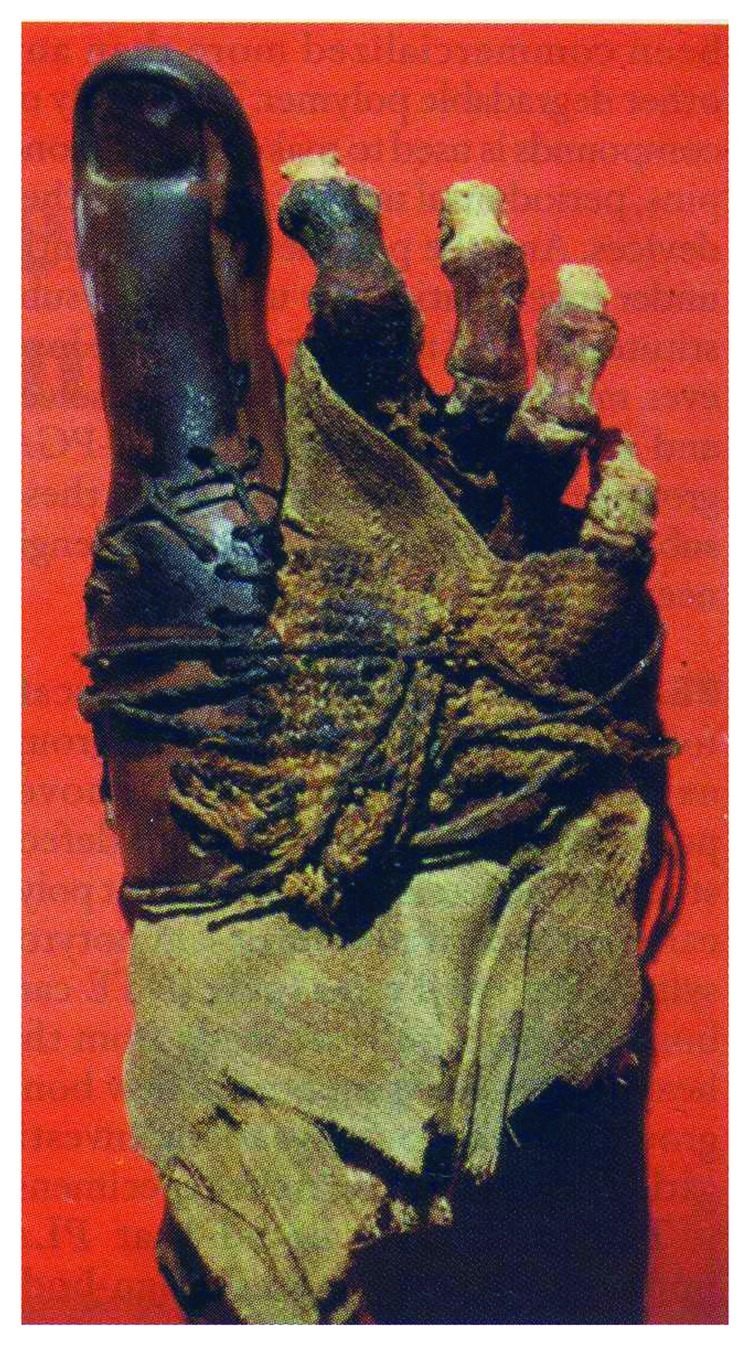
Figure 2. A wooden prosthesis of a hallux of a mummy. Reprinted from reference 197 with permission.
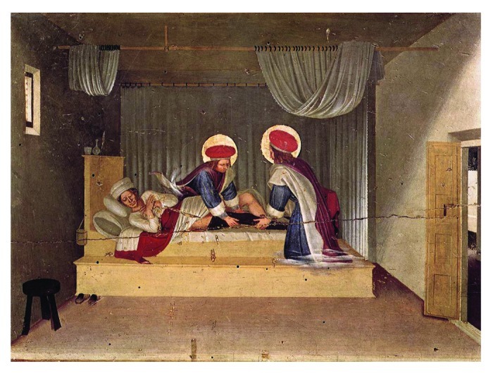
Figure 3. Fra Angelico (ca. 1395–1455) “The Healing of Justinian by Saint Cosmas and Saint Damian” (approx. 1439) is exhibited at Museo di San Marco, Florence, Italy.
Historically, a selection of the materials was based on their availability and an ingenuity of the individual making and applying the prosthetic.198 Archaeological findings exhibited in museums showed that materials used to replace missing human bones and teeth included animal or human (from corpses) bones and teeth, shells, corals, ivory (elephant tusk), wood (Fig. 2), as well as some metals (gold or silver). For instance, the father of Western medicine Hippocrates (ca. 460 BC–370 BC) apparently used gold wire and linen thread for ligatures in the repair of bone fractures. Aulus Cornelius Celsus (ca. 25 BC–50 AD) recommended the filling of large cavities with lint, lead and other substances before attempting extraction to prevent the tooth from breaking under the pressure of the instrument. This may have been the beginning of filling materials for carious teeth. The Etruscans learned to substitute missing teeth with bridges made from artificial teeth carved from the bones of oxen, while in ancient Phoenicia loose teeth were bound together with gold wires for tying artificial ones to neighboring teeth. Popp states that ancient Egyptians also made artificial ears, noses and eyes.199 The Chinese recorded the first use of dental amalgam to repair decayed teeth in the year 659 AD, while in Americas the pre-Columbian civilizations used gold sheets to heal cranial cavities following trepanation.200 Besides, while excavating Mayan burial sites in Honduras in 1931, archeologists found a fragment of mandible of Mayan origin, dating from about 600 AD. This mandible, which is considered to be that of a woman in her twenties, had three tooth-shaped pieces of shell placed into the sockets of three missing lower incisor teeth. In 1970, a Brazilian dental academic Prof. Amadeo Bobbio studied the mandible specimen and took a series of radiographs. He noted compact bone formation around two of the implants, which led him to conclude that the implants were placed during life.201 This may be the first recorded use of dental implants. More to the point, an iron dental implant in a corpse dated ~200 AD was found in Europe. This implant, too, was described as properly bone integrated.202
In middle ages, one of the first scientific descriptions of congenital and acquired defects of the maxilla and their treatments was given by Ambroise Paré (151 –1590) in his Dix livres de la chirurgie, avec le magasin des instrumens necessaires à icelle, published in Paris in 1564. He specifically described defects of the palate with bone destruction caused by arquebus shots, stab wounds or syphilitic gumma, describing also the accompanying speech deficiency and giving general principles of treatment. He used a flat, vaulted, metallic plate in gold or silver with a sponge attached to it. The sponge was introduced into the defect, where it expanded with readily absorbed nasal and oral secretions, thus holding the obturator base in position. Paré is also credited with having prepared artificial teeth from bones and ivory. In the 17th century, a piece of dog skull was successfully transplanted into the damaged skull of a Dutch duke.
In the 18th century, a common method to replace teeth was the homologous transplantation of teeth in humans. John Hunter (1728–1793) investigated in his pioneering work the effect of transplantation not only at a clinical level (he claimed, that homologous transplanted teeth lasted for years in the host) but also performed animal experimental work on the fate of transplants, thereby setting the basis for a scientific approach on transplantation medicine.203 Besides, various restorative materials might be used for feeling defects, capping exposed pulps and dental cavities. Currently they include zinc orthophosphate, zinc polyacrylate (polycarboxylate), various amalgams, glass ionomer cement of complicated formulations, calcium hydroxide, etc.; however, this is another story. Within the scope of this review, calcium orthophosphate-based formulations will be considered only. According to the available literature, Dr. Junius E. Cravens (1844–1920) from the US proffered creative concepts in pulp capping in the 1870s. He had the opinion that dentin-like material would be the best to keep the pulp vital. Therefore, Cravens used a calcium orthophosphate powder, which was mixed with lactic acid to low viscosity. The result was a soluble calcium lactic orthophosphate, which was applied onto the exposed pulp tissue.204 This pulp-capping agent was brought to the market by the S.S. White company with the trade name “Lacto-Phosphate of Lime” (Fig. 4). Sugar-containing formulations were known as well.205 To the best of my findings, the study by Cravens might be considered as the first mentioning on artificial calcium orthophosphate-based biocomposites and hybrid biomaterials (reviewed in reference 206). Thus, one might claim that the grafting history of calcium orthophosphates started in 1870s.
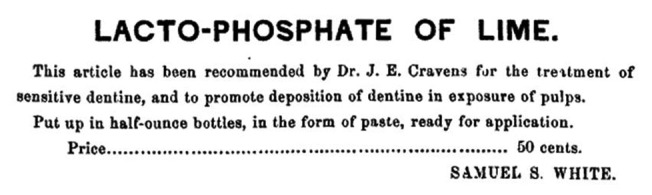
Figure 4. An advertisement of the S.S. White company for “Lacto-Phosphate of Lime” 1873. Reprinted from Dent. Cosmos 1873, 15, 683.
One must stress that in the past, many implantations failed due to various infections, which tended to be exacerbated in the presence of implants, since they provided a region inaccessible to the body’s immunologically competent cells. Thus, the use of implantable biomaterials did not become practical until the advent of an aseptic surgical technique developed by a British surgeon Sir Joseph Lister (1827–1912) in the 1860s. Furthermore, there was a lack of knowledge about a toxicity of the selected materials. The exact chemical composition of the normal calcified tissues of mammals has been known since, at least, 1870 sec, which might be concluded from both Table 238 and the following citations: “The bones and teeth of animals contain large amounts of calcium phosphate, together with some carbonate and fluoride” (ref. 55, p. 188) and “Calcium phosphate is also the chief inorganic constituent of bones, forming about 80 per cent of burnt bones; the other constituents being magnesium phosphate, calcium carbonate, and calcium fluoride” (ref. 55, p. 202). Those data were updated in 1894.207 However, the apatitic structure of the calcium orthophosphates of bones and teeth has been established by X-ray diffraction in 1926208 and confirmed in 1932209 and 1933.210 Since that time, bone mineral has been frequently identified as HA. In this frame, application of calcium orthophosphates as artificial grafts appears to be logical due to their similarity with the mineral phases of bones and teeth.
Table 2. The chemical composition of two bone samples taken from a publication of 187138.
| Commercial bone-ash | Pure ox bone-ash | |
|---|---|---|
| Moisture and volatile matter |
6.70 |
1.86 |
| Siliceous matter |
9.69 |
0.51 |
| Oxide of iron |
0.58 |
0.17 |
| Lime |
43.37 |
52.46 |
| Magnesia |
1.14 |
1.02 |
| Phosphoric acid |
33.68 |
39.55 |
| Carbonic acid, alkalies, and other substances undetermined |
4.84 |
4.43 |
| Total | 100.00 | 100.00 |
To conclude this topic, one should stress that the performance of living tissues is the result of millions years of evolution, while the performance of acceptable artificial substitutions those humankind has designed to repair damaged tissues are only a few decades old. This explains the greatest differences between them. To get the historical perspective on the development of artificial grafts prepared from other materials, the interested readers are referred to the special literature.6,8-11,211-214
Calcium Orthophosphates as Bone Graft Substitutes: A Historical Perspective
Historically, plaster of Paris (calcium sulfate) was the first widely tested artificial bioceramics. For example, according to Wikipedia, the free encyclopedia, literature dating back to 975 AD notes that calcium sulfate was useful for setting broken bones. However, those were ex vivo applications. According to the available literature, by the end of the 19th century, surgeons already used plaster of Paris as a bone-filling substitute.215 Nevertheless, it was a famous German surgeon Themistocles Gluck (1853–1942), who, among his range of contributions, on 20 May 1890 performed the first well documented ivory (virtually, pure biological apatite) knee replacement bedded in a calcium sulfate based cement, which was followed by a total wrist replacement in another patient three weeks later.216 Later in 1890, Gluck presented a further case of a total knee replacement to the Berlin Medical Society: at only 35 days after operation, the patient was pain free with active knee flexion and extension. All the joint arthroplasties performed by Gluck were remarkably successful in the short-term; however, all ultimately failed because of chronic infections.217,218 After the abovementioned case of lacto-phosphate of lime (Fig. 4), this seems to be the second well-documented grafting application of calcium orthophosphates.
However, in the aforementioned cases, either the biomedical applications of biologically produced calcium orthophosphates (Gluck) or dental applications, not requiring any surgery (Cravens) have been described. According to both the electronic databases and previous reviews on the subject,6,8-11,211-214 the first attempt to implant a laboratory produced calcium orthophosphate (it was TCP) as an artificial material to repair surgically created defects in rabbit bones was performed in 19207 by an US surgeon Fred Houdlette Albee (1876–1945), who invented bone grafting219 and some other advances in orthopedic surgery. The researchers injected either 0.5 or 1 c.c. of 5% slurries of TCP in distilled water (which was then sterilized for three successive days in the Arnold sterilizer, at 60°C) into surgically created radial bone gaps of rabbits, leaving the periosteum intact.7 Radiographic analysis revealed that the TCP injected defect demonstrated more rapid bone growth and union than the control. The average length of time for bony union with TCP was 31 d, compared with 41 d for the controls. No appreciable bone growth was stimulated by injecting TCP beneath the periosteum in non-defective radii or into subcutaneous tissues. Although this seems to be the first scientific study on use of an artificially prepared calcium orthophosphate for in vivo repairing of bone defects, it remains unclear whether that TCP was a precipitated or a ceramic material and whether it was in a powder or a granular form. Unfortunately, the researchers published nothing further on this topic. In 1927, Hey Groves (1872–1944) described pure ivory hip hemi-arthroplasty for fracture.220 In 1931, Murray also reported an acceleration of healing following implantation of calcium salts composed of 85% TCP and 15% CaCO3 in canine long bone defects.221,222
At the beginning of the 1930s, the classic osteoinductive phenomenon was defined well by Huggins,223 who demonstrated that autoimplantation of transitional epithelium of the urinary bladder to abdominal wall muscle in dogs provoked ectopic bone formation. A bit later, Levander demonstrated that crude alcoholic extracts of bones induced a new bone formation when injected into muscle tissue.224,225
Simultaneously, in the 1930s, Haldeman and Moore,226 Stewart,227 Key228 and Shands229 discovered the fact that only certain types of calcium orthophosphates mentioned in Table 1 really influence the bone healing process. Namely, Haldeman and Moore implanted various calcium orthophosphates such as MCP and DCP (it remains unclear whether they were in hydrated or anhydrous forms), TCP, as well as calcium glycerophosphate as dry powdered salts into 0.5 to 1.0 cm defects in radii of 17 rabbits, while the opposite side served as control. Radiographic analysis demonstrated that in no case did the presence of MCP, DCP or calcium glycerophosphate had a favorable influence delayed healing compared with control, while the presence of TCP at the site of the fracture appeared to favor the union.226 Furthermore, Key228 suggested that “if a defect in bone could be filled by a non-irritating, slowly soluble mass, which was porous and which contained calcium phosphate and carbonate in a form in which they could be resorbed, it would be reasonable to expect osteoblasts to invade the mass, utilize the calcium, and build new bone which would replace the mass of calcium and cause the bone to be restored to its original form. The ideal material would appear to be rather dense cancellous bone from which a large percentage of the organic material had been removed” (ref. 228, p. 176). However, Key found that “Neither calcium phosphate and carbonate in the proportions in which they occur in bone, nor bone powder, made by removing the organic matter from bone, appear to stimulate osteogenesis of bone when implanted in a bone defect” (ref. 228, p. 184). Stewart227 concluded that “1. Lime salts and boiled bone when placed into a bone defect with either traumatized muscle or fascia do not serve as a source of available calcium resulting in supersaturation of connective tissue and regeneration of missing bone. 2. Live bone chips placed in bone defects regenerate the missing bone” (ref. 227, p. 871). Shands229 also reported conflicting effects of several calcium salts [calcium glycerophosphate, a mixture of TCP (3 parts) and CaCO3 (1 part), bone ash and calcium gluconate] on bone repair. Namely, in defects in the ulna of dogs, the investigated calcium salts appeared to stimulate bone formation, while in operations upon the spine, calcium glycerophosphate did not stimulate bone formation and appeared rather to exert an inhibiting influence. In 1948, Schram and Fosdick confirmed the fact that only certain types of calcium orthophosphates influence the bone healing process.230 Similar conclusions were obtained in 1951 by Ray and Ward.231
Due to the reasons mentioned in the abstract, more recent historical events are reported very briefly. Namely, in 1950 the history of calcium orthophosphate cements was started.232 The author of that important publication investigated mixtures of both oxides and hydroxides of various metals with aqueous solutions of orthophosphoric acid and discovered a number of cold-setting formulations. For example, he found that CaO, sintered at 1100°C, did not set in H3PO4, while that in liquid containing 9.6% CaO was found to set after ~12 h in presence of H3PO4.232 The latter mixture might be considered as the first prototype of self-setting calcium orthophosphate cements (reviewed in refs. 233 and 234). The next publication on this subject appeared in 1975235; however, the real history of the self-setting calcium orthophosphate cements started in 1982.236,237
The modern history of ACP34 started in 1955,238 while more than 20 years afterwards the first dental application of a calcium orthophosphate (erroneously described as TCP) in surgically created periodontal defects239 and the use of dense HA cylinders for immediate tooth root replacement240 were reported. According to the available databases, the first paper with the term “bioceramics” in the abstract was published in 1971,241 while with that in the title were published in 1972.242,243 However, application of the ceramic materials as prostheses had been known before.244-247 On April 26, 1988, the first international symposium on bioceramics was held in Kyoto, Japan.
The history of calcium orthophosphate based biocomposites and hybrid biomaterials (reviewed in ref. 206) started in 1981, while that of calcium orthophosphate coatings (reviewed in refs. 248 and 249) started in 1980.250 An extensive commercialization of the dental and surgical applications of calcium orthophosphate (mainly, HA) bioceramics occurred in the 1980s, largely due to the pioneering efforts by Jarcho251-254 in the USA, de Groot255-257 in Europe and Aoki258-261 in Japan. Shortly afterwards HA has become a bioceramic of reference in the field of calcium orthophosphates for biomedical applications. Preparation and biomedical applications of apatites derived from sea corals (coralline HA)262-264 and bovine bone265 have been reported at the same time,266 while the history of biphasic, triphasic and multiphasic calcium orthophosphates (reviewed in ref. 267) started in 1985.268,269 Finally yet importantly, the history of nanodimensional and nanocrystalline calcium orthophosphates (reviewed in refs. 270 and 271) started in 1994.272-276
To get further details on a recent history of calcium orthophosphates, bioceramics and biomaterials in general, the interested readers are referred to other reviews on the subject.6,8-11,211-214
Conclusions
The reported historical findings clearly demonstrate that the subject of calcium orthophosphates have been investigated for almost two and a half centuries. Furthermore, the substantial amount of both scientific facts and experimental approaches has been known for very many decades and, in fact, the considerable quantity of relatively recent investigations on calcium orthophosphates is just either a further development of the earlier studies or a rediscovery of the already forgotten knowledge. Nevertheless, first commercially produced bone grafts and other types of biomaterials consisting of calcium orthophosphates appeared just a few decades ago.251-261 Furthermore, the biomedical properties of even the most advanced formulations are still unsatisfactory and incomparable with those of natural bones and teeth. However, the advantages and obvious benefits of calcium orthophosphates remain both an inspiration and a hope of many researchers and clinicians. Hopefully, the reported historical overview will keep encouraging both natural scientists and clinicians to rethink and reinvestigate the already forgotten and poorly known facts and approaches in further development of calcium orthophosphates and calcium orthophosphate-based formulations.
Footnotes
As a mineral species, apatite was first recognized by the father of German geology Abraham Gottlob Werner (1750 – 1817) in 1786 and named by him from the ancient Greek απατάω (apatao) – “to mislead” or “to deceive,” because it had previously been mistaken for other minerals, such as beryl, tourmaline, chrysolite, amethyst, fluorite, etc. Currently, apatite is the name for a group of minerals with the same crystallographic structure and does not indicate one chemical composition. That is why the term “calcium apatite” is used in this review.
Johan Gottlieb Gahn (1745–1818) was a Swedish chemist and metallurgist, who discovered manganese in 1774.
Carl Wilhelm Scheele (1742 – 1786) was a German-Swedish pharmaceutical chemist, who discovered oxygen (although Joseph Priestley published his findings first), as well as identified molybdenum, tungsten, barium and chlorine before Humphry Davy.
Previously published online: www.landesbioscience.com/journals/biomatter/article/21340
References
- 1.Dorozhkin SV. Calcium orthophosphates in nature, biology and medicine. Materials. 2009;2:399–498. doi: 10.3390/ma2020399. [DOI] [Google Scholar]
- 2.Dorozhkin SV. Calcium orthophosphates: occurrence, properties, biomineralization, pathological calcification and biomimetic applications. Biomatter. 2011;1:121–64. doi: 10.4161/biom.18790. [DOI] [PMC free article] [PubMed] [Google Scholar]
- 3.Davy H. The Bakerian lecture: On some new phenomena of chemical changes produced by electricity, particularly the decomposition of the fixed alkalies, and the exhibition of the new substances which constitute their bases; and on the general nature of alkaline bodies. Philos Trans R Soc Lond B Biol Sci. 1808;98:1–44. doi: 10.1098/rstl.1808.0001. [DOI] [Google Scholar]
- 4.Davy H. Electro-chemical researches on the decomposition of the earths; with observations on the metals obtained from the alkaline earths, and on the amalgam procured from ammonia. Philos Trans R Soc Lond B Biol Sci. 1808;98:333–70. doi: 10.1098/rstl.1808.0023. [DOI] [Google Scholar]
- 5.A paper of the honourable Robert Boyl’s, depoſited with the secretaries of the Royal Society, Octob. 14. 1680. and opened ſince his death; being an account of his making the phosphorus, etc. Phil Trans. 1693;17:583–4. doi: 10.1098/rstl.1693.0002. [DOI] [Google Scholar]
- 6.Driskell TD. Early history of calcium phosphate materials and coatings. In: Characterization and performance of calcium phosphate coatings for implants. ASTM STP 1196. Horowitz E, Parr JE., eds. Philadelphia: American Society for Testing and Materials, 1994: 1-9. [Google Scholar]
- 7.Albee FH. assisted by Morrison HF. Studies in bone growth triple calcium phosphate as a stimulus to osteogenesis. Ann Surg. 1920;71:32–9. doi: 10.1097/00000658-192001000-00006. [DOI] [PMC free article] [PubMed] [Google Scholar]
- 8.Shackelford JF. Bioceramics – an historical perspective. Mater Sci Forum. 1999;293:1–4. doi: 10.4028/www.scientific.net/MSF.293.1. [DOI] [Google Scholar]
- 9.Hulbert SF, Hench LL, Forbers D, Bowman LS. History of bioceramics. Ceram Int. 1982;8:131–40. doi: 10.1016/0272-8842(82)90003-7. [DOI] [Google Scholar]
- 10.Hulbert SF, Hench LL, Forbers D, Bowman LS. History of bioceramics. In: Ceramics in Surgery. Vincenzini P, ed. Amsterdam: Elsevier, 1983: 3-29. [Google Scholar]
- 11.Shepperd J. The early biological history of calcium phosphates. In: Fifteen years of clinical experience with hydroxyapatite coatings in joint arthroplasty. Epinette JA, Manley MT, eds. France: Springer, 2004: 3-8. [Google Scholar]
- 12.Roscoe HE, Schorlemmer C. A treatise on chemistry. Volume I: The non-metallic elements. London: Macmillan and Co., 1881: 751. [Google Scholar]
- 13.Hands, S. The Monthly Review, or Literary Journal by Several Hands. Vol XLII. London: The Strand, 1770: 584. [Google Scholar]
- 14.Macquer PJ, Keir J. A dictionary of chemistry. Vol III. London: The Strand, 1777: 666. [Google Scholar]
- 15.de Born. Catalogue methodique et raisonné de la collection des fossils de mlle. Alberti: Aux Dépens de J.V. Degen, 1790: 504. [Google Scholar]
- 16.Crell L. Chemische Annalen für die Freunde der Naturlehre, Arznengelahrtheit, Haushaltungskunst und Manufakturen. Leipzig: Müllerschen Buchhandlung, 1789: 568. [Google Scholar]
- 17.Lavoisier AL. Elements of chemistry, in a new systematic order, containing all the modern discoveries. Translated from the French, by Robert Kerr. Edinburgh: printed for William Creech; and sold in London by G.G. & J. Robinsons, and T. Kay, 1790: 511. [Google Scholar]
- 18.Perthensis E. Universal dictionary of the arts, Sciences, literature, &c. intended to supersede the use of other books of reference. Vol XVII. Edinburgh: Printed by John Brown, Anchor Close, for the proprietors, and sold by all the booksellers in the United Kingdom, 1816: 720. [Google Scholar]
- 19.Fourcroy AF. A general system of chemical knowledge; and its application to the phenomena of nature and art. In eleven volumes. Vol III. London: The Strand, 1804: 472. [Google Scholar]
- 20.Nisbet W. A general dictionary of chemistry, containing the leading principles of the science, in regard to facts, experiments, and nomenclature. For the use of students. London: Printed for S. Highley (successor to the late Mr. John Murray), 1805: 415. [Google Scholar]
- 21.Murray J. A system of chemistry in four volumes. Vol I. Edinburgh: Constable & Co. Edinburgh, 1806: 592. [Google Scholar]
- 22.Bache F. A system of chemistry for the use of students of medicine. Philadelphia: William Fry Printer, 1819: 624. [Google Scholar]
- 23.Good JM, Olinthus G, Newton B. Pantologia: A new cyclopædia, comprehending a complete series of Essays, Treatises, and Systems, alphabetically arranged; with a general dictionary of arts, sciences, and words: the whole presenting a distinct survey of Human Genius, Learning, and Industry. Vol IX. London: Sherwood, Neelt and Jones, 1813. [Google Scholar]
- 24.Encyclopædia Britannica or, a dictionary of arts, sciences, and miscellaneous literature; enlarged and improved. Vol V. Edinburgh: Archibald Constable and Company, 1810: 797. [Google Scholar]
- 25.Muhlenberg WF. Address in Hygiene. In: Transactions of the medical society of the state of Pennsylvania, at its thirty-third annual session, held at Titusville, May 10, 11 and 12, 1832. Vol XIV. Philadelphia: Times printing house, 1832: 81-102. [Google Scholar]
- 26.Berzelius J. Untersuchungen über die Zusammensetzung der Phosphorsäure, der phosphorigen Säure und ihrer Salze. Ann Phys. 1816;53:393–446. doi: 10.1002/andp.18160530805. [DOI] [Google Scholar]
- 27.Berzelius J. Ueber basische phosphorsaure Kalkerde. Justus Liebigs Ann Chem. 1845;53:286–8. doi: 10.1002/jlac.18450530212. [DOI] [Google Scholar]
- 28.Baruel M. Analysis of a double phosphate of lead and lime. J Franklin Inst. 1838;25:343. doi: 10.1016/S0016-0032(38)91002-6. [DOI] [Google Scholar]
- 29.Smith JD. Ueber die Zusammensetzung verschiedener Arten von südamerikanischem Guano, nebst der Beschreibung einer neuen Methode, Ammoniak zu bestimmen, so wie Kalk und Magnesia, wenn sie an Phosphorsäure gebunden sind, zu trennen. J Prakt Chem. 1845;35:277–91. doi: 10.1002/prac.18450350140. [DOI] [Google Scholar]
- 30.Mitscherlich E. Lehrbuch der Chemie. Erster Band. Die Metalloïde. Berlin: Vierte Auflage, 1844: 609. [Google Scholar]
- 31.On the method by which the phosphate and carbonate of lime is introduced into the organs of plants. J Franklin Inst. 1849;48:156. doi: 10.1016/0016-0032(49)90039-3. [DOI] [Google Scholar]
- 32.Wilson G. Chemistry. Edinburgh: William and Robert Chambers, 1850: 316. [Google Scholar]
- 33.Brande WT, Taylor AS. Chemistry. Philadelphia: Blanchard and Lea, 1863: 696. [Google Scholar]
- 34.Dorozhkin SV. Amorphous calcium orthophosphates: nature, chemistry and biomedical applications. Int J Mater Chem. 2012;2:19–46. doi: 10.1039/c9tb01976f. [DOI] [PubMed] [Google Scholar]
- 35.Morfit C. On Colombian guano; and certain peculiarities in the chemical behavior of “bone phosphate of lime”. J Franklin Inst. 1855;30:325–9. doi: 10.1016/0016-0032(55)90343-4. [DOI] [Google Scholar]
- 36.Warington R. CXIX. On a curious change in the composition of bones taken from the guano. Mem Proc Chem Soc. 1843;2:223–6. doi: 10.1039/mp8430200223. [DOI] [Google Scholar]
- 37.Warington R., Jr. XXVII. – Researches on the phosphates of calcium, and upon the solubility of tricalcic phosphate. J Chem Soc. 1866;19:296–318. doi: 10.1039/js8661900296. [DOI] [Google Scholar]
- 38.Warington R. X. – On the solubility of the phosphates of bone-ash in carbonic water. J Chem Soc. 1871;24:80–3. doi: 10.1039/js8712400080. [DOI] [Google Scholar]
- 39.Warington R. XL. – On the decomposition of tricalcic phosphate by water. J Chem Soc. 1873;26:983–9. doi: 10.1039/js8732600983. [DOI] [Google Scholar]
- 40.Fresenius R. Ueber die Bestimmung der Phosphorsäure im Phosphorit nebst Mittheilung der Analysen des Phosphorits und Staffelits aus dem Lahnthal. Z Anal Chem. 1867;6:403–9. doi: 10.1007/BF01347651. [DOI] [Google Scholar]
- 41.Lorah JR, Tartar HV, Wood L. A basic phosphate of calcium and of strontium and the adsorption of calcium hydroxide by basic calcium phosphate and by tricalcium phosphate. J Am Chem Soc. 1929;51:1097–106. doi: 10.1021/ja01379a015. [DOI] [Google Scholar]
- 42.Hassall A. On the detection and preservation of crystalline deposits of uric acid, urate of ammonia, phosphate of lime, triple phosphate, oxalate of lime, and other salts. Lancet. 1852;59:466–7. doi: 10.1016/S0140-6736(02)41306-2. [DOI] [Google Scholar]
- 43.Abel FA. XVIII. – On the occurrence of considerable deposits of crystallized phosphate of lime in teak-wood. J Chem Soc. 1862;15:91–3. doi: 10.1039/js8621500091. [DOI] [Google Scholar]
- 44.Stammer C. Bestimmung kohlensauren Kalkes neben phosphorsaurem Kalk. Z Anal Chem. 1863;2:96–7. doi: 10.1007/BF01430170. [DOI] [Google Scholar]
- 45.Roussin Z. Prfung des Wismuthsubnitrats auf eine Verfälschung mit Kalkphosphat. Z Anal Chem. 1868;7:511. doi: 10.1007/BF01334947. [DOI] [Google Scholar]
- 46.Janovsky JV. Ueber die verschiedenen Methoden der Phosphorsäure-Bestimmung neben Eisenoxyd, Thonerde, Kalk und Magnesia. Z Anal Chem. 1872;11:153–67. doi: 10.1007/BF01588404. [DOI] [Google Scholar]
- 47.Pellet H. Die Zusammensetzung des Niederschlags, welcher durch Ammoniak aus sauren Lösungen von Phosphorsäure, Baryt, Kalk und Magnesia gefällt wird. Z Anal Chem. 1882;21:261. doi: 10.1007/BF01356431. [DOI] [Google Scholar]
- 48.Stokvis BJ, Salkowski E, Smith WG. Ueber die Löslichkeitsverhältnisse des phosphorsauren Kalks im Harn. Zeitschrift für Analytische Chemie. 1884;23:273–274. doi: 10.1007/BF01360506. [DOI] [Google Scholar]
- 49.Ott A. Die Löslichkeitsverhältnisse des phosphorsauren Kalks im Harn. Z Anal Chem. 1886;25:279–80. doi: 10.1007/BF01335431. [DOI] [Google Scholar]
- 50.Kennepohl G. Zur Bestimmung von Eisenoxyd und Thonerde neben Kalk und Phosphorsäure. Z Anal Chem. 1889;28:343. doi: 10.1007/BF01375987. [DOI] [Google Scholar]
- 51.Immendorff H, Reitmair O. Zur Bestimmung des Kalks in Gegenwart von Phosphorsäure, Eisen, Thonerde und Mangan. Zeitschrift für Analytische Chemie. 1892;31:313–6. doi: 10.1007/BF01344959. [DOI] [Google Scholar]
- 52.Fingerling G, Grombach A. Eine neue Modifikation der Bestimmung der zitratlöslichen Phosphorsäure in den Futterkalken nach Petermann. Z Anal Chem. 1907;46:756–60. doi: 10.1007/BF01301337. [DOI] [Google Scholar]
- 53.Schulze B. Untersuchung des phosphorsauren Futterkalkes. Z Anal Chem. 1911;50:126–7. doi: 10.1007/BF01371221. [DOI] [Google Scholar]
- 54.Hinden F. Anreicherungsmethode zur Bestimmung der Phosphorsäure in phosphorsäurearmen Kalksteinen. Z Anal Chem. 1915;54:214–6. doi: 10.1007/BF01470848. [DOI] [Google Scholar]
- 55.Roscoe HE, Schorlemmer C. A treatise on chemistry. Volume II: Metals. Part 1. London: Macmillan and Co., 1879, 504 pp. [Google Scholar]
- 56.Duncan A. The Edinburgh new dispensatory. Worcester: From the press of Isaiah Thomas, Jun., sold by him in Worcester and Springfield, [and 2 others], 1805: 710. [Google Scholar]
- 57.Fresenius R. Zur Analyse der Superphosphate. Z Anal Chem. 1868;7:304–9. doi: 10.1007/BF01334869. [DOI] [Google Scholar]
- 58.Chesshire JA, Hughes J, Sutton F, Sibson A. Ueber die Bestimmung des Betrags an “reducirten” Phosphaten in Superphosphaten. Zeitschrift für Analytische Chemie. 1870;9:524–527. [Google Scholar]
- 59.Williams CP. On the solubility of some forms of phosphate of lime. J Franklin Inst. 1871;92:419–23. doi: 10.1016/0016-0032(71)90318-8. [DOI] [Google Scholar]
- 60.Rümpler A. Ueber eisen- und thonerdehaltige Superphosphate und deren analytische Untersuchung. Z Anal Chem. 1873;12:151–63. doi: 10.1007/BF01462997. [DOI] [Google Scholar]
- 61.Albert H, Siegfried L. Beiträge zur Werthbestimmung der Superphosphate. Z Anal Chem. 1877;16:182–8. doi: 10.1007/BF01356063. [DOI] [Google Scholar]
- 62.Albert H, Siegfried L. Beiträge zur Werthbestimmung der Superphosphate. Z Anal Chem. 1879;18:220–4. doi: 10.1007/BF01345375. [DOI] [Google Scholar]
- 63.Pavec A. Zur maassanalytischen Bestimmung der Phosphorsäure im Superphosphat und Spodium mittelst Uranlösung. Z Anal Chem. 1879;18:360–1. doi: 10.1007/BF01345415. [DOI] [Google Scholar]
- 64.Mohr C. Ein maassanalytisches Bestimmungsverfahren der in Rohphosphaten und Superphosphaten enthaltenen Phosphorsäure mit Uran bei Gegenwart von Eisenoxyd. Z Anal Chem. 1880;19:150–3. doi: 10.1007/BF01340917. [DOI] [Google Scholar]
- 65.Erlenmeyer E, Wattenberg H, Wein E, Rösch L, Lehmann J, Johnson SW, et al Zur Analyse der Superphosphate. Zeitschrift für Analytische Chemie. 1880;19:243–246. [Google Scholar]
- 66.Meyer CF. Weitere Mittheilungen über das Zurückgehen der eisen- und thonerdehaltigen Superphosphate – Berichtigung. Z Anal Chem. 1880;19:309–11. doi: 10.1007/BF01340995. [DOI] [Google Scholar]
- 67.Drewsen S. Zur Bestimmung der löslichen Phosphorsäure in Superphosphaten. Z Anal Chem. 1881;20:54–7. doi: 10.1007/BF01374606. [DOI] [Google Scholar]
- 68.Mollenda A. Eine neue Methode zur maassanalytischen Bestimmung der Phosphorsäure in den Superphosphaten. Z Anal Chem. 1883;22:155–9. doi: 10.1007/BF01338046. [DOI] [Google Scholar]
- 69.Phillips WB. Rate of reversion in superphosphates prepared from red navassa rock. J Am Chem Soc. 1884;6:224–8. doi: 10.1021/ja02131a020. [DOI] [Google Scholar]
- 70.Wagner P. Eine neue Methode zur Feststellung des Handelswerthes der Superphosphate. Z Anal Chem. 1886;25:272–8. doi: 10.1007/BF01335427. [DOI] [Google Scholar]
- 71.Emmerling A. Eine Methode zur Bestimmung der wasserlöslichen Phosphorsäure in Superphosphaten auf maassanalvtischem Wege. Z Anal Chem. 1887;26:244–7. doi: 10.1007/BF01385150. [DOI] [Google Scholar]
- 72.Stoklasa J. Bestimmung des Wassers in den Superphosphaten. I. Z Anal Chem. 1890;29:390–7. doi: 10.1007/BF01367207. [DOI] [Google Scholar]
- 73.Crispo D. Belgische Methode zur Bestimmung der in Wasser löslichen Phosphors[UNKNOWN ENTITY &adie;]ure in den Superphosphaten. Z Anal Chem. 1891;30:301–3. doi: 10.1007/BF01592043. [DOI] [Google Scholar]
- 74.Güssefeld O. Eine Schüttelmaschine für die Analyse von Superphosphaten. Z Anal Chem. 1892;31:556. doi: 10.1007/BF01345062. [DOI] [Google Scholar]
- 75.Keller A. Eine Schüttelmaschine für Superphosphaten. Z Anal Chem. 1893;32:590–1. doi: 10.1007/BF01369030. [DOI] [Google Scholar]
- 76.Kalmann W, Meissels K. Eine Methode zur maassanalytischen Schätzung der wasserlöslichen Phosphors[UNKNOWN ENTITY &adie;]ure in Superphosphaten. Z Anal Chem. 1894;33:764–6. doi: 10.1007/BF01335960. [DOI] [Google Scholar]
- 77.Glaser C. Zur maassanalytischen Bestimmung der wasserlöslichen Phosphorsäure in Superphosphaten. Z Anal Chem. 1895;34:768–9. doi: 10.1007/BF01595963. [DOI] [Google Scholar]
- 78.Seib O. Bestimmung der zitratlöslichen Phosphorsäure in Superphosphaten. Z Anal Chem. 1905;44:397–8. doi: 10.1007/BF01329880. [DOI] [Google Scholar]
- 79.Cameron FK, Bell JM. The phosphates of calcium, III; superphosphate. J Am Chem Soc. 1906;28:1222–9. doi: 10.1021/ja01975a016. [DOI] [Google Scholar]
- 80.Wells HG. Pathological Calcification. J Med Res. 1906;14:491–525. [PMC free article] [PubMed] [Google Scholar]
- 81.C. Vitreous phosphate of lime. J Franklin Inst. 1877;104:315. doi: 10.1016/0016-0032(77)90209-5. [DOI] [Google Scholar]
- 82.Church AH. New analyses of certain mineral arseniates and phosphates. 1. Apatite; 2. Arseniosiderite; 3. Childrenite; 4. Ehlite; 5. Tyrolite; 6. Wavellite. J Chem Soc. 1873;26:101–11. doi: 10.1039/js8732600101. [DOI] [Google Scholar]
- 83.L, WJ. On a fine specimen of apatite from Tyrol, lately in the possession of Mr. Samuel Henson. Nature. 1883;27:608–9. doi: 10.1038/027608a0. [DOI] [Google Scholar]
- 84.Reichardt E. Ueber neutralen phosphorsauren Kalk, Darstellung und Löslichkeit desselben. Z Anal Chem. 1872;11:275–7. doi: 10.1007/BF01588458. [DOI] [Google Scholar]
- 85.Mohr C. Ueber die quantitative Bestimmung der zurückgegangenen Phosphorsäure und der Phosphorsäure im Dicalciumphosphat. Z Anal Chem. 1884;23:487–91. doi: 10.1007/BF01360583. [DOI] [Google Scholar]
- 86.Glaser C. Bemerkungen zu der Abhandlung des Herrn Carl Mohr über die quantitative Bestimmung der zurückgegangenen Phosphorsäure und der Phosphorsäure im Dicalciumphosphat. Z Anal Chem. 1885;24:180. doi: 10.1007/BF01366673. [DOI] [Google Scholar]
- 87.Hutchings WM. Occurrence of apatite in slag. Nature. 1887;36:460. doi: 10.1038/036460a0. [DOI] [Google Scholar]
- 88.Hilgenstock G. Eine neue Verbindung von P2O5 und CaO. Stahl und Eisen. 1883;3:498. [Google Scholar]
- 89.Hilgenstock G. Das vierbasische Kalkphosphat und die Basicitätsstufe des Silicats in der Thomas-Schlacxke. Stahl und Eisen. 1887;7:557–60. [Google Scholar]
- 90.Scheibler C. Ueber die Herstellung reicher Kalkphosphate in Verbindung mit einer Verbesserung des Thomasprocesses. Ber Dtsch Chem Ges. 1886;19:1883–93. doi: 10.1002/cber.18860190258. [DOI] [Google Scholar]
- 91.Tzschucke H. Versuch einer directen Bestimmung der Phosphorsäure als dreibasisch phosphorsauren Kalk. Angew Chem. 1888;1:383–5. doi: 10.1002/ange.18880011303. [DOI] [Google Scholar]
- 92.Georgievics GV. Über das Verhalten des Tricalciumphosphats gegen Kohlensäure und Eisenhydroxyd. Monatsh Chem. 1891;12:566–81. doi: 10.1007/BF01538628. [DOI] [Google Scholar]
- 93.Cameron FK, Hurst LA. The action of water and saline solutions upon certain slightly soluble phosphates. J Am Chem Soc. 1904;26:885–913. doi: 10.1021/ja01998a001. [DOI] [Google Scholar]
- 94.Cameron FK, Seidell A. The action of water upon the phosphates of calcium. J Am Chem Soc. 1904;26:1454–63. doi: 10.1021/ja02001a007. [DOI] [Google Scholar]
- 95.Cameron FK, Seidell A. The phosphates of calcium. I. J Am Chem Soc. 1905;27:1503–12. doi: 10.1021/ja01990a005. [DOI] [Google Scholar]
- 96.Cameron FK, Bell JM. The phosphates of calcium. II. J Am Chem Soc. 1905;27:1512–4. doi: 10.1021/ja01990a006. [DOI] [Google Scholar]
- 97.Cameron FK, Bell JM. The phosphates of calcium. IV. J Am Chem Soc. 1910;32:869–73. doi: 10.1021/ja01925a003. [DOI] [Google Scholar]
- 98.Cameron FK, McCaughey WJ. Apatite and spodiosite. J Phys Chem. 1911;15:463–70. doi: 10.1021/j150122a002. [DOI] [Google Scholar]
- 99.Mebane WM, Dobbins JT, Cameron FK. The solubility of the phosphates of calcium in aqueous solutions of sulfur dioxide. J Phys Chem. 1929;33:961–9. doi: 10.1021/j150301a001. [DOI] [Google Scholar]
- 100.Bassett H., Jr. Beiträge zum Studium der Calciumphosphate. I. Die Hydrate der Calcium-Hydroorthophosphate. Z Anorg Chem. 1907;53:34–48. doi: 10.1002/zaac.19070530104. [DOI] [Google Scholar]
- 101.Bassett H., Jr. Beiträge zum Studium der Calciumphosphate. II. Die Einwirkung von Ammoniakgas auf Calcium-Hydroorthophosphate. Z Anorg Chem. 1907;53:49–62. doi: 10.1002/zaac.19070530105. [DOI] [Google Scholar]
- 102.Bassett H., Jr. Beiträge zum Studium der Calciumphosphate. III. Das System CaO – P2O5 – H2O. Z Anorg Chem. 1908;59:1–55. doi: 10.1002/zaac.19080590102. [DOI] [Google Scholar]
- 103.Bassett H., Jr. The phosphates of calcium. Part IV. The basic phosphates. J Chem Soc. 1917;111:620–42. doi: 10.1039/ct9171100620. [DOI] [Google Scholar]
- 104.Norton TH, Newman HE. LV. - On a soluble compound of hydrastine with monocalcium phosphate. J Am Chem Soc. 1897;19:838–40. doi: 10.1021/ja02084a017. [DOI] [Google Scholar]
- 105.Rolfe BH. Autunite (hydrated uranium-calcium phosphate) Lancet. 1911;177:766. doi: 10.1016/S0140-6736(01)62347-X. [DOI] [Google Scholar]
- 106.Withers WA, Field AL. A conductivity study of the reaction between calcium nitrate and dipotassium phosphate in dilute solution. J Am Chem Soc. 1915;37:1091–105. doi: 10.1021/ja02170a010. [DOI] [Google Scholar]
- 107.Wendt GL. An electrometric study of the neutralization of phosphoric acid by calcium hydroxide. J Am Chem Soc. 1923;45:881–7. doi: 10.1021/ja01657a003. [DOI] [Google Scholar]
- 108.Shipley PG, Kramer B, Howland J. Studies upon Calcification in vitro. Biochem J. 1926;20:379–87. doi: 10.1042/bj0200379. [DOI] [PMC free article] [PubMed] [Google Scholar]
- 109.Benjamin HR. The forms of the calcium and inorganic phosphorus in human and animal sera II. The nature and significance of the filtrable, adsorbable calcium-phosphorus complex. J Biol Chem. 1933;100:57–78. [Google Scholar]
- 110.Náray-Szabó S. The structure of apatite (CaF)Ca4(PO4)3. Z Kristallogr. 1931;75:387–98. [Google Scholar]
- 111.Hendricks SB, Hill WL, Jacob KD, Jefferson ME. Structural characteristics of apatite-like substances and composition of phosphate rock and bone as determined from microscopical and X-ray diffraction. Ind Eng Chem. 1931;23:1413–8. doi: 10.1021/ie50264a025. [DOI] [Google Scholar]
- 112.Clark NA. The system P2O5–CaO–H2O and the recrystallization of monocalcium phosphate. J Phys Chem. 1931;35:1232–8. doi: 10.1021/j150323a005. [DOI] [Google Scholar]
- 113.Lugg JWH. A study of aqueous salt solutions in equilibrium with solid secondary calcium phosphate at 40° C. Trans Faraday Soc. 1931;27:297–309. doi: 10.1039/tf9312700297. [DOI] [Google Scholar]
- 114.Bredig MA, Franck HH, Fülnder H. Beiträge zur Kenntnis der Kalk-Phosphorsäure-Verbindungen. Z Elektrochem. 1932;38:158–64. [Google Scholar]
- 115.Trömel G. Beiträge zur Kenntnis des Systems Kalziumoxyd-Phosphorpentoxyd. Mitt. Kaiser-Wilhelm-Inst. Eisenforsch Düsseldorf. 1932;14:25–34. [Google Scholar]
- 116.Nagelschmidt G. A new calcium silicophosphate. J Chem Soc. 1937:865–7. doi: 10.1039/jr9370000865. [DOI] [Google Scholar]
- 117.Hill WL, Hendricks SB, Jefferson ME, Reynolds DS. Phosphate fertilizers by calcination process. Composition of defluorinated phosphate. Ind Eng Chem. 1937;29:1299–304. doi: 10.1021/ie50335a023. [DOI] [Google Scholar]
- 118.Marshall HL, Reynolds DS, Jacob KD, Tremearne TH. Phosphate fertilizers by calcination process. Reversion of defluorinated phosphate at temperatures below 1400 °C. Ind Eng Chem. 1937;29:1294–8. doi: 10.1021/ie50335a022. [DOI] [Google Scholar]
- 119.Curtis HA, Copson RL, Brown EH, Pole GR. Fertilizer from rock phosphate conversion by fusion and treatment with water vapor. Ind Eng Chem. 1937;29:766–71. doi: 10.1021/ie50331a009. [DOI] [Google Scholar]
- 120.Moore LA. Activation of dicalcium phosphate for the chromatographic determination of carotene. Ind Eng Chem. 1942;14:707–8. [Google Scholar]
- 121.Elsenberger S, Lehrman A, Turner WD. The basic calcium phosphates and related systems. Some theoretical and practical aspects. Chem Rev. 1940;26:257–96. doi: 10.1021/cr60084a008. [DOI] [Google Scholar]
- 122.Elmore KL, Farr TD. Equilibrium in the system calcium oxide–phosphorus pentoxide–water. Ind Eng Chem. 1940;32:580–6. doi: 10.1021/ie50364a030. [DOI] [Google Scholar]
- 123.Shear MJ, Kramer B. Composition of bone. III. Physicochemical mechanism. J Biol Chem. 1928;79:125–45. [Google Scholar]
- 124.Trömel G, Möller H. Die Bildung schwer löslicher Calciumphosphate aus wäßriger Lösung und die Beziehungen dieser Phosphate zur Apatitgruppe. Z Anorg Allg Chem. 1932;206:227–40. doi: 10.1002/zaac.19322060212. [DOI] [Google Scholar]
- 125.Larson HWE. Preparation and properties of mono-, di-, and tricalcium phosphates. Ind Eng Chem Anal Ed. 1935;7:401–6. doi: 10.1021/ac50098a016. [DOI] [Google Scholar]
- 126.Kirwan R. Elements of mineralogy. Second edition with considerable improvements and additions. Vol I. Earth and stones. London: The Strand, 1794: 510. [Google Scholar]
- 127.Murray J. A system of chemistry. In four volumes. Vol III. Edinburgh: Printed for Longman, Hurst, Rees & Orme, 1809: 712. [Google Scholar]
- 128.Jameson R. A system of mineralogy, in which minerals are arranged according to the natural history method. Third edition, enlarged and improved. Vol. II. London: Hurst, Robinson & Co., 1820: 632. [Google Scholar]
- 129.Lowry D. Conversations on mineralogy. Philadelphia: Uriah Hunt, 1822: 332. [Google Scholar]
- 130.Shaler NS. Nature and origin of phosphate of lime. Science. 1889;13:144–8. doi: 10.1126/science.ns-13.316.144. [DOI] [PubMed] [Google Scholar]
- 131.Graber H. XV. Diopsid und Apatit von Zöptau. Tschermaks Mineral Petrogr Mitt. 1895;14:265–70. [Google Scholar]
- 132.Church AH. Apatite in Ceylon. Nature. 1901;63:464. doi: 10.1038/063464b0. [DOI] [Google Scholar]
- 133.Klein C. Brushite from the island of Mona (between Haiti and Puerto Rico). Sitzber K Preuss Aka 1901;720-725. [Google Scholar]
- 134.Wolff JE, Palache C. Apatite from Minot, Maine. Proc Am Acad Arts Sci. 1902;37:517–28. doi: 10.2307/20021696. [DOI] [Google Scholar]
- 135.Wolff JE, Palache C. On apatite from Minot, Maine. Zeitschrift für Krystallographie und Mineralogie. 1902;36:438–48. [Google Scholar]
- 136.de Mercey N. Sur des gites de phosphate de chaux de la Craie à Bélemnites, formés avant le soulèvement du Bray. C R Hebd Seances Acad Sci. 1902;135:1137–8. [Google Scholar]
- 137.Zimanyi K. On the green apatite from Malmberget in Sweden. Zeitschrift für Krystallographie und Mineralogie. 1904;39:505–19. [Google Scholar]
- 138.Fraps GS. Availability of phosphoric acid of the soil. J Am Chem Soc. 1906;28:823–34. doi: 10.1021/ja01973a002. [DOI] [Google Scholar]
- 139.Fourcroy, AF. Elements of natural history, and of chemistry. Vol IV. London: Printed for G.G.J. and J. Robinson, Pater-Noster-Row, 1788: 465. [Google Scholar]
- 140.Tilloch A. The philosophical magazine: comprehending the various branches of science, the liberal and fine arts, agriculture, manufactures, and commerce. Vol II. London: Ricbabo Taylor and Co., 1798: 442. [Google Scholar]
- 141.The New-York magazine, or literary repository. New York: T. and J. Swords, 1797: 672. [Google Scholar]
- 142.Parr B. The London medical dictionary; including under distinct heads every branch of medicine, viz. anatomy, physiology, and pathology, the practice of physic and surgery, therapeutics, and materia medica; with whatever relates to medicine in natural philosophy, chemistry and natural history. Vol I. Philadelphia: Mitchell, Ames, and White, 1809: 786. [Google Scholar]
- 143.Parkes S. The chemical catechism, with notes, illustrations, and experiments. London: Lackington, Allen, and Co., 1812: 562. [Google Scholar]
- 144.Davy H. Conversations on chemistry, in which the elements of that science are familiarly explained and illustrated by experiments and plates. London: Didnep’s Press, 1814: 383. [Google Scholar]
- 145.Wilson J. Lectures on the structure and physiology of the parts composing the skeleton, and on the diseases of the bones and joints of the human body, preceded by some observations on the influence of the brain and nerves. London: Burgess and Hill, 1820: 410. [Google Scholar]
- 146.Nasse H. Ueber die Bestandtheile der Knochen in einigen Krankheiten. J Prakt Chem. 1842;27:274–80. doi: 10.1002/prac.18420270142. [DOI] [Google Scholar]
- 147.Levy M. Chemische Untersuchungen über osteomalacische Knochen. Zeitschrift für physiologische Chemie. 1895;19:239–270. [Google Scholar]
- 148.Aeby C. Ueber die Constitution des phosphorsauren Kalkes der Knochen. J Prakt Chem. 1872;5:308–11. doi: 10.1002/prac.18720050127. [DOI] [Google Scholar]
- 149.Aeby C. Ueber die nähern Bestandtheile des Knochenphosphates. J Prakt Chem. 1873;6:169–71. doi: 10.1002/prac.18730060117. [DOI] [Google Scholar]
- 150.Aeby C. Ueber die Metamorphose der Knochen. J Prakt Chem. 1873;7:37–44. doi: 10.1002/prac.18730070105. [DOI] [Google Scholar]
- 151.Wibel F. Die Constitution des Knochenphosphates, insbesondere die Existenz und Bildung einer basischen Verbindung (Ca3P2O8+xCaO) J Prakt Chem. 1874;9:113–32. doi: 10.1002/prac.18740090106. [DOI] [Google Scholar]
- 152.Weiske H. Beitrag zur Knochenanalyse. Zeitschrift für Physiologische Chemie. 1883;7:474–8. [Google Scholar]
- 153.Kramer B, Shear MJ. Composition of bone. IV. Primary calcification. J Biol Chem. 1928;79:147–60. [Google Scholar]
- 154.Jones HB. II. – On the occurrence of deposits of crystallised phosphate of lime in human urine. J Chem Soc. 1862;15:8–15. doi: 10.1039/js8621500008. [DOI] [Google Scholar]
- 155.Hassal, Phosphorsaurer Kalk im Urinsediment. Z Anal Chem. 1862;1:125. doi: 10.1007/BF01429956. [DOI] [Google Scholar]
- 156.Moss, Ueber das Vorkommen und die Bedeutung phosphorsaurer Kalkconcremente im Stamme des Gehörnerven. Arch Psychiatr Nervenkr. 1879;9:122–8. doi: 10.1007/BF01802987. [DOI] [Google Scholar]
- 157.Tereg A. Das Verhalten der Calciumphosphate im Organismus der Fleischfresser. Pflüger, Archiv für die Gesammte Physiologie des Menschen und der Thiere. Pflügers Archiv Eur J Physiology. 1883;32:122–70. doi: 10.1007/BF01628854. [DOI] [Google Scholar]
- 158.The monthly magazine; or, British register. Vol. 27, Part I. London: Printed for Richard Phillips, 1809: 736. [Google Scholar]
- 159.Pemberton CR. A practical treatise on various diseases of the abdominal viscera. Third edition, revised and corrected. London: W. Bulmer and Co., 1814: 201. [Google Scholar]
- 160.Under the care of Mr. Canton. Stone in the bladder of a boy for upwards of three years; lithotomy; extraction of a large oval calculus of phosphate of lime; recovery. Lancet. 1856;68:650–1. doi: 10.1016/S0140-6736(02)60383-6. [DOI] [Google Scholar]
- 161.Virchow R. Untersuchungen über die Entwicklung des Schädelgrundes im gesunden und krankhaften Zustände und über den Einfluss derselben auf Schädelform, Gesichtsbildung und Gehirnbau. Berlin: Druck und Verlag von Georg Reimer, 1857: 128. [Google Scholar]
- 162.Jones SSt. Thomas’s hospital.: Lateral lithotomy; removal of large stone composed of oxalate of lime covered with phosphates; recovery. Lancet. 1882;119:781–2. doi: 10.1016/S0140-6736(02)21974-1. [DOI] [Google Scholar]
- 163.Hill B. University college hospital.: Two cases of calculus of oxalate and phosphate of lime removed by lithotrity at a single sitting; singularly long duration of symptoms, with remarks upon an improved clover’s evacuating bottle. Lancet. 1883;121:59–60. [Google Scholar]
- 164.Hotz G. Phosphorsäure- und Kalkstoffwechsel bei Osteomalacie unter dem Einfluss der Phosphortherapie. Zeitschrift für Experimentelle Pathologie und Therapie. 1906;3:605–32. doi: 10.1007/BF02622083. [DOI] [Google Scholar]
- 165.Gaßmann T, Chemische Untersuchungen von gesunden und rachitischen Knochen. Zeitschrift für physiologische Chemie 1910;70:161-170.
- 166.Zuckmayer F. Uber die Frauenmilch der ersten Laktationszeit und den Einfluss einer Kalk- und Phosphorsurezulage auf ihre Zusammensetzung. Pflugers Arch Gesamte Physiol Menschen Tiere. 1914;158:209–18. doi: 10.1007/BF01681134. [DOI] [Google Scholar]
- 167.Meigs EB, Blatherwick NR, Cary CA, Woodward TE. Further contributions to the physiology of phosphorus and calcium metabolism of dairy cows. J Biol Chem. 1919;40:469–500. [Google Scholar]
- 168.de Wesselow OLV. The calcium and inorganic phosphorus content of the maternal blood during pregnancy and lactation. Lancet. 1922;200:227–8. doi: 10.1016/S0140-6736(01)15147-0. [DOI] [Google Scholar]
- 169.Schneider JZ. The calcium to phosphorus ratio as related to mineral metabolism. Int J Orthodontia Oral Surg. Radiography. 1930;16:277–85. [Google Scholar]
- 170.Fisher T. A singular case of extensive deposit of phosphate of lime in the lungs. Lancet. 1901;157:244–5. doi: 10.1016/S0140-6736(01)87196-8. [DOI] [Google Scholar]
- 171.Moody DWK. Note on a case of a large concretion composed mainly of calcium phosphate found inside the fistulous channel in a case of fistula in ano. Lancet. 1913;181:684. doi: 10.1016/S0140-6736(01)02723-4. [DOI] [Google Scholar]
- 172.Zuckmayer F. Beitrag zur Aufnahme und Verwertung von Kalk und Phosphorsäure durch den Darm. Pflugers Arch Gesamte Physiol Menschen Tiere. 1912;148:225–56. doi: 10.1007/BF01680786. [DOI] [Google Scholar]
- 173.Blühdorn K. Untersuchungen des Kalk- und Phorphorsäurestoffwechsels bei Verabfolgung großer Gaben von Kalk und Natriumphosphat. Z Kinderheilkd. 1921;29:43–55. doi: 10.1007/BF02222361. [DOI] [Google Scholar]
- 174.Herbst O. Calcium und Phosphor beim Wachstum am Ende der Kindheit. Z Kinderheilkd. 1913;7:161–92. doi: 10.1007/BF02088390. [DOI] [Google Scholar]
- 175.Lowy R. The necessity of normal calcium phosphorus metabolism. Int J Orthodontia. Oral Surgery and Radiography. 1930;16:947–50. doi: 10.1016/S0099-6963(30)90093-2. [DOI] [Google Scholar]
- 176.Barrio NG. Comparative studies in the chemistry of blood and cerebrospinal fluid. II. Calcium, magnesium, and phosphorus. J Lab Clin Med. 1923;9:54–6. [Google Scholar]
- 177.Wang CC, Felsher AR. The effect of hemolysis on the calcium and inorganic phosphorus content of serum and plasma. J Lab Clin Med. 1925;10:269–72. [Google Scholar]
- 178.Nitschke A. Darstellung Einer den Calciumgehalt und Einer den Phosphatgehalt des Serum Senkenden Substanz - II. Mitteilung. Nachweis in Milz und Lymphknoten. Klin Wochenschr. 1929;8:794. doi: 10.1007/BF01737705. [DOI] [Google Scholar]
- 179.Lowenberg C, Mattice MR. Note on the determination of inorganic phosphate of serum and spinal fluid on the supernatant fluid from calcium estimation. J Lab Clin Med. 1930;15:598–600. [Google Scholar]
- 180.Krasnow F, Karshan M, Krejci LE. The determination of calcium and phosphorus in saliva. J Lab Clin Med. 1932;17:1148–52. [Google Scholar]
- 181.Timpe O. Das Verhalten des Calcium- und Phosphorspiegels des Serums in der Schwangerschaft und bei Osteomalacie unter der Einwirkung von Vigantol. Arch Gynakol. 1931;146:240–7. doi: 10.1007/BF01971214. [DOI] [Google Scholar]
- 182.Klauder JV, Brown H. Study of the calcium-phosphorus ratio in the serum of syphilitic pregnant women. Am J Obstet Gynecol. 1931;22:60–4. [Google Scholar]
- 183.Lierle DM, Sage RA. A study of the calcium, phosphorus and potassium of the serum and spinal fluid in asthma. J Allergy. 1932;3:325–31. doi: 10.1016/S0021-8707(32)90228-7. [DOI] [Google Scholar]
- 184.Schmidt CLA, Greenberg DM. Occurrence, transport and regulation of calcium, magnesium and phosphorus in animal organism. Physiol Rev. 1935;15:297–434. [Google Scholar]
- 185.Huggins C. The composition of bone and the function of the bone cell. Physiol Rev. 1937;17:119–43. [Google Scholar]
- 186.Beneke W. On the physiology and pathology of the phosphate and oxalate of lime, and their relation to the formation of cells. Part I. Lancet. 1851;57:431–4. doi: 10.1016/S0140-6736(02)75533-5. [DOI] [Google Scholar]
- 187.Beneke W. On the physiology and pathology of the phosphate and oxalate of lime, and their relation to the formation of cells. Part II. Lancet. 1851;57:668–700. doi: 10.1016/S0140-6736(02)74493-0. [DOI] [Google Scholar]
- 188.Blanc H. On the treatment of phthisis by the phosphate of lime and the juice of raw meat. Lancet. 1874;103:831–2. doi: 10.1016/S0140-6736(02)48787-9. [DOI] [Google Scholar]
- 189.Fessenden RA. Cancer and secondary rays from calcium phosphate. Lancet. 1913;182:1804. doi: 10.1016/S0140-6736(01)79919-9. [DOI] [Google Scholar]
- 190.Thomas S, Assi P, Marycel B, Correa M, Liberato W, Brito V. Yttrium 90-hydroxyapatite, a new radioisotope for chronic synovitis in hemophilia. Haemophilia. 2008;14:77. [Google Scholar]
- 191.Chinol M, Vallabhajosula S, Goldsmith SJ, Klein MJ, Deutsch KF, Chinen LK, et al. Chemistry and biological behavior of samarium-153 and rhenium-186-labeled hydroxyapatite particles: potential radiopharmaceuticals for radiation synovectomy. J Nucl Med. 1993;34:1536–42. [PubMed] [Google Scholar]
- 192.Argüelles MG, Berlanga ISL, Torres EA. Preparation of 153Sm-particles for radiosynovectomy. J Radioanal Nucl Chem. 1999;240:509–11. doi: 10.1007/BF02349404. [DOI] [Google Scholar]
- 193.O’Duffy EK, Clunie GP, Lui D, Edwards JC, Ell PJ. Double blind glucocorticoid controlled trial of samarium-153 particulate hydroxyapatite radiation synovectomy for chronic knee synovitis. Ann Rheum Dis. 1999;58:554–8. doi: 10.1136/ard.58.9.554. [DOI] [PMC free article] [PubMed] [Google Scholar]
- 194.Sterling A. The value of phosphorus and calcium in asthma, hay fever, and allied diseases. J Lab Clin Med. 1928;13:997–1005. [Google Scholar]
- 195.De Franciscis G. [Bicalcium phosphate as adjunct in the feeding of domestic animals] Boll Soc Ital Biol Sper. 1953;29:1794–6. [PubMed] [Google Scholar]
- 196.Ratner BD. A history of biomaterials. In: Biomaterials science: an introduction to materials in medicine. Ratner BD, Hoffman AS, Schoen FJ, Lemons JE, eds. 2nd Ed. San Diego: Academic Press, 2004: 10-19. [Google Scholar]
- 197.Jacoby M. Custom-made biomaterials. Chem Eng News. 2001;79:30–5. doi: 10.1021/cen-v079n006.p030. [DOI] [Google Scholar]
- 198.Huebsch N, Mooney DJ. Inspiration and application in the evolution of biomaterials. Nature. 2009;462:426–32. doi: 10.1038/nature08601. [DOI] [PMC free article] [PubMed] [Google Scholar]
- 199.Popp H. Zur Geschichte der Prosthesen. Med Welt. 1939;13:961–4. [Google Scholar]
- 200.Ring ME. Dentistry: an illustrated history. New York: Harry N. Abrams, 1992: 320. [Google Scholar]
- 201.Bobbio A. The first endosseous alloplastic implant in the history of man. Bull Hist Dent. 1972;20:1–6. [PubMed] [Google Scholar]
- 202.Crubézy E, Murail P, Girard L, Bernadou JP. False teeth of the Roman world. Nature. 1998;391:29. doi: 10.1038/34067. [DOI] [PubMed] [Google Scholar]
- 203.Hunter J. The natural history of the human teeth: explaining their structure, use, formation, growth, and diseases. London: Printed for J. Johnson, Nº 72. St. Paul’s Church-yard, 1771: 191. [Google Scholar]
- 204.Cravens JE. Lacto-phosphate of lime; pathology and treatment of exposed dental pulps and sensitive dentine. Dent Cosmos. 1876;18:463–469. [PMC free article] [PubMed] [Google Scholar]
- 205.Pendleton LW. The lacto-phosphate of lime. Transactions of the Maine Medical Association. 1873;4:313–8. [Google Scholar]
- 206.Dorozhkin SV. Biocomposites and hybrid biomaterials based on calcium orthophosphates. Biomatter. 2011;1:3–56. doi: 10.4161/biom.1.1.16782. [DOI] [PMC free article] [PubMed] [Google Scholar]
- 207.Gabriel S. Chemische Untersuchungen über die Mineralstoffe der Knochen und Zähne. Zeitschrift für physiologische Chemie. 1894;18:257–303. [Google Scholar]
- 208.de Jong WF. La substance minérale dans les os. Recueil des Travaux Chimiques des Pays-Bas. 1926;45:445–8. doi: 10.1002/recl.19260450613. [DOI] [Google Scholar]
- 209.Henschen C, Straumann R, Bucher R. Ergebnisse röntgenspektrographischer Untersuchungen am Knochen - I. Mitteilung. Krystallitbau des anorganischen und des organischen Knochens. Deutsche Zeitschrift für Chirurgie. 1932;236:485–514. doi: 10.1007/BF02793286. [DOI] [Google Scholar]
- 210.Bredig MA. Zur Apatitstruktur der anorganischen Knochen- und Zahnsubstanz. Hoppe Seylers Z Physiol Chem. 1933;216:239–43. doi: 10.1515/bchm2.1933.216.5-6.239. [DOI] [Google Scholar]
- 211.Weinberger BW. An introduction to the history of dentistry with medical and dental chronology and bibliographic data. Two volumes. St. Louis: The C. V. Mosby Company, 1948: 992. [Google Scholar]
- 212.Baden E. Prosthetic therapy of congenital and acquired clefts of the palate: an historical essay. J Hist Med Allied Sci. 1955;10:290–301. doi: 10.1093/jhmas/X.3.290. [DOI] [PubMed] [Google Scholar]
- 213.Ratner BD, Bryant SJ. Biomaterials: where we have been and where we are going. Annu Rev Biomed Eng. 2004;6:41–75. doi: 10.1146/annurev.bioeng.6.040803.140027. [DOI] [PubMed] [Google Scholar]
- 214.Dammaschke T. The history of direct pulp capping. J Hist Dent. 2008;56:9–23. [PubMed] [Google Scholar]
- 215.Dreesmann H. Ueber Knochenplombierung. Beitr Klin Chir. 1892;9:804–10. [Google Scholar]
- 216.Gluck T. Referat über die durch das moderne chirurgische. Langenbecks Archiv für Klinische Chirurgie. 1891;41:187–239. [Google Scholar]
- 217.Muster D. Themistocles Gluck, Berlin 1890: a pioneer of multidisciplinary applied research into biomaterials for endoprostheses. Bull Hist Dent. 1990;38:3–6. [PubMed] [Google Scholar]
- 218.Eynon-Lewis NJ, Ferry D, Pearse MF. Themistocles Gluck: an unrecognised genius. BMJ. 1992;305:1534–6. doi: 10.1136/bmj.305.6868.1534. [DOI] [PMC free article] [PubMed] [Google Scholar]
- 219.Albee FH. Bone-graft surgery. London: W.B. Saunders Company, 1915: 417. [Google Scholar]
- 220.Hey Groves EW. Some contributions to the reconstructive surgery of the hip. Br J Surg. 1927;14:486–517. doi: 10.1002/bjs.1800145506. [DOI] [Google Scholar]
- 221.Murray CR. Delayed and non-union in fractures in the adult. Ann Surg. 1931;93:961–7. doi: 10.1097/00000658-193105000-00001. [DOI] [PMC free article] [PubMed] [Google Scholar]
- 222.Murray CR. The modern conception of bone formation and its relation to surgery. J Dent Res. 1931;11:837–45. doi: 10.1177/00220345310110060201. [DOI] [Google Scholar]
- 223.Huggins C. The formation of bone under the influence of epithelium of the urinary tract. Arch Surg. 1931;22:377–408. doi: 10.1001/archsurg.1931.01160030026002. [DOI] [Google Scholar]
- 224.Levander G. On the formation of new bone in bone transplantation. Acta Chir Scand. 1934;74:425–6. [Google Scholar]
- 225.Levander G. A study of bone regeneration. Surg Gynecol Obstet. 1938;67:705–14. [Google Scholar]
- 226.Haldeman KO, Moore JM. Influence of a local excess of calcium and phosphorus on the healing of fractures. An experimental study. Arch Surg. 1934;29:385–96. doi: 10.1001/archsurg.1934.01180030050004. [DOI] [Google Scholar]
- 227.Stewart WJ. Experimental bone regeneration using lime salts and autogenous grafts as sources of available calcium. Surg Gynecol Obstet. 1934;59:867–71. [Google Scholar]
- 228.Key JA. The effect of a local calcium depot on osteogenesis and healing of fractures. J Bone Joint Surg. 1934;16:176–84. [Google Scholar]
- 229.Shands AR., Jr. Studies in bone formation: the effect of the local presence of calcium salts on osteogenesis. J Bone Joint Surg. 1937;19:1065–76. [Google Scholar]
- 230.Schram WR, Fosdick LS. Stimulation of healing in long bones by use of artificial material. J Oral Surg (Chic) 1948;6:209–17. [PubMed] [Google Scholar]
- 231.Ray RD, Ward AA., Jr. A preliminary report on studies of basic calcium phosphate in bone replacement. Surg Forum. 1951;3:429–34. [PubMed] [Google Scholar]
- 232.Kingery WD. II, Cold-setting properties. J Am Ceram Soc. 1950;33:242–6. doi: 10.1111/j.1151-2916.1950.tb14172.x. [DOI] [Google Scholar]
- 233.Dorozhkin SV. Calcium orthophosphate cements and concretes. Materials. 2009;2:221–91. doi: 10.3390/ma2010221. [DOI] [Google Scholar]
- 234.Dorozhkin SV. Self-setting calcium orthophosphate formulations: cements, concretes, pastes and putties. Int J Mater Chem. 2011;1:1–48. [Google Scholar]
- 235.Driskell TD, Heller AL, Koenigs JF. Dental treatments. US Patent No. 3913229. October 21, 1975.
- 236.LeGeros RZ, Chohayeb A, Shulman A. Apatitic calcium phosphates: possible dental restorative materials. J Dent Res. 1982;61(Spec. Iss.):343. [Google Scholar]
- 237.Brown WE, Chow LC. A new calcium phosphate setting cement. J Dent Res. 1983;62(Spec. Iss.):672. [Google Scholar]
- 238.Robinson RA, Watson ML. Crystalcollagen relationships in bone as observed in the electron microscope. III. Crystal and collagen morphology as a function of age. Ann N Y Acad Sci. 1955;60:596–628. doi: 10.1111/j.1749-6632.1955.tb40054.x. [DOI] [PubMed] [Google Scholar]
- 239.Nery EB, Lynch KL, Hirthe WM, Mueller KH. Bioceramic implants in surgically produced infrabony defects. J Periodontol. 1975;46:328–47. doi: 10.1902/jop.1975.46.6.328. [DOI] [PubMed] [Google Scholar]
- 240.Denissen HW, de Groot K. Immediate dental root implants from synthetic dense calcium hydroxylapatite. J Prosthet Dent. 1979;42:551–6. doi: 10.1016/0022-3913(79)90253-1. [DOI] [PubMed] [Google Scholar]
- 241.Blakeslee KC, Condrate RA., Sr. Vibrational spectra of hydrothermally prepared hydroxyapatites. J Am Ceram Soc. 1971;54:559–63. doi: 10.1111/j.1151-2916.1971.tb12207.x. [DOI] [Google Scholar]
- 242.Garrington GE, Lightbody PM. Bioceramics and dentistry. J Biomed Mater Res. 1972;6:333–43. doi: 10.1002/jbm.820060106. [DOI] [PubMed] [Google Scholar]
- 243.Cini L, Sandrolini S, Paltrinieri M, Pizzoferrato A, Trentani C. [Bioceramic materials for replacement purposes. Preliminary note] Chir Organi Mov. 1972;60:423–30. [PubMed] [Google Scholar]
- 244.Rivault MA. [Evolution, conception and technology of fixed prosthesis made of ceramic and metal] Rev Fr Odontostomatol. 1966;13:1367–402. [PubMed] [Google Scholar]
- 245.Dumont A, Appel M, Favard E. [Multiple prostheses made of ceramics on metal. Soldering and artifacts of the junction] Ann Odontostomatol (Lyon) 1968;25:231–40. [PubMed] [Google Scholar]
- 246.Hulbert SF, Young FA, Mathews RS, Klawitter JJ, Talbert CD, Stelling FH. Potential of ceramic materials as permanently implantable skeletal prostheses. J Biomed Mater Res. 1970;4:433–56. doi: 10.1002/jbm.820040309. [DOI] [PubMed] [Google Scholar]
- 247.Hench LL, Splinter RJ, Allen WC, Greenlee TK. Bonding mechanisms at the interface of ceramic prosthetic materials. J Biomed Mater Res. 1971;2:117–41. doi: 10.1002/jbm.820050611. [DOI] [Google Scholar]
- 248.Paital SR, Dahotre NB. Calcium phosphate coatings for bio-implant applications: materials, performance factors, and methodologies. Mater Sci Eng Rep. 2009;66:1–70. doi: 10.1016/j.mser.2009.05.001. [DOI] [Google Scholar]
- 249.León B, Jansen JA, eds. Thin calcium phosphate coatings for medical implants. New York: Springer, 2009: 326. [Google Scholar]
- 250.Ducheyne P, Hench LL, Kagan A, 2nd, Martens M, Bursens A, Mulier JC. Effect of hydroxyapatite impregnation on skeletal bonding of porous coated implants. J Biomed Mater Res. 1980;14:225–37. doi: 10.1002/jbm.820140305. [DOI] [PubMed] [Google Scholar]
- 251.Jarcho M, Bolen CH, Thomas MB, Bobick J, Kay JF, Doremus RH. Hydroxylapatite synthesis and characterization in dense polycrystalline form. J Mater Sci. 1976;11:2027–35. doi: 10.1007/BF02403350. [DOI] [Google Scholar]
- 252.Jarcho M, O’Connor JR, Paris DA. Ceramic hydroxylapatite as a plaque growth and drug screening substrate. J Dent Res. 1977;56:151–6. doi: 10.1177/00220345770560020901. [DOI] [PubMed] [Google Scholar]
- 253.Jarcho M, Salsbury RL, Thomas MB, Doremus RH. Synthesis and fabrication of β-tricalcium phosphate ceramics for potential prosthetic applications. J Mater Sci. 1979;14:142–50. doi: 10.1007/BF01028337. [DOI] [Google Scholar]
- 254.Jarcho M. Calcium phosphate ceramics as hard tissue prosthetics. Clin Orthop Relat Res. 1981;157:259–78. [PubMed] [Google Scholar]
- 255.Rejda BV, Peelen JGJ, de Groot K. Tri-calcium phosphate as a bone substitute. J Bioeng. 1977;1:93–7. [PubMed] [Google Scholar]
- 256.de Groot K. Bioceramics consisting of calcium phosphate salts. Biomaterials. 1980;1:47–50. doi: 10.1016/0142-9612(80)90059-9. [DOI] [PubMed] [Google Scholar]
- 257.de Groot K, ed. Bioceramics of calcium phosphate. Boca Raton: CRC Press, 1983: 146. [Google Scholar]
- 258.Aoki H, Kato KM, Ogiso M, Tabata T. Studies on the application of apatite to dental materials. J Dent Eng. 1977;18:86–9. [Google Scholar]
- 259.Kato K, Aoki H, Tabata T, Ogiso M. Biocompatibility of apatite ceramics in mandibles. Biomater Med Devices Artif Organs. 1979;7:291–7. doi: 10.3109/10731197909117584. [DOI] [PubMed] [Google Scholar]
- 260.Akao M, Aoki H, Kato K. Mechanical properties of sintered hydroxyapatite for prosthetic applications. J Mater Sci. 1981;16:809–12. doi: 10.1007/BF02402799. [DOI] [Google Scholar]
- 261.Akao M, Aoki H, Kato K, Sato A. Dense polycrystalline β-tricalcium phosphate for prosthetic applications. J Mater Sci. 1982;17:343–6. doi: 10.1007/BF00591468. [DOI] [Google Scholar]
- 262.Roy DM, Linnehan SK. Hydroxyapatite formed from coral skeletal carbonate by hydrothermal exchange. Nature. 1974;247:220–2. doi: 10.1038/247220a0. [DOI] [PubMed] [Google Scholar]
- 263.Holmes RE. Bone regeneration within a coralline hydroxyapatite implant. Plast Reconstr Surg. 1979;63:626–33. doi: 10.1097/00006534-197905000-00004. [DOI] [PubMed] [Google Scholar]
- 264.Elsinger EC, Leal L. Coralline hydroxyapatite bone graft substitutes. J Foot Ankle Surg. 1996;35:396–9. doi: 10.1016/S1067-2516(96)80058-5. [DOI] [PubMed] [Google Scholar]
- 265.Shipman P, Foster G, Schoeninger M. Burnt bones and teeth: an experimental study of color, morphology, crystal structure and shrinkage. J Archaeol Sci. 1984;11:307–25. doi: 10.1016/0305-4403(84)90013-X. [DOI] [Google Scholar]
- 266.LeGeros RZ, LeGeros JP. Calcium phosphate bioceramics: past, present, future. Key Eng Mater. 2003;240-242:3–10. doi: 10.4028/www.scientific.net/KEM.240-242.3. [DOI] [Google Scholar]
- 267.Dorozhkin SV. Biphasic, triphasic and multiphasic calcium orthophosphates. Acta Biomater. 2012;8:963–77. doi: 10.1016/j.actbio.2011.09.003. [DOI] [PubMed] [Google Scholar]
- 268.Anuta DA, Richardson D. Biphasic hydroxyapatite/beta-tricalcium phosphate granules bound in polymerized methyl methacrylate: bone substitute studies. Transactions of the Annual Meeting of the Society for Biomaterials in conjunction with the Interna. 1985;8:62. [Google Scholar]
- 269.Moore DC, Chapman MW, Manske DJ. Evaluation of a new biphasic calcium phosphate ceramic for use in grafting long bone diaphyseal defects. Transactions of the Annual Meeting of the Society for Biomaterials in conjunction with the Interna. 1985;8:160. [Google Scholar]
- 270.Dorozhkin SV. Nanosized and nanocrystalline calcium orthophosphates. Acta Biomater. 2010;6:715–34. doi: 10.1016/j.actbio.2009.10.031. [DOI] [PubMed] [Google Scholar]
- 271.Dorozhkin SV. Nanodimensional and nanocrystalline apatites and other calcium orthophosphates in biomedical engineering, biology and medicine. Materials. 2009;2:1975–2045. doi: 10.3390/ma2041975. [DOI] [PMC free article] [PubMed] [Google Scholar]
- 272.Layrolle P, Lebugle A. Characterization and reactivity of nanosized calcium phosphate prepared in anhydrous ethanol. Chem Mater. 1994;6:1996–2004. doi: 10.1021/cm00047a019. [DOI] [Google Scholar]
- 273.Cui FZ, Wen HB, Zhang HB, Ma CL, Li HD. Nanophase hydroxyapatite-like crystallites in natural ivory. J Mater Sci Lett. 1994;13:1042–4. [Google Scholar]
- 274.Li YB, de Wijn J, Klein CPAT, de Meer SV, de Groot K. Preparation and characterization of nanograde osteoapatite-like rod crystals. J Mater Sci Mater Med. 1994;5:252–5. doi: 10.1007/BF00122393. [DOI] [Google Scholar]
- 275.Li YB, de Groot K, de Wijn J, Klein CPAT, de Meer SV. Morphology and composition of nanograde calcium phosphate needle-like crystals formed by simple hydrothermal treatment. J Mater Sci Mater Med. 1994;5:326–31. doi: 10.1007/BF00058956. [DOI] [Google Scholar]
- 276.Shirkhanzadeh M. X-ray diffraction and Fourier transform infrared analysis of nanophase apatite coatings prepared by electrocrystallization. Nanostruct Mater. 1994;4:677–84. doi: 10.1016/0965-9773(94)90020-5. [DOI] [Google Scholar]