Abstract
The main aim of the present investigation was to study the retention and separation of eight nucleotides with the use of seven stationary phases in RP HPLC mode. Two octadecyl columns were used; however, aminopropyl, alkylamide, cholesterol, alkyl-phosphate, and phenyl were also studied. Special attention was paid to the mobile phase buffer pH, since it appears that this factor is very influential in the case of chromatographic separation of nucleotides. The retention of nucleotides was greater for mobile phase pH 4.0 in comparison with pH 7.0 (except for octadecyl and phenyl packing). This is a consequence of protonization of polar groups present on the stationary phase surface. It was proved that aminopropyl, alkyl phosphate, alkylamide packing materials are not suitable for the resolution of nucleotides. On the other hand, cholesterol and phenyl stationary phases are alternatives for conventional octadecyl phases. Application of cholesterol packing allows separation of small, polar nucleotides, which is impossible to achieve in the case of octadecyl phase. Moreover, a phenyl support allows separation of nucleotides in a shorter time in comparison with octadecyl packing.
Keywords: High-performance liquid chromatography, Nucleotides, Stationary phases, Separation, Retention mechanism
Introduction
Nucleotides are essential for living organisms and are the building blocks of nucleic acids [1, 2]. Moreover, these compounds are driving forces for cell growth and control changes of energy. Nucleotides take part in many cellular and intercellular processes, playing important regulatory functions [1–3].
Their determination is of great importance in the fields of chemistry, biochemistry, medicine, genetics, and environment. Determination of nucleotide content in the cell provides information for understanding cellular energy metabolism [1, 3, 4]. Their presence and quantity has to be controlled for such purposes as cardiovascular system monitoring [3], determination biomarkers of oxidative stress [4], microorganisms [5], cellular extracts study [2, 6, 7], erythocytes [3], baby foods and human milk determination [8, 9], food investigation [10, 11], and human cerebrospinal fluid analysis [12].
Many analytical methods have been developed to determine neutral and ionized nucleotides in complex biological matrices [3–15]; however, reversed-phase high-performance liquid chromatography (RP HPLC) and ion-exchange chromatography (IEC) became the most popular. IEC seems to be more suitable, since it should be used for the determination of charged analytes, such as nucleotides. However, IEC has one great disadvantage, namely column [2, 6, 7, 16]. Column packings are poorly reproducible, less stable and providing long separation times than in the case of RP HPLC [2, 6, 7, 16]. RP columns offer higher efficiency and greater versatility in comparison with IEC [2, 6, 7]. Nucleotides are very polar compounds, due to the presence of multiple phosphate groups [1]. For this reason, these compounds are retained inside conventional columns to a lower extent, especially when RP HPLC mode is used. Ion-pairing mode has been employed to circumvent the poor retention of nucleotides [4–10]. However, the mobile phases employed for IP RP HPLC reduce the stationary phase efficiency during shorter period of time, than in the case of RP HPLC.
On the other hand, although many different packing materials may be used as stationary phases for RP HPLC, generally one type is used for nucleotides analysis. Octadecyl column is the most commonly used in the determination of these compounds [3–16]. In some cases, this phase, however, does not provide satisfactory resolution of nucleotides, depending on the composition of the analyzed sample (the type and number of nucleotides) and biological matrix (serum, food, cell extract, cerebrospinal fluid, microorganisms). Moreover, the peak shapes of nucleotides are often asymmetric [5–11]. For this reason, the improvement in analysis of nucleotides by RP HPLC is of great importance. The simplest way to achieve it is to change the mobile phase composition or stationary phase type.
The main aim of the present investigation was to study the retention and separation of eight nucleotides with the use of seven stationary phases in RP HPLC mode. Conventional octadecyl column was chosen; however, also aminopropyl, alkylamide, cholesterol, alkyl-phosphate, and phenyl were studied. They pose different structural properties and each of them may take part in various types of interactions with nucleotides. The special attention was paid to the mobile phase buffer pH, since such systematic studies concerning the influence of this parameter on the retention of nucleotides on specific packing materials were not performed ever before. The attempt to predict the complex interactions taking place during chromatographic process was done. On the other hand, the systematic study performed during present investigation allowed for the selection of the best stationary phase for the separation of nucleotides.
Materials and methods
Materials
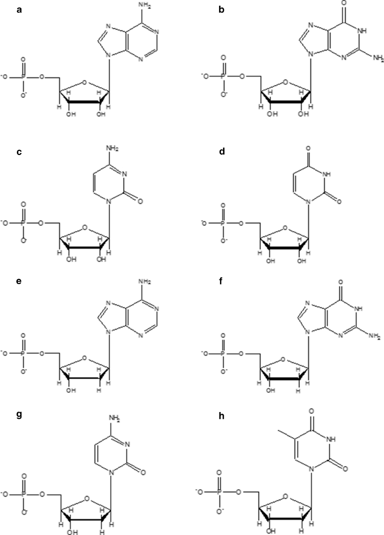
Standards of 2′-deoxyadenosine 5′-monophosphate disodium salt (DA), 2′-deoxycytidine 5′-monophosphate sodium salt (DC), 2′-deoxyguanosine 5′-monophosphate sodium salt hydrate (DG), thymidine 5′-monophosphate disodium salt hydrate (DT), uridine 5′-monophosphate disodium salt (U), adenosine 5′-monophosphate disodium salt (A), cytidine 5′-monophosphate (C), and guanosine 5′-monophosphate disodium salt hydrate (G) were purchased from Sigma-Aldrich (Gillingham, Dorset, UK). Schematic structures of analyzed nucleotides are given in Fig. 1. The stock solutions of standards were prepared by dissolving a weighed amount of each nucleotide in deionized water. Concentrations of nucleotides used for retention studies were in the range of 10 to 30 μg ml−1. The highest concentration was used only when retention factor values exceeded 3. For the rest, chromatographic runs concentration was equal 10 μg ml−1.
Fig. 1.
Structures of nucleotides used in investigations (for shortcuts, see “Materials and methods” section): a u, b c, c g, d a, e dc, f dg, g da, h dt
For the preparation of mobile phases, methanol and acetonitrile of gradient grade purity (J.T. Baker, Deventer, Holland) and deionized water from Milli-Q system (Millipore, El Paso, TX, USA) were used. Ammonium acetate (CH3COONH4), ammonium formate (HCOONH4), potassium dihydrogen phosphate (KH2PO4), dipotassium hydrogen phosphate (K2HPO4), orthophosphoric acid (H3PO4), acetic acid (CH3COOH) were also used for the preparation of mobile phase buffer. All of these reagents were of high purity and were purchased from Sigma-Aldrich (Gillingham, Dorset, UK).
Apparatus and analysis conditions
The UltiMate® 3000 Binary Rapid Separation LC (Dionex, Sunnyvale, CA, USA) ultra high-performance liquid chromatography system equipped with a diode-array detector was used in the investigation. Chromeleon 7 program was utilized for the data collection.
In the current study, seven HPLC columns have been used: two octadecyl ones (SG-C18A, SG-C18B), cholesterol (SG-CHOL), alkylamide (SG-AP), phospho-alkyl (SG-PC10), phenyl (SG-Ph), and aminopropyl (SG-NH2). All of them were home-made according to procedures described earlier in details for SG-C18 [17], SG-CHOL [18], SG-AP [19], SG-PC10 [20], SG-Ph [21], SG-NH2 [18, 19]. Detailed characteristics of stationary phases were presented in Table 1. The received packing materials were prepared with the same batch of silica gel Kromasil®. They were next packed into 125 mm × 4.6 mm I.D. stainless-steel columns with the use of home-made apparatus equipped with Haskel pump (Burbank, CA, USA) under constant pressure.
Table 1.
Characteristics of stationary phases used in the investigations
| Type of stationary phasea | Shortcut | Proportional part carbon P C [%] | |
|---|---|---|---|
| I modification step | II modification step | ||
| Octadecyl | SG-C18A | 7.55 | – |
| Octadecyl, end-capped | SG-C18B | 17.79 | – |
| Aminopropyl | SG-NH2 | 4.47 | – |
| Alkylamide | SG-AP | 4.47 | 11.46 |
| Cholesterol | SG-CHOL | 4.47 | 17.82 |
| Alkyl-phosphate | SG-PC10 | 2.83 | 8.43 |
| Phenyl | SG-Ph | 11.75 | – |
aColumn dimensions, 125 × 4.6 mm; silica particle size, 5 μm; pore diameter, 100 Å
Elution was carried out with mixtures of buffer and methanol or acetonitrile. Chromatographic elution was carried out with isocratic conditions in a organic solvent range between 5 % v/v and 20 % v/v. For each column, at least four different mobile phase compositions were used. The flow rate was 0.5 ml min−1. The detection wavelength was selected as λ = 254 nm. Injection volume was 1 μl. The temperature of autosampler and column was 20 °C. The void (t 0) of the column was measured by the injection of methanol.
Four different mobile phase pH levels were tested: 4.0, 5.0, 6.0, and 7.0; 30 mM HCOONH4 and 30 mM CH3COONH4 were used for pH 4.0 and 5.0. In the case of pH 6.0 and 7.0, 30 mM KH2PO4/K2HPO4 buffer was studied. The pH of HCOONH4 and CH3COONH4 solutions was adjusted with the use of 1 M solution of proper acid: acetic or formic. Phosphate buffer was obtained by mixing KH2PO4 and K2HPO4 solutions to receive pH 6.0 or 7.0.
Results and discussion
Stationary phase selection
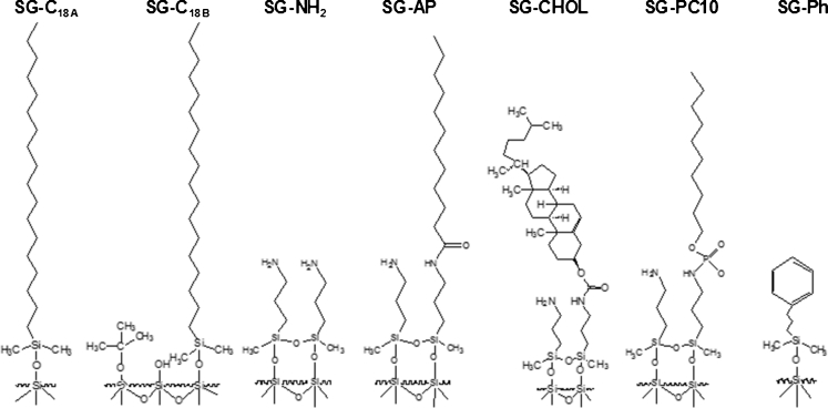
Stationary phases were selected on the basis of their structure. The goal of the study was to test variety of HPLC columns to choose the best one for the analysis of polar nucleotides. Two octadecyl columns were used, differing in the carbon load (Table 1). One has 7.6 % C and other has 17.6 % C on silica surface. On the other hand, very polar aminopropyl packing was also tested. This phase is the starting material for the synthesis of alkylamide, cholesterol or alkyl-phosphate stationary phases (Fig. 2). SG-AP and SG-CHOL differ with alkyl chain and large cholesterol molecule (Fig. 2). In the case of SG-AP and SG-PC10 the difference is connected with the type of middle group localized in the ligand: amide and phosphate ones (Fig. 2). Phenyl packing material was also studied as it seemed to be interesting material. Phenyl molecules may interact with nucleotides by π–π interactions, which are connected with stacking, commonly observed for adjacent nucleotides in DNA.
Fig. 2.
Schemes of stationary phases studied in nucleotides analysis and separation
All stationary phases synthesized on the same batch of silica gel were prepared. The detailed characteristics of packing materials were carried out, due to their home-made character (Table 1). Carbon content was analyzed with the use of elemental analysis. It allows predicting a contribution of given ligands on a surface of stationary phase and as a consequence in the chromatographic process (Table 1). The lowest carbon content was observed for polar SG-NH2. SG-AP has similar carbon load as SG-Ph, although they poses various structures (Table 1). The highest and similar percentage part of carbon may be noticed for hydrophobic SG-C18B and SG-CHOL (Table 1).
Mobile phase selection
The first step of mobile phase optimization was the utilization of mixtures consisting of water and organic solvents. However, the peak shape was asymmetric and retention factor (k) values were low. Polar character of nucleotides leads to the necessity of buffer utilization in the mobile phase. The concentration of the buffer was chosen on the basis of our earlier experience, it was equal to 30 mM [22]. Utilization of lower concentrations of mobile phase buffers caused very asymmetric peak shapes (f AS between 1.6 and 3.3). The great care was taken in the preparation of mobile phases, in particular regarding pH value adjustment. The pH of the mobile phase will affect the resolution of chromatographically determined nucleotides, for this reason we have tested four different pH. In the case of pH 4.0 and 5.0, HCOONH4 and CH3COONH4 were investigated, however better results, in terms of peak symmetry, were obtained for the second one. The neutral pH of the mobile phase was obtained with the use of phosphate buffer KH2PO4/K2HPO4. The 30 mM KH2PO4, adjusted to pH 6.0 and 7.0 with 1 M KOH was also tested; however, the peaks were more asymmetric than in the case of KH2PO4/K2HPO4. One of the main goals of the present study was to simplify the mobile phase composition, therefore non ion-pairing reagent was used.
Methanol was used as a mobile phase organic modifier. It was selected on the basis of lower elution strength in comparison with acetonitrile.
Retention study
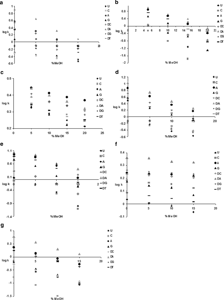
The retention of nucleotides was investigated as a function of methanol content in the mobile phase. These studies were performed for each compound, stationary phase and four different mobile phase pH. The most interesting and important effects were shown in the present manuscript. Exemplary dependencies of log k as a function of organic modifier content (φ) for pH 5.0 are presented in Fig. 3.
Fig. 3.
Dependencies of log k vs. percentage of methanol in mobile phase (φ) for stationary phases: A SG-C18A, B SG-C18B, C SG-NH2, D SG-AP, E SG-CHOL, F SG-PC10, G SG-Ph. Mobile phase consisted of mixtures of methanol and 30 mM CH3COONH4, pH 5. Detailed chromatographic conditions and abbreviations of stationary phases names may be found in the “Materials and methods” section
It was observed that with increasing of methanol percentage, k values for nucleotides were reduced. On the other hand, very low (not higher than 20 % v/v) content of methanol was needed to assure retention of nucleotides. It was observed in the case of all pH of mobile phase buffers. The greatest retention was observed for two DA, DG deoxynucleotides and A (Figs. 1 and 3), among which DA was retained with the highest extent. k values for U and C are low and in some cases (15 or 20 % v/v) they are eluted in the dead volume of the column. It is connected with the structure of these nucleotides (Fig. 1) and consequently with their polarity. It should be noticed that for polar packing materials, SG-NH2 and SG-PC10 (Fig. 3c, f), both U and C are retained with greater extent in comparison with, e.g., G or DT. These phenomenon is strictly connected with the structure and polarity of supports used in the study.
Figure 3a and b presents results of nucleotides retention study for two octadecyl columns. log k values for SG-C18A are lower than for the other column. It is a consequence of the difference in carbon load for both packing materials, which equals 10 % (Table 1). Octadecyl packing interact with analyzed compounds mainly by hydrophobic interactions, therefore with the increase of alkyl chains content on the silica support, nucleotides will be retained stronger inside the column.
Hydrophilic nucleotides were also analyzed with the use of SG-NH2 (Fig. 2). Log k values for all of nucleotides analyzed with the use of SG-NH2 are lower than for both octadecyl columns since reversed-phase conditions were applied (Fig. 3a, b, c).
Aminopropyl groups are present also on SG-AP and SG-CHOL surfaces (Fig. 2). Results obtained for SG-AP material were presented in Fig. 3d. Surprisingly, log k values are higher than for SG-C18A and lower than in the case of SG-C18B. SG-AP poses alkyl chains in its structure, similar as SG-C18B (Fig. 2). Alkyl chains in the case of SG-AP consist of 12 carbon atoms (Fig. 2), they are shorter than for SG-C18. For this reason, hydrophobic interactions will be not as significant as in the case of SG-C18. However, hydrogen bonds between aminopropyl and amide groups and nucleotides will improve log k values (Fig. 3d). Therefore, both interaction types will control final result of chromatographic analysis of nucleotides on SG-AP: hydrophobic ones and hydrogen bonding.
Results of nucleotides retention studies on SG-CHOL were shown in Fig. 3e. Log k values are higher than in the case of SG-C18B (Fig. 3b and e). This stationary phase may interact with nucleotides by hydrophobic, hydrogen bond and π–π interactions (Fig. 2). Moreover, SG-C18B and SG-CHOL have similar carbon content on their surface (Table 1), therefore hydrophobic interactions will be at least as significant as in the case of SG-C18B. Still, log k values are higher in the case of SG-CHOL. This proves the signification of polar interactions participation in the retention mechanism of nucleotides.
A different situation occurs in the case of SG-PC10 (Fig. 3f). The negatively charged phosphate groups are present in the structure of SG-PC10 (Fig. 2). Moreover, SG-PC10 has similar carbon content as SG-C18A (Table 1), but nucleotides are retained to a lower extent. log k values are very low as a consequence of repulsion forces between negatively charged nucleotides and stationary phase surface (Fig. 3f).
An interesting situation was observed for SG-Ph column (Fig. 3g), which has similar carbon content as SG-AP (Table 1). Nucleotides were retained on SG-Ph but not as strong as in the case of SG-AP. On the other hand, log k values for SG-Ph are comparable to these obtained for SG-C18A. SG-Ph may retain nucleotides mainly by π–π interactions. The aromatic rings of nucleotide bases are positioned parallel to each other in RNA or DNA, as a consequence of π-stacking. During the chromatographic process on SG-Ph, similar π-stacking occurs.
The influence of the mobile phase pH
The retention of nucleotides was investigated not only as a function of methanol content in the mobile phase, but also as a function of pH of mobile phase buffer. The pH will influence not only nucleotides retention, but also the stationary phase surface [21]. Table 2 presents k values determined for all nucleotides and packing materials in the case of four different pH. Data collected in Table 2 were appointed for 5 % v/v of methanol in the mobile phase. This composition was selected on the basis of k, because for higher content of organic modifier polar, some of nucleotides (C, U, DC) were eluted in the dead volume.
Table 2.
The retention factor k values for all of stationary phases and nucleotides used in the study for the different pH of mobile phase buffer. Experimental conditions: 95 % v/v of 30 mM CH3COONH4 (for pH 4.0 and 5.0) or 30 mM KH2PO4/K2HPO4 (for pH 6.0 and 7.0), 5 % v/v of methanol, flow rate 0.5 ml min−1
| pH | k | |||||||
|---|---|---|---|---|---|---|---|---|
| U | C | A | G | DC | DA | DG | DT | |
| SG-C18A | ||||||||
| 7 | t 0 | t 0 | 0.44 | 0.08 | t 0 | 1.47 | 0.55 | 0.28 |
| 6 | 0.12 | 0.13 | 1.27 | 0.40 | 0.31 | 3.11 | 1.23 | 0.79 |
| 5 | 0.44 | 0.41 | 1.99 | 0.67 | 0.80 | 4.44 | 1.68 | 1.22 |
| 4 | 0.33 | 0.48 | 2.00 | 0.68 | 1.41 | 4.48 | 1.75 | 1.26 |
| SG-C18B | ||||||||
| 7 | 0.66 | 0.38 | 7.55 | 3.07 | 1.10 | 9.86 | 7.76 | 4.83 |
| 6 | 0.90 | 0.44 | 7.62 | 3.17 | 1.14 | 10.03 | 7.97 | 4.92 |
| 5 | 1.07 | 0.58 | 7.76 | 3.31 | 1.19 | 10.17 | 8.28 | 5.28 |
| 4 | 1.10 | 0.69 | 7.72 | 3.48 | 1.28 | 10.21 | 8.34 | 5.34 |
| SG-NH2 | ||||||||
| 7 | 0.25 | 0.74 | 0.66 | 0.57 | 0.75 | 0.77 | 0.86 | 0.66 |
| 6 | 1.24 | 1.99 | 1.96 | 1.57 | 2.19 | 2.14 | 2.11 | 1.39 |
| 5 | 2.11 | 2.22 | 2.78 | 2.14 | 2.44 | 2.75 | 2.56 | 2.19 |
| 4 | 3.37 | 2.96 | 4.14 | 4.94 | 2.75 | 4.56 | 6.78 | 4.17 |
| SG-AP | ||||||||
| 7 | t 0 | t 0 | 0.31 | 0.06 | t 0 | 0.87 | 0.35 | 0.15 |
| 6 | 0.12 | 0.16 | 1.06 | 0.42 | 0.38 | 2.21 | 1.04 | 0.62 |
| 5 | 0.59 | 0.57 | 4.16 | 1.83 | 0.77 | 5.45 | 2.70 | 2.00 |
| 4 | 0.61 | 0.60 | 4.26 | 1.85 | 0.86 | 5.55 | 2.87 | 2.02 |
| SG-CHOL | ||||||||
| 7 | 0.05 | 0.03 | 4.28 | 1.65 | 0.45 | 5.64 | 5.00 | 3.83 |
| 6 | 0.58 | 0.28 | 6.10 | 2.16 | 0.84 | 7.48 | 6.78 | 4.32 |
| 5 | 1.07 | 0.65 | 7.42 | 3.39 | 1.34 | 9.65 | 7.97 | 5.58 |
| 4 | 1.09 | 0.69 | 7.45 | 3.45 | 1.35 | 9.71 | 8.00 | 5.65 |
| SG-PC10 | ||||||||
| 7 | 0.27 | 0.52 | 0.62 | 0.56 | 0.67 | 0.93 | 0.79 | 0.42 |
| 6 | 0.69 | 1.13 | 1.29 | 1.07 | 1.36 | 1.77 | 1.37 | 0.85 |
| 5 | 0.97 | 1.42 | 1.71 | 1.22 | 1.41 | 1.72 | 1.73 | 1.10 |
| 4 | 1.06 | 1.57 | 1.83 | 1.34 | 1.67 | 2.00 | 2.35 | 1.87 |
| SG-Ph | ||||||||
| 7 | t 0 | t 0 | 0.57 | 0.13 | t 0 | 2.03 | 0.73 | 0.56 |
| 6 | 0.11 | 0.02 | 1.18 | 0.30 | 0.20 | 3.00 | 1.14 | 0.97 |
| 5 | 0.17 | 0.08 | 1.37 | 0.36 | 0.32 | 3.58 | 1.22 | 1.07 |
| 4 | 0.17 | 0.13 | 1.40 | 0.37 | 0.45 | 3.66 | 1.21 | 1.06 |
Results for two octadecyl phases present one interesting tendency. In the case of SG-C18B, the influence of pH is negligible. The retention becomes greater with the decrease of pH, but the difference is small (about 0.1–0.2) (Table 2). A similar trend was observed for SG-C18A; however, in the case of this packing the difference of k for each mobile phase pH is significant (0.4–0.8). Such effect concerns all nucleotides. In the case of SG-C18B, the end-capping process was applied during the synthesis (Fig. 2). Most of the residual silanols were blocked with the use of end-capping reagent, therefore, the surface is more hydrophobic and consequently not influenced by the pH changes, in comparison with SG-C18A. On the contrary, SG-C18A poses greater population of free silanols on the support surface, and probably they are responsible for changes of nucleotides retention in various pH. These polar groups will get the partial negative charge when the pH of mobile phase buffer will be increased (from 4.0 to 7.0). Phosphate groups in the nucleotides carries negative charge, therefore k will be lower for pH 7.0 in comparison with pH 4.0. It is a consequence of electrostatic repulsion between residual silanols and nucleotides at basic pH. Furthermore, it should be noticed that differences for k values between pH 4.0 and 5.0 are small.
The most significant influence of the mobile phase pH on nucleotides retention (among all stationary phases used in the study) was noticed for SG-NH2 (Table 2). The retention factor values were increasing with the increase of pH to a great extent. Differences of k values between pH 7.0 and 4.0 equal about 4 (for A, G, DA, DT) or even 5 (DG) (Table 2). Amine groups present at the outer layer of support undergo protonization process during the decrease of pH of the buffer. Positively charged stationary phase surface attract negatively charged nucleotides; consequently, electrostatic attraction and ionic interactions are the basic ones in the retention mechanisms of these compounds on SG-NH2.
The pH of the mobile phase buffer influences the retention of nucleotides also in the case of SG-AP. The largest k differences were observed between pH 7.0 and 5.0. SG-AP posses aminopropyl groups, which will be protonized in the low pH of a buffer (Fig. 2). The impact of protonization and electrostatic interactions is not as significant as in the case of SG-NH2, because some of –NH2 groups were blocked with alkylamide ligands. It may be concluded that for low pH (4.0) of the mobile phase buffer, the retention mechanism of nucleotides is of mixed nature. The hydrophobic and ionic interactions will be responsible for chromatographic behavior of analyzed compounds.
Similar effects were observed also for SG-CHOL (Table 2). Log k values increase with the decrease of mobile phase pH; however, this effect is not as significant as in the case of SG-NH2 or SG-AP (Table 2). SG-CHOL poses large cholesterol molecule bonded to aminopropyl groups (Fig. 2). This molecule will eclipse the access of negatively charged nucleotides to positively charged amino groups. The size of straight alkylamide chains bonded to silica surface is smaller than cholesterol, therefore aminopropyl ligands are not eclipsed in the case of SG-AP (Fig. 2). Consequently, nucleotides analyzed on SG-AP are more pH-sensitive than for SG-CHOL.
Another interesting effect may be observed when data for SG-CHOL and SG-C18B will be compared. In the case of pH 7.0 and 6.0, the retention of nucleotides is greater for octadecyl phase in comparison with SG-CHOL (Table 2). This proves the influence of hydrophobic interactions on nucleotide behavior inside the chromatographic column. However, the situation is diverse when the mobile phase buffer becomes more acidic (pH equals 5.0 and 4.0). k values are higher for SG-CHOL in comparison with SG-C18B for polar nucleotides (U, C, DC). Retention becomes similar for both packing materials in the case of G, DG, DT, while for A and DA k remains greater for SG-C18B. This is another proof of the impact of protonization on the retention of nucleotides in the case of RP HPLC. Nucleotides are retained inside the chromatographic column with the same strength as for SG-C18B, when electrostatic attraction begins to play an important role during the chromatographic process.
The influence of the change made in the mobile phase buffer is not significant for SG-PC10 (Table 2). Two opposite effects take place during the chromatographic determination of nucleosides on this stationary phase. The first one is connected with electrostatic repulsion between negatively charged nucleotides and phosphate groups localized in the stationary phase ligands (Fig. 2). However, SG-PC10 is another stationary phase with aminopropyl groups, which carry a positive charge when the pH is acidic. Therefore, the k value for each nucleotide will be greater for pH 4.0 in comparison with pH 7.0. The retention is a resultant of electrostatic repulsion, attraction (only in acidic mobile phase buffer), and moreover hydrophobic interactions (alkyl chain).
SG-Ph possesses in its structure two kinds of active centers: residual silanols and phenyl molecules. Only silanol groups may be influenced by changes made in the mobile phase pH buffer. The situation is similar to SG-C18B, for which the influence of mobile phase pH was not significant. In the case of SG-Ph, the carbon load is lower in comparison with SG-C18B (Table 1); however, phenyl molecules can eclipse residual silanols. The percentage part of carbon on the support surface is similar for SG-Ph and SG-AP. The k value is greater in the case of SG-AP in comparison with SG-Ph (Table 2). This effect proves that although nucleotides may interact with stationary phase surface by hydrophobic, electrostatic and π–π interactions, the last type has the lower impact.
Separation of nucleotides mixture
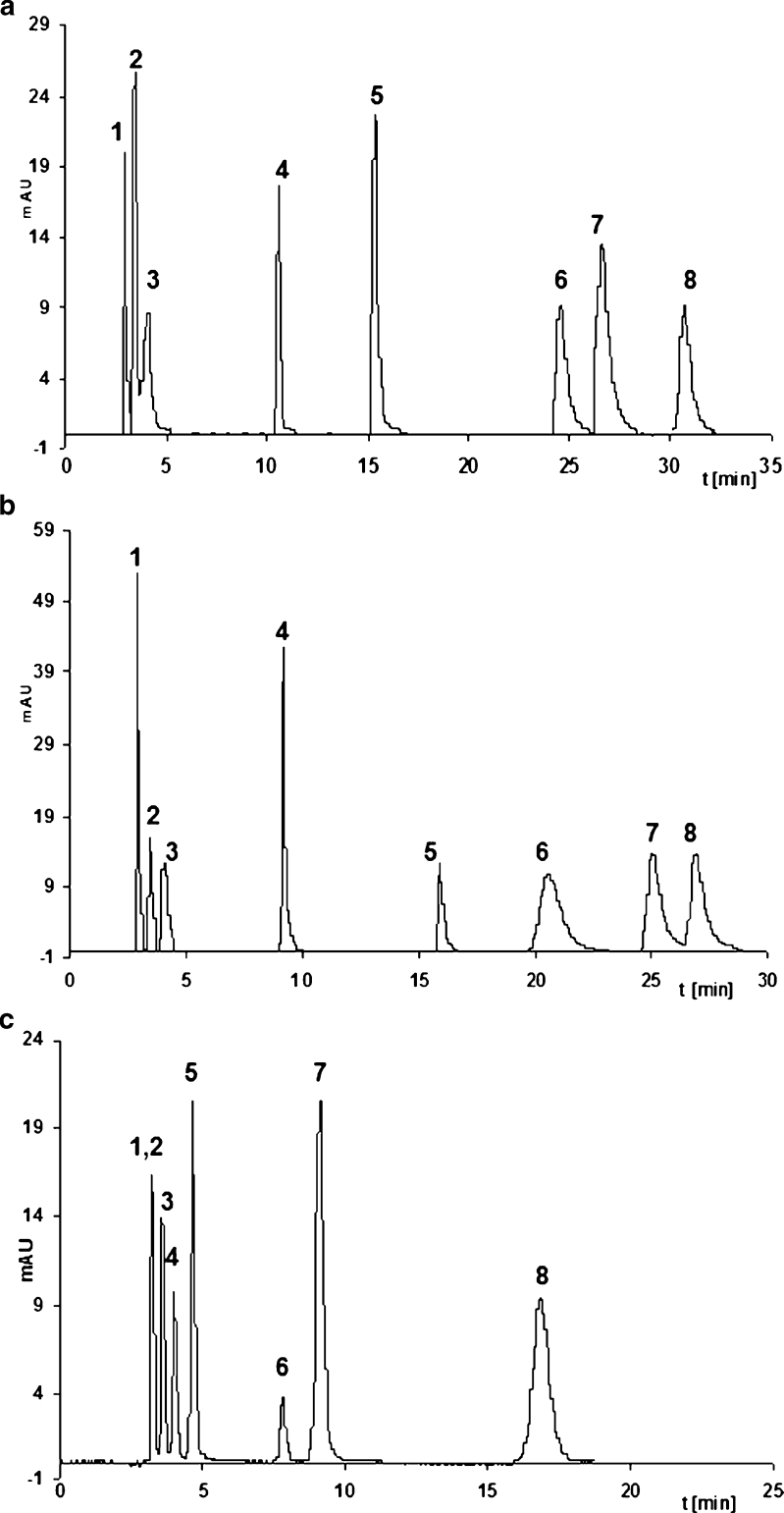
All of these considerations, concerning retention mechanism, would be useless if they would not lead to the separation of nucleotides. Complete separation of all eight nucleotides was impossible for most of stationary phases because of peak bordering. The best asymmetry for analyzed compounds was obtained for SG-C18B, SG-CHOL, and SG-Ph. Consequently, the best results of separation were achieved for these packing materials. Exemplary chromatograms are presented in Fig. 4. It may be observed that SG-Ph fails in the separation of polar nucleotides, such as U, C, and DC (Fig. 4a and c). Moreover, these nucleotides were eluted at void time for SG-C18B. An opposite situation concerns SG-CHOL (Fig. 4b), for which all three nucleotides were separated. Moreover, complete separation of all nucleotides for SG-CHOL was possible, although the time of analysis was long and some of the peaks were tailing (SG-CHOL) (Fig. 4b). On the other hand, both the time of separation and asymmetry factor were similar for SG-CHOL and SG-C18B (Fig. 4a, b). Furthermore, complete separation of analyzed compounds was impossible. For this reason, it should be concluded that SG-CHOL is more suitable in the analysis of nucleotides in comparison with conventional octadecyl phase. Utilization of SG-Ph provided a short time of analysis (Fig. 4c). Additionally, asymmetry factor for all peaks was satisfactory and equal 1.0–1.1. On the other hand, SG-Ph do not resolve polar nucleotides (C, U), similar to SG-C18B. SG-CHOL and SG-Ph are interesting substitutes for commonly used octadecyl phases. SG-CHOL should be used when U, C, DC have to be separated. SG-Ph is a good alternative to assure short time of separation with symmetric peaks.
Fig. 4.
Chromatograms of nucleotides mixture separation for: a SG-C18B (5/95 % v/v MeOH/CH3COONH4, pH 5), b SG-CHOL (100 % v/v 30 mM CH3COONH4, pH 5), c SG-Ph (5/95 % v/v MeOH/phosphate buffer, pH 6). Notation: 1 C, 2 U, 3 DC, 4 G, 5 DT, 6 A, 7 DG, 8 DA. Nucleotides concentration was in the range of 5 to 10 μg ml−1
Conclusions
The present study provided an interesting results concerning the retention mechanism and separation of nucleotides with the use various stationary phases. Several types of interactions take place during the chromatographic process and consequently, it has a mixed nature. The most important interactions are hydrophobic, electrostatic, and π…π. It appeared that pH of mobile phase buffer is a very powerful parameter, which may improve the final result of nucleotides determination with the use of HPLC. It concerns especially stationary phases with polar aminopropyl ligands. One of the most important findings of the present investigation concern results of separation. It may be pointed out that aminopropyl, alkyl-phosphate, alkylamide packing materials are not suitable for the resolution of nucleotides. On the other hand, cholesterol and phenyl stationary phases may be alternatively used instead of octadecyl phases. Application of cholesterol packing allows separation of small, polar nucleotides, what is impossible to achieve in the case of octadecyl phase. Moreover, phenyl support allows separation of nucleotides in a shorter period of time in comparison with octadecyl packing; however, C and U stay unseparated.
Acknowledgments
The authors are grateful to Foundation for Polish Science for Parent-Bridge programme (POMOST/2011-3/9), cofinanced from European Union, Regional Development Fund.
Abbreviations
- A
Adenosine 5′-monophosphate disodium salt
- C
Cytidine 5′-monophosphate
- DA
2′-Deoxyadenosine 5′-monophosphate disodium salt
- DC
2′-Deoxycytidine 5′-monophosphate sodium salt
- DG
2′-Deoxyguanosine 5′-monophosphate sodium salt hydrate
- DT
Thymidine 5′-monophosphate disodium salt hydrate
- G
Guanosine 5′-monophosphate disodium salt hydrate
- SG-AP
Alkylamide stationary phase
- SG-C18
Octadecyl stationary phase
- SG-CHOL
Cholesterol stationary phase
- SG-NH2
Aminopropyl stationary phase
- SG-PC10
Phosphoalkyl stationary phase
- SG-Ph
Phenyl stationary phase
- U
Uridine 5′-monophosphate disodium salt
Contributor Information
S. Studzińska, Phone: +48-56-6114308, FAX: +48-56-6114837, Email: skowalska@o2.pl
B. Buszewski, Email: bbusz@chem.umk.pl
References
- 1.Alberts B, Johnson A, Lewis J, Raff M, Roberts K, Walter P. Molecular biology of the cell. New York: Garland Science; 2002. [Google Scholar]
- 2.Kochanowski N, Blanchard F, Cacan R, Chirat F, Guedon E, Marc A, Goergen J-L. Anal Biochem. 2006;348:243–251. doi: 10.1016/j.ab.2005.10.027. [DOI] [PubMed] [Google Scholar]
- 3.Yeung P, Ding L, Casley WL. J Pharm Biomed Anal. 2008;47:377–382. doi: 10.1016/j.jpba.2008.01.020. [DOI] [PubMed] [Google Scholar]
- 4.Bolin C, Cardozo-Pelaez F. J Chromatogr B. 2007;856:121–130. doi: 10.1016/j.jchromb.2007.05.034. [DOI] [PMC free article] [PubMed] [Google Scholar]
- 5.Ganzera M, Vrabl P, Worle E, Burgstaller W, Stuppner H. Anal Biochem. 2006;359:132–140. doi: 10.1016/j.ab.2006.09.012. [DOI] [PubMed] [Google Scholar]
- 6.Qian T, Cai Z, Yang MS. Anal Biochem. 2004;325:77–84. doi: 10.1016/j.ab.2003.10.028. [DOI] [PubMed] [Google Scholar]
- 7.Cichna M, Raab M, Daxecker H, Griesmacher A, Muller MM, Markl P. J Chromatogr B. 2003;787:381–391. doi: 10.1016/S1570-0232(02)01007-3. [DOI] [PubMed] [Google Scholar]
- 8.Viñas P, Campillo N, Férez Melgarejo G, Vasallo I, López-García I, Hernández-Córdoba M. J Chromatogr A. 2010;1217:7323–7330. doi: 10.1016/j.chroma.2010.09.058. [DOI] [PubMed] [Google Scholar]
- 9.Viñas P, Campillo N, López-García I, Martínez-López S, Vasallo I, Hernández-Córdoba M. J Agric Food Chem. 2009;57:7245–7249. doi: 10.1021/jf901726e. [DOI] [PubMed] [Google Scholar]
- 10.Hernández-Cázares AS, Aristoy MC, Toldrá F. Meat Sci. 2011;87:125–129. doi: 10.1016/j.meatsci.2010.09.010. [DOI] [PubMed] [Google Scholar]
- 11.Yamaoka N, Kudo Y, Inazawa K, Inagawa S, Yasuda M, Mawatari K, Nakagomi K, Kaneko K. J Chromatogr B. 2010;878:2054–2060. doi: 10.1016/j.jchromb.2010.05.044. [DOI] [PubMed] [Google Scholar]
- 12.Czarnecka J, Cieślak M, Komoszyński M. J Chromatogr B. 2005;822:85–90. doi: 10.1016/j.jchromb.2005.05.026. [DOI] [PubMed] [Google Scholar]
- 13.Brown PR, Robb CS, Geldart SE. J Chromatogr A. 2002;965:163–173. doi: 10.1016/S0021-9673(01)01561-8. [DOI] [PubMed] [Google Scholar]
- 14.Willems AV, Deforce DL, Van Peteghem CH, Van Bocxlaer JF. Electrophoresis. 2005;26:1221–1253. doi: 10.1002/elps.200410278. [DOI] [PubMed] [Google Scholar]
- 15.Ohyama K, Fujimoto E, Wada M, Kishikawa N, Ohba Y, Akiyama S, Nakashima K, Kuroda N. J Sep Sci. 2005;28:767–773. doi: 10.1002/jssc.200500030. [DOI] [PubMed] [Google Scholar]
- 16.Yang M, Butler M. Biotechnol Prog. 2000;16:751–759. doi: 10.1021/bp000090b. [DOI] [PubMed] [Google Scholar]
- 17.Buszewski B, Cendrowska I, Krupczyńska K, Gadzała-Kopciuch RM. J Liquid Chromatogr Rel Technol. 2003;26:737–750. doi: 10.1081/JLC-120018418. [DOI] [Google Scholar]
- 18.Buszewski B, Jezierska-Świtała M, Kaliszan R, Wojtczak A, Albert K, Bochmann S, Matyska MT, Pesek JJ. Chromatographia. 2001;53:204–212. doi: 10.1007/BF02490329. [DOI] [Google Scholar]
- 19.Gadzała-Kopciuch RM, Buszewski B. J Sep Sci. 2003;26:1273–1283. doi: 10.1002/jssc.200301428. [DOI] [Google Scholar]
- 20.Bocian SZ, Nowaczyk A, Buszewski B (2012) Anal Bioanal Chem. doi:10.1007/s00216-012-6134-0 [DOI] [PMC free article] [PubMed]
- 21.Gadzała-Kopciuch R, Kluska M, Wełniak M, Buszewski B. Mat Chem Phys. 2005;89:228–234. doi: 10.1016/j.matchemphys.2004.03.022. [DOI] [Google Scholar]
- 22.Kowalska S, Krupczyńska K, Buszewski B. J Sep Sci. 2005;28:1502–1511. doi: 10.1002/jssc.200400044. [DOI] [PubMed] [Google Scholar]


