Abstract
Shrimp is an important commodity in international trade accounting for 15 % in terms of value of internationally traded seafood products which reached $102.00 billion in 2008. Aquaculture contributes to over 50 % of global shrimp production. One of the major constraints faced by shrimp aquaculture is the loss due to viral diseases like white spot syndrome, yellow head disease, and Taura syndrome. There are several examples of global spread of shrimp diseases due to importation of live shrimp for aquaculture. Though millions of tonnes of frozen or processed shrimp have been traded internationally during the last two decades despite prevalence of viral diseases in shrimp producing areas in Asia and the Americas, there is no evidence of diseases having been transmitted through shrimp imported for human consumption. The guidelines developed by the World Animal Health Organisation for movement of live animals for aquaculture, frozen crustaceans for human consumption, and the regulations implemented by some shrimp importing regions in the world are reviewed.
Keywords: Shrimp, Aquaculture, White spot syndrome virus, Yellow head virus, Taura syndrome virus, Risk analysis, International trade, Transboundary movement
Introduction
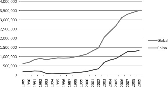
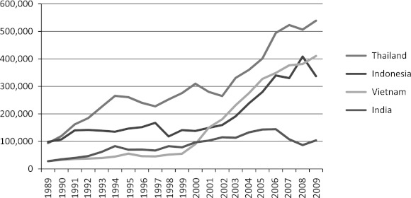
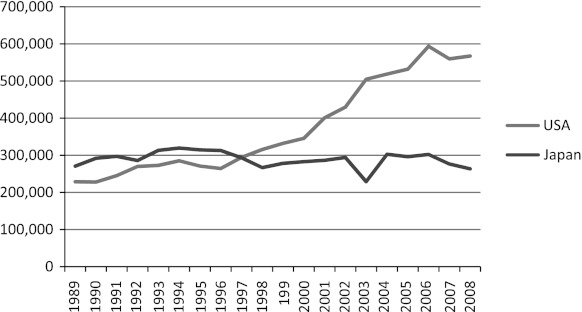
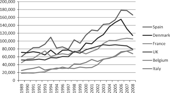
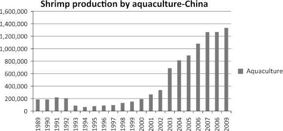
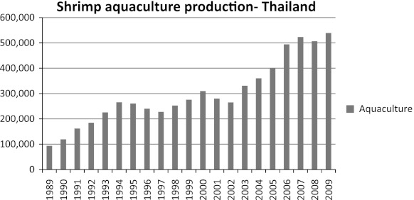
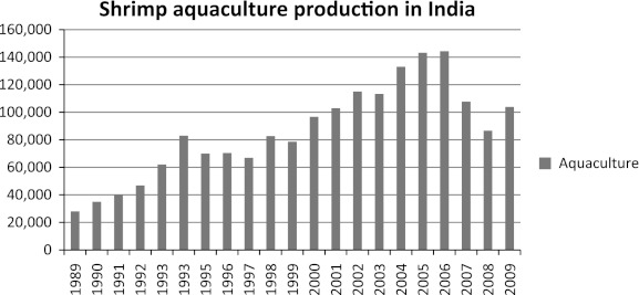
Shrimp is an important commodity in international trade and during 2009, the global production by capture was 3.17 million tonnes and by aquaculture, 3.5 million tonnes. China is the largest producer of aquacultured shrimp (1.33 million tonnes) followed by Thailand, Vietnam, Indonesia, Ecuador, Mexico, India, and Bangladesh (Figs. 1, 2). Shrimp accounted for 15 % in terms of value of internationally traded seafood products which reached $102.00 billion in 2008 [7]. United States is the major importer followed by Japan (Fig. 3). In 2008, USA imported 0.57 million tonnes valued at $4.1 billion and Japan imported 0.26 million tonnes valued at $2.4 billion. Other major importers include Spain, Denmark, France, UK, Belgium, and Italy (Fig. 4).
Fig. 1.
Global and Chinese shrimp aquaculture production (tonnes)
Fig. 2.
Shrimp aquaculture production in some major producing countries (tonnes)
Fig. 3.
Shrimp imports into USA and Japan (tonnes)
Fig. 4.
Other major importers of shrimp
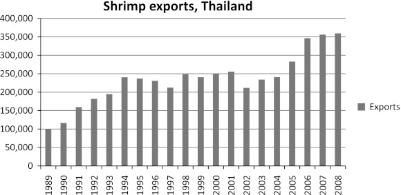
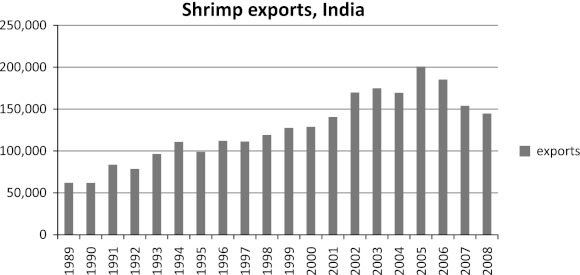
The impact of diseases on shrimp production in different countries is shown in Figs. 5, 6, 7. In China, there was a drop in production in 1992 due mainly to outbreaks of white spot syndrome and this continued till 2000 (Fig. 5). In Thailand, there was drop in production in 1995 that continued till 1997 and there was another drop in 2003 (Fig. 6). Shrimp production in India dropped in 1995 and extended till 2000 (Fig. 7). The rebounding of production in most cases is due to the introduction of Pacific whiteleg shrimp, Penaeus vannamei into Asia. The culture of this species reached a total of 1.8 million tonnes outside America in 2008. This accounted for 80.7 % of the global aquaculture production of this species and 40.7 % of the production of all cultured crustaceans outside America. India has permitted culture of P. vannamei only in 2008. The fall in shrimp production in India in 2007 is attributed to continued disease problems and conversion of some shrimp ponds into catfish ponds. Since the outbreak of white spot disease in 1994–1995, shrimp farmers have been trying various modifications of culture systems including acclimatizing P. monodon to fresh water [19]. Driven mostly by the demand for Pangasius in domestic market, some of these fresh water ponds have moved from shrimp to catfish (Figs. 8, 9).
Fig. 5.
Shrimp aquaculture production in China (tonnes)
Fig. 6.
Shrimp aquaculture production in Thailand (tonnes)
Fig. 7.
Shrimp aquaculture production in India (tonnes)
Fig. 8.
Shrimp exports from Thailand (tonnes)
Fig. 9.
Shrimp exports from India (tonnes)
Viral Diseases of Shrimp
Shrimp production by aquaculture has been seriously impacted by diseases. In Asia, during the early 1990s, there were large scale mortalities in shrimp culture ponds in most major producing countries due to white spot syndrome virus (WSSV) and the loss has been estimated to be around $6.0 billion. The World Animal Health Organisation (OIE) uses all the three criteria indicated in Table 1 to list the diseases and the OIE member countries have responsibility to notify OIE when there are outbreaks of these diseases. The OIE listed viral diseases of shrimp are indicated in Table 2. Of these, WSSV, a double stranded (ds) DNA virus has caused massive losses in both Asia and the Americas. The first outbreaks were recorded in Asia. Horizontal transmission of the virus by ingestion (e.g., cannibalism, predation) of infected tissue has been well demonstrated and other routes confirmed are egg-associated and waterborne transmission. The virus can replicate in several decapod crustaceans and presence of virus in non-decapod crustaceans such as Artemia salina, copepods as well as other aquatic animals such as bivalves, polychaete worms, aquatic arthropods including insect larvae has been observed [5]. The infectious dose is not yet determined. Persistent and lifelong infections have been demonstrated and viral loads can be very low in persistent infections and often undetectable by most diagnostic tests [15]. The virus can be inactivated in less than 1 min at 60 °C [14].
Table 1.
Office International des Epizooties (OIE) criteria for listing of aquatic animal diseases [15]
| Criteria | Parameters | |
|---|---|---|
| A | Consequences | The disease has been shown to cause significant production losses at a national or multinational (zonal or regional) level or The disease has been shown to or scientific evidence indicates that it is likely to negatively affect wild aquatic animal populations or The agent is of public health concern |
| B | Spread | Infectious etiology of the disease is proven or An infectious agent is strongly associated with the disease, but the etiology is not yet known and Likelihood of international spread, including via live animals, their products or fomites and Several countries or countries with zones may be declared free of the disease based on the general surveillance principles outlined in chapter 1.4. of the Aquatic Code |
| C | Diagnosis | A repeatable and robust means of detection/diagnosis exists |
Table 2.
Office International des Epizooties (OIE) listed viral diseases of shrimp [15]
| Disease | Causative agent | Host range | Geographical distribution |
|---|---|---|---|
| White spot disease | White spot syndrome virus, a dsDNA virus | Various decapod crustacea | Asia, America, Middle East, Europe, Africa |
| Yellow head disease | Yellow head virus and Gill associated virus (GAV), ssRNA virus (Yellow head virus complex has six genotypes and GAV belongs to genotype 2) | Various penaeid species | Asia, Pacific, and Australia |
| Infectious hypodermal and hematopoietic necrosis (IHHN) | IHHN virus, a ssDNA virus | Various penaeid species | Worldwide |
| Taura syndrome | Taura syndrome virus, a ssRNA virus | Various penaeid species | Americas, parts of Asia |
| Infectious myonecrosis | Infectious myonecrosis virus (IMNV), a dsRNA virus | Penaeus vannamei | Americas, parts of Asia |
Shrimp mortality due to yellow head disease was first reported in Thailand in 1991 and presently, six genotypes are recognized of which only genotype 1 is associated with mass mortalities and is highly virulent. Gill associated virus (GAV) present in Australia belongs to genotype 2 and other genotypes are found in healthy shrimp in Africa and Mexico [14]. Transmission appears to be commonly by horizontal route (ingestion of infected tissue) and for GAV vertical transmission from both male and female, probably through surface contamination or infection of tissue surrounding the fertilized egg has been reported. The economic loss in Thailand due to this disease outbreak in 1991 is estimated to be $0.5 billion [10].
Taura syndrome virus (TSV) outbreaks in P. vannamei were first reported in Ecuador in 1992, but it is suspected by the farmers in this region to have been present since mid-80s [23]. Based on the sequence of the gene encoding the largest and the dominant structural protein, VP1, four genotypic groups of the virus have been recognized: (a) the American group (b) the Southeast Asian group (c) the Belize group, and (d) Venezuela group [14]. Natural and experimental infections have been demonstrated in several species of penaeid prawns. Transmission is mainly by horizontal route and most often, the disease occurs at nursery or grow-out phase during 14–40 days of stocking. TSV has been reported to have been introduced into Chinese Taipei in 1999 through infected P. vannamei and has spread with the movement of broodstock and larvae into China, Thailand, Malaysia, and Indonesia, where epizootics with high mortality rates have been reported (OIE). The economic loss due to TSV in Americas during 1991/1992 is estimated to be $1–2 billion and the outbreak in Asia in 1999 has been estimated to cause losses of about $0.5–1 billion [10].
Infectious hypodermal and hematopoietic necrosis virus (IHHNV) was reported from P. stylirostris and P. vannamei in the Americas during the early 80s and is believed to have been introduced through importation of live experimental stocks of P. monodon from Asia. The first recorded epizootic caused >90 % mortality in juvenile and subadult P. stylirostris in a super intensive raceway system in Hawaii [10]. Though the presence of the virus can be demonstrated in several penaeid shrimp from all over the world, acute epizootics and mass mortalities have been reported only in P. stylirostris, being more common in juveniles and subadults at about 35 days or more after stocking. In P. vannamei, the virus causes “runt deformity syndrome” (RDS) characterized by reduced, irregular growth and cuticular deformities. Though this does not cause mortality, the commercial loss can be very high. P. monodon can carry IHHNV without any disease symptoms, but P. indicus and P. merguensis have been reported to be refractory to infection [6]. A large portion of IHHNV genome has been inserted into the genome of P. monodon in some geographical locations such as Indo-Pacific, Australia, and East Africa [14]. The complete nucleic acid sequence is available for IHHNV from Hawaii, China, and India [18]. The sequence of the Hawaii strain is very close to the South Asian strain supporting the argument that IHHNV was imported to America from Asia. The economic loss due to this virus in the Americas during 1981 is estimated to be $0.5–1 billion [10].
Infectious myonecrosis virus (IMNV) was first reported from farmed P. vannamei from Brazil in 2004 [17] and the disease is characterized by necrosis of striated muscles, particularly in the distal abdominal segment and tail fan. The affected muscles show whitish discolouration. Sudden mortality may follow stressful events such as capture by cast-net or sudden changes in environmental temperature or salinity. Experimental infections have been reported in P. stylirostris and P. monodon [14]. In 2006, outbreaks due to IMNV occurred in farmed P. vannamei in East Java, Indonesia and the nucleotide sequence shows very high identity with that of IMNV from Bazil [21]. The virus is suspected to have entered Indonesia through import of P. vannamei broodstock from Brazil. The estimated economic loss due to this virus in Brazil is $100–200 million [10].
Disease Transmission Risk from Shrimps Exported for Human Consumption
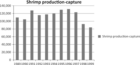
The spread of many of the shrimp viruses globally has been mainly due to the importation of live infected animals for aquaculture. The suggestion by some authors [10, 11] that the spread could occur through importation of commodity shrimp in international trade has not been scientifically validated [6]. Some examples of such global spread are discussed in this review. Thailand experienced outbreaks of yellow head disease in 1991 and the virus has been characterized as a single stranded RNA virus assigned to a new viral family, Roniviridae and genus Okavirus [27]. Six genotypes of the virus have been recognized and the Type 1 has been reported to be responsible for outbreaks in Thailand. The gill associated virus found in Australia has been reported to belong to genotype 2 [15]. A study of 58 geographical isolates of virus belonging to this complex revealed that the Type 1 that causes rapid shrimp mortalities is found only in Thailand [28]. Avirulent types of YHV have recently been reported in shrimp farms growing P. vannamei in Mexico, but sequencing of genes encoding structural proteins of these viruses indicated that YHV types in Mexico differ significantly from Type 1 genotype causing shrimp mortalities in Asia [2]. This clearly shows that though YHV carrying frozen shrimp has been in international trade for over two decades and have been imported by USA, Japan, and Australia, there has been no transmission of Type 1 YHV through this commodity. This supports the view that commodity shrimp in international trade poses negligible or no risk of transmitting shrimp pathogens. It has been argued that imported commodity shrimp may be reprocessed and effluents from such processing plants may pose a threat to natural shrimp populations in an importing country [11]. However, this has not been supported by any scientific evidence. YHV containing shrimp have been detected in US markets [11], but no infection due to genotype 1 of YHV has ever been reported in the natural populations of shrimp in the Americas. Further, though YHV has been causing mortalities in Thailand and YHV containing shrimp has been handled by processing establishments in Thailand, the natural shrimp population does not seem to have been affected as indicated by shrimp production by capture fisheries during 1989–1999 (Fig. 10). These further provide support for the argument that the risk of disease transmission through commodity shrimp in international trade is insignificant [6].
Fig. 10.
Shrimp production by capture in Thailand during 1989–1999
White spot disease was first documented in literature in 1994 from Japan [24], but there are reports of mass mortalities due to this disease in China in 1992. The disease spread very rapidly in Asia and caused serious losses to shrimp aquaculture industry in the region since the mid nineties and the cumulative loss has been estimated to be over $10 billion [14, 20]. The spread of this virus from Asia to America and Europe has been due to movement of live crustaceans or due to feeding of broodstock with shrimp carcasses from Asia in aquaculture facilities. Stentiford and Lightner [22] analyzed the cause of white spot disease in shrimp farms in Europe (Greece, Italy, Spain, and Turkey) during 1995–2001. Case notes of outbreak in Spain suggest that the disease followed feeding of wild P. japonicus broodstock with carcasses of P. monodon imported from Asia. The outbreak in Italy was associated with importation of postlarvae from Turkey and the outbreak in Greece was due to importation of postlarvae from Taiwan. WSSV is a virus with very broad host range and can be detected in crabs without any symptoms. In fact, a virus designated B2 that is morphologically similar to WSSV was described in Mediterranean crab Carcinus mediterraneus much before WSSV outbreaks were reported in Asia [13]. However, in experimental infection studies, WSSV was not able to develop in Mediterranean crab C. menas and this led Corbel and colleagues [4] to suggest that bacilliform viruses morphologically similar to WSSV found in Mediterranean crabs might be either different viruses or at least different strains.
Recent studies suggest that genetic elements with high degree of similarity to WSSV open reading frames (ORF) can be detected in shrimp [6]. A cDNA clone ED72 from Australian shrimp contained 1 kb fragment with 54 % amino acid identity and E value of 10−63 to ORF167 of WSSV. It has been suggested that WSSV-like sequences may be ancestral viral sequences integrated into shrimp genome. No outbreaks of WSSV have ever been reported from shrimp in Australia and this data suggests that WSSV-shrimp association has been very ancient and has been existing globally. The Japanese investigators have constructed a bacterial artificial chromosome library that has about 3X coverage of the 2,000 Mbp kuruma shrimp (Marsupenaeus japonicus) genome and detected seven ORFs that show homology with predicted proteins of WSSV [9]. This further suggests that Japanese shrimp also have long history of association with WSSV with the viral gene segments inserted into the host chromosome. WSSV can often be detected in wild shrimp tested in many parts of the world [3, 8, 12, 23], but this has not caused any significant decline in natural populations of these shrimp as evident from capture fisheries data from Asia since the mid nineties [6]. It is possible that aquaculture systems provided the environment in which WSSV outbreaks could be triggered. Molecular studies have confirmed that even in aquaculture systems, it is possible to find WSSV positive shrimp growing normally [25, 26] and outbreaks are related to environmental stresses [16].
Import Risk Analysis and Regulations Related to International Trade
The European Union, a major importer of shrimp adopted the EC Council Directive 2006/88/EC introducing controls for aquaculture animals and products thereof. Three shrimp diseases caused by WSSV, YHV, and TSV are listed in this regulation. WSSV is listed as a non-exotic pathogen (recognizing its reported occurrence in farms in Southern Europe), while YHV and TSV are listed as exotic. Animal health certification is required for live crustaceans for farming, for the ornamental trade and for release into the wild (restocking). Council Regulation (EC) No 1251/2008 deals with conditions and certification requirements for implementation of Council Directive 2006/88/EC and lays down a list of vector species that can carry the listed pathogens.
According to Article 2 of EC Council Directive 2006/88/EC, wild aquatic animal harvests for direct entry into food chain (e.g., crustaceans from capture fisheries) are not covered and there are no restrictions on them. However, wild crustaceans caught for use in aquaculture or if they are fed in holding facilities for short periods before sale for consumption would be covered. Article 19 deals with aquaculture animals and products thereof placed on market for human consumption without further processing. There are no restrictions on items packed in retail-sale packages that comply with provisions for packaging and labeling provided for in regulation (EC) No 853/2004. Article 18 of the EC Council Directive 2006/88/EC applies for aquaculture animals and products thereof placed on market for further processing before human consumption. In this case, for processing in a member state, zone or compartment declared free of a disease, the aquaculture animals and products should comply with one of the following conditions: (a) originate from another member state, zone, or compartment declared free of the disease in question, (b) they are processed in an authorized establishment under conditions that prevent the spreading of disease, and (c) they are dispatched as unprocessed or processed products. If the aquaculture animals or products thereof are to be temporarily stored at the place of processing, they must originate from another member state, zone, or compartment declared free of the disease in question or they are stored in centers (purification/dispatch) where effluent is subject to treatment reducing the risk of transmitting diseases to natural waters to an acceptable level.
Biosecurity Australia [1] carried out import risk analysis for prawn and prawn products. The pathogenic agents considered included WSSV, YHV, TSV and additionally, for unfrozen products, necrotising hepatopancreatitis bacterium (NHPB). The risk analysis concluded that importation of prawn and prawn products could be permitted subject to compliance with the following risk management measures: (a) sourcing all uncooked prawn product from a country or zone determined to the satisfaction of Australian government authorities to be free of WSSV, YHV, and TSV, and in addition, NHPB if the product is not frozen (i.e., the product is chilled) or (b) having the head and shell removed (the last shell segment and tail fans permitted), and each imported batch held on arrival in Australia under quarantine control, tested (polymerase chain reaction test as per OIE Manual of Diagnostic Tests for Aquatic Animals, or equivalent and a sampling regimen that would provide 95 % confidence of detecting the agent if present at 5 % prevalence) and found to be free of WSSV and YHV or (c) being highly processed that is with the head and shell removed (the last shell segment and tail fans permitted) and coated for human consumption by being breaded (crumbed) or battered or marinated or (d) being cooked in premises approved by and under the control of an appropriate Competent Authority (CA) in the exporting country to a minimum time and temperature standard where all the protein in the prawn meat is coagulated and no uncooked meat remains [1].
Biosecurity Australia [1] recommended that uncooked prawns be accompanied by health certificate issued by the CA in the exporting country, attesting that the prawns had been inspected, processed, and graded in premises approved by and under the control of the CA, were free from visible lesions associated with infectious disease and are fit for human consumption. Since use of uncooked prawn as bait was identified as a major risk factor, it was recommended that the uncooked prawns imported for human consumption that are not considered to be highly processed be marked with the words ‘for human consumption only’ and ‘not to be used as bait or feed for aquatic animals’.
The OIE Aquatic Animal Health Code [15] provides guidelines for performing import risk analysis. Regarding international trade, the OIE Aquatic Animal Heath Code provides guidance on the responsibilities of the exporting and importing countries. According to Article 5.1.3, the exporting country should on request supply the following to importing country: (a) information on the aquatic animal health situation and national aquatic animal health information systems to determine whether the country is free or has zones or compartments free from OIE listed diseases, including the regulations and procedures to maintain the free status, (b) regular and prompt information on the occurrence of listed diseases, (c) details of countries ability to apply measures to control and prevent OIE listed diseases, (d) information on the structure of the CA and the authority that they exercise, and (e) technical information, particularly on biological tests applied in all parts of the country. The CA of the exporting country is accountable for the certification used in international trade and should have official procedures for the authorization of certifying officials, defining their functions and duties, as well as conditions of oversight and accountability. The CA should ensure that relevant instructions and training are provided to certifying officials and monitor the activities of the certifying officials to verify their integrity and impartiality.
The importing country should comply with OIE standards and if the measures adopted are stricter than OIE standards, they should be based on import risk analysis. The animal health certification demanded should not include requirement for exclusion of pathogenic agents or diseases present in the importing country and not subject to official control program except when the pathogenic agent in the exporting country is of significantly higher pathogenicity and/or has a larger host range. The animal health certificate should not include pathogenic agent or diseases that are not listed by OIE unless the importing country demonstrates risk through import risk analysis carried out according to OIE Aquatic Animal Health Code.
Article 5.3.2 of this Code deals with criteria to assess safety of aquatic animals or products thereof for retail trade for human consumption from country, zone, or compartment not declared free of a disease. The criteria specify that the aquatic animal or product should be prepared and packaged for retail trade for human consumption and either only a small amount of waste is generated by the consumer or the pathogenic agent is not present in the waste generated by the consumer [15].
Conclusions
Some viral pathogenic agents such as WSSV, TSV, and YHV pose significant health risk for aquacultured crustaceans and there is evidence that WSSV and TSV have spread globally though import of live shrimp or products for aquaculture purposes. Presently, there is no confirmed evidence for transmission of disease risk through commodity shrimp in international market, though several viral agents have been detected in frozen shrimp in international trade. Viral pathogens causing mortalities in aquaculture systems have been detected in wild shrimp populations in Asia, but this has not caused any significant decline in their populations as indicated by data on shrimp production by capture fisheries in these countries around disease peaks, but there is a general decline in shrimp production by capture due to negative impact of bottom trawling operations. Some shrimp importing countries or regions (e.g., EU) have adopted regulations to protect domestic populations of crustaceans by adopting measures with respect of import of aquaculture animals. There is a need to improve awareness about the risk of spread of diseases through importation of live animals for aquaculture and this calls for strengthening capabilities in implementing quarantine, surveillance, and disease diagnosis in developing countries that are major producers of aquaculture shrimp.
References
- 1.Biosecurity Australia. Generic import risk analysis report for prawns and prawn products. Biosecurity Australia, Canberra, Australia. 2009; http://www.daff.gov.au/ba/ira/final-animal/prawns. Accessed Jan 2012.
- 2.Cedano-Thomas Y, de la Rosa-Velez J, Bonami JR, Vargas-Albores F. Gene expression kinetics of the yellow head virus in experimentally infected Litopenaeus vannamei. Aquacult Res. 2010;41:1432–1443. doi: 10.1111/j.1365-2109.2009.02434.x. [DOI] [PMC free article] [PubMed] [Google Scholar]
- 3.Chakraborty A, Otta SK, Joseph B, Sanath K, Hossain MS, Karunasagar I, Venugopal MN, Karunasagar I. Prevalence of white spot syndrome virus (WSSV) in wild crustaceans along the coast of India. Curr Sci. 2002;82:1392–1397. [Google Scholar]
- 4.Corbel V, Zuprizal, Shi Z, Huang C, Sumartono, Arcier JM, Bonami JR. Experimental infection of European crustaceans with white spot syndrome virus (WSSV) J Fish Dis. 2001;24:377–382. doi: 10.1046/j.1365-2761.2001.00302.x. [DOI] [Google Scholar]
- 5.Escabedo-Bonilla CM, Alday-Sanz V, Wille M, Sorgeloos P, Pensaert MB, Nauwynck HJ. A review on the morphology, molecular characterization, morphogenesis and pathogenesis of white spot syndrome virus. J Fish Dis. 2008;31:1–18. doi: 10.1111/j.1365-2761.2007.00877.x. [DOI] [PubMed] [Google Scholar]
- 6.Flegel TW. Review of disease transmission risks from prawn products exported for human consumption. Aquaculture. 2009;290:179–189. doi: 10.1016/j.aquaculture.2009.02.036. [DOI] [Google Scholar]
- 7.FAO. The State of the World Fisheries and Aquaculture 2010. Rome: FAO; 2010. p. 197.
- 8.Hossain MS, Chakraborty A, Joseph B, Otta SK, Karunasagar I, Karunasagar I. Detection of new host for white spot syndrome virus of shrimp using nested polymerase chain reaction. Aquaculture. 2001;198:1–11. doi: 10.1016/S0044-8486(00)00571-8. [DOI] [Google Scholar]
- 9.Koyama T, Asakawa S, Katagiri T, Shimizu A, Fagutano FF, Mavichak R, Santos MD, Fuji K, Sakamoto T, Kitakado T, Kondo H, Shimizu N, Aoki T, Hirono I. Hyper-expansion of large DNA segments in the genome of kuruma shrimp Marsupenaeus japonicus. BMC Genomics. 2010;11:141. doi: 10.1186/1471-2164-11-141. [DOI] [PMC free article] [PubMed] [Google Scholar]
- 10.Lightner DV. Virus diseases of farmed shrimp in the Western Hemisphere (the Americas): a review. J Invert Pathol. 2011;106:110–130. doi: 10.1016/j.jip.2010.09.012. [DOI] [PMC free article] [PubMed] [Google Scholar]
- 11.Lightner DV, Redman RM, Poulos BT, Nunan LM, Mari JL, Hasson KW. Risk of spread of penaeid shrimp viruses in the Americas by the international movement of live and frozen shrimp. Rev Sci Tech-Off Int Epizoot. 1997;16:146–160. doi: 10.20506/rst.16.1.1010. [DOI] [PubMed] [Google Scholar]
- 12.Lo CF, Ho CH, Peng SE, Chen CH, Hsu HC, Chiu YL, Chang CF, Liu KF, Su MS, Wang CH, Kou GH. White spot syndrome baculovirus detected in cultured and captured shrimp, crabs and other arthropods. Dis Aquat Org. 1996;27:215–225. doi: 10.3354/dao027215. [DOI] [Google Scholar]
- 13.Mari J, Bonami JR. Les infections virales du crabe Carcinus mediterraneus Czerniavski. In: Vivares CP, Bonami JR, Jasper E, editors. Pathology in marine aquaculture. Special publication 9. Bredene: European Aquaculture Society; 1986. p. 283–293.
- 14.OIE (Office International des Epizooties). Manual of diagnostic tests for aquatic animal diseases. 5th edn. Paris: Office International des Epizooties; 2009. p. 383.
- 15.OIE. Aquatic Animal Health Code 2011. http://www.oie.int/international-standard-setting/aquatic-code/access-online/. Accessed Jan 2012.
- 16.Peng SE, Lo CF, Lin SC, Chen LL, Chang YS, Liu KF, Su MS, Kou GH. Performance of WSSV infected and WSSV-negative postlarvae in culture ponds. Dis Aquat Org. 2001;46:165–172. doi: 10.3354/dao046165. [DOI] [PubMed] [Google Scholar]
- 17.Poulos BT, Tang PFJ, Pantoja CR, Bonami JB, Lightner DV. Purification and characterization of infectious myonecrosis virus of penaeid shrimp. J Gen Virol. 2006;87:987–996. doi: 10.1099/vir.0.81127-0. [DOI] [PubMed] [Google Scholar]
- 18.Rai P, Safeena MP, Karunasagar I, Karunasagar I. Complete nucleic acid sequence of Penaeus stylirostris densovirus (PstDNV) from India. Virus Res. 2011;158:37–45. doi: 10.1016/j.virusres.2011.03.005. [DOI] [PubMed] [Google Scholar]
- 19.Ramanathan N, Padmavathy P, Francis T, Athithan S. Culture of Penaeus monodon in fresh water ponds. Indian J Fish. 2004;51:303–310. [Google Scholar]
- 20.Sanchez-Martinez JG, Aguirre-Guzman G, Mejia-Ruiz H. White spot syndrome virus in cultured shrimp: a review. Aquacult Res. 2007;38:1339–1354. doi: 10.1111/j.1365-2109.2007.01827.x. [DOI] [Google Scholar]
- 21.Senapin S, Phewsaiya K, Briggs M, Flegel TW. Outbreaks of infectious myonecrosis virus (IMNV) in Indonesia confirmed by genome sequencing and use of an alternative RT-PCR detection method. Aquaculture. 2007;266:32–38. doi: 10.1016/j.aquaculture.2007.02.026. [DOI] [Google Scholar]
- 22.Stentiford GD, Lightener DV. Cases of white spot disease in European shrimp farms. Aquaculture. 2011;319:302–306. doi: 10.1016/j.aquaculture.2011.06.032. [DOI] [Google Scholar]
- 23.Stentiford GD, Bonami JR, Alday-Sanz V. A critical review of the susceptibility of crustaceans to Taura syndrome, Yellow head disease and White spot disease and implications of inclusion of these diseases in European legislation. Aquaculture. 2009;291:1–17. doi: 10.1016/j.aquaculture.2009.02.042. [DOI] [Google Scholar]
- 24.Takahashi Y, Itami T, Kondo M, Maeda M, Fujii R, Tomonaga S, Supamattaya K, Boonyaratpalin S. Electron microscopic evidence of bacilliform virus infection in kuruma shrimp (Penaeus japonicus) Fish Pathol. 1994;29:121–125. doi: 10.3147/jsfp.29.121. [DOI] [Google Scholar]
- 25.Tsai MF, Kou GH, Liu HC, Liu KF, Chang CF, Peng SE, Hsu HC, Wang CH, Lo CF. Long term presence of white spot syndrome virus (WSSV) in a cultivated shrimp population without disease outbreaks. Dis Aquat Org. 1999;38:107–114. doi: 10.3354/dao038107. [DOI] [Google Scholar]
- 26.Umesha KR, Bob Kennedy MD, Manja Naik B, Venugopal MN, Karunasagar I, Karunasagar I. High prevalence of dual and triple viral infections in black tiger shrimp ponds in India. Aquaculture. 2006;258:91–96. doi: 10.1016/j.aquaculture.2006.04.003. [DOI] [Google Scholar]
- 27.Walker PJ, Bonami JR, Boonsaeng V, Chang PS, Cowley JA, Enjuanes L, Flegel TW, Lightner DV, Loh PC, Snijder EJ, Tang K. Family Roniviridae. In: Fauqyet CM, Mayo MA, Maniloff J, Desselberger U, Ball LA, editors. Virus taxonomy. Eighth report of the International Committee on Taxonomy of Viruses. Amsterdam: Elsevier; 2005. pp. 973–977. [Google Scholar]
- 28.Wijegoonawardane PKM, Cowley JA, Phan T, Hodgson RAJ, Nielsen L, Kiatpathomachi W, Walker PJ. Genetic diversity in yellow head Nidovirus complex. Virology. 2008;380:213–225. doi: 10.1016/j.virol.2008.07.005. [DOI] [PMC free article] [PubMed] [Google Scholar]


