Abstract
A 64-year-old man with a known history of diabetes and hypertension presented to the Accident and Emergency Department with a 2-day history of sudden decreased vision in the right eye. Temporal arteritis was suspected with an elevated erythrocyte sedimentation rate (71 mm/h), and oral prednisolone was started immediately. Four days later, the patient's right eye vision deteriorated from 0.6 to 0.05, with a grade-4 relative afferent pupillary defect and ophthalmoplegia. Computed tomography showed a contrast-enhancing orbital apex mass in the right orbit abutting the medial and lateral portions of the optic nerve with extension to the posterior ethmoid and sphenoid sinuses. A transethmoidal biopsy was performed which yielded septate hyphae suggestive of Aspergillus infection. Ten days later, the patient's right eye vision further deteriorated to hand movement with total ophthalmoplegia. MRI of the orbit showed suspicion of cavernous sinus thrombosis. A combined lateral orbitotomy and transethmoidal orbital apex drainage and decompression were performed to eradicate the orbital apex abscess. Drained pus cultured Aspergillus. The patient was prescribed systemic voriconazole for a total of 22 weeks. The latest MRI scan, performed 8 months after surgery, showed residual inflammatory changes with no signs of recurrence of the disease. To our knowledge, this is the first case report which describes the use of a combined open and endoscopic approach for orbital decompression and drainage in a case of orbital aspergillosis. We believe the combined approach gives good exposure to the orbital apex, and allows the abscess in this region to be adequately drained.
Key words: Orbital aspergillosis, Lateral orbitotomy, Transethmoidal orbital apex drainage
Introduction
Orbital aspergillosis is a rare yet potentially fatal clinical entity in immunocompetent patients. The close proximity between the orbit and the middle cranial fossa provides a direct pathway for intracranial extension of the disease; the mortality rate can be over 50% [1]. It can mimic many other orbital conditions and a high index of clinical suspicion is required for early diagnosis and appropriate treatment in these patients. Management of orbital aspergillosis comprises systemic antifungal agents and surgical treatment that could range from limited debridement to orbital exenteration. We report a case of orbital aspergillosis in an immunocompetent patient with impending intracranial invasion.
Case Report
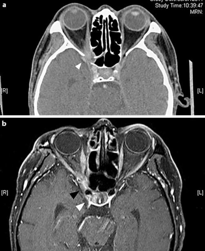
A 64-year-old man with a known history of well-controlled diabetes mellitus and hypertension presented to the Accident and Emergency Department with a 2-day history of sudden decreased vision in the right eye. Temporal arteritis was suspected with an elevated erythrocyte sedimentation rate (71 mm/h), and oral prednisolone 1 mg/kg was prescribed immediately. When the patient was seen by the ophthalmologist 4 days after the start of the oral steroid, right eye vision had deteriorated from 0.6 to 0.05. He had a concomitant grade-4 relative afferent pupillary defect and ophthalmoplegia, but there was no optic disc swelling. Computed tomography showed a contrast-enhancing orbital apex mass in the right orbit abutting the medial and lateral portions of the optic nerve with extension to the posterior ethmoid and sphenoid sinuses; the lamina papyracea was also eroded (fig. 1a). In view of the rapid deterioration of the patient's ocular condition and the unknown nature of the orbital apex mass, an endoscopic ethmoidal examination was performed which excluded ethmoidal sinusitis. Transethmoidal orbital apical biopsy yielded pus (fig. 2a). Inflamed orbital apical tissue was sent for histopathology which showed septate hyphae branching at right angles that was highly suggestive of Aspergillus. Grocott's staining highlighted the fungus (fig. 3a, b). Further deterioration of right eye vision to hand movement and total ophthalmoplegia occurred. MRI of the orbit showed the continued presence of the previously identified orbital apex mass but with suspicion of cavernous sinus thrombosis at 10 days following the endoscopic drainage and biopsy.
Fig. 1.
a Axial contrast CT scan of the orbit showing a right orbital apex mass (white arrow) abutting the right posterior ethmoid and sphenoid sinuses. Lamina papyracea was eroded. b Axial T1 contrast MRI scan (3 months after surgery) showing residual inflammatory and infective changes (black arrow) at the right orbital apex just anterior to the cavernous sinus (white arrow).
Fig. 2.
a Endoscopic view of the right orbital apex. Pus was released near the orbital apex (black arrow). 1: anterior wall of sphenoid sinus; 2: lamina papyracea; 3: pituitary fossa of the sphenoid sinus; 4: periorbita. b Endoscopic decompression of the right medial and inferior orbital walls. Medial rectus (black arrow) and intraconal fat (white arrow) exposed.
Fig. 3.
a Microscopy photos of the right orbital apex lesion, for frozen sections, showing fungal hyphae (black arrow). Grocott's staining. ×600 magnification. b Microscopic photos of the right orbital apex lesion biopsy specimen showing fungal hyphae with regular caliber and occasional septae (white arrow) and spore formation (black arrow). Grocott's staining. ×600 magnification.
To eradicate the orbital apex abscess, a combined lateral orbitotomy and transethmoidal orbital apex drainage and decompression were performed (fig. 2b). Drained pus cultured A. fumigatus. The patient was prescribed systemic voriconazole and right eye vision remained at hand movement with improving ophthalmoplegia 1 month after surgery. Systemic workup including blood tests for autoimmune markers, HIV, immunoglobulin and paraprotein, and thorax/abdominal CT and PET scans for the source of infection were all unremarkable. A follow-up MRI scan 3 months after surgery showed improvement of the inflammation at the right orbital apex (fig. 1b). Oral voriconazole was continued for a total of 22 weeks and was then stopped in view of his stable clinical condition. The latest MRI scan, performed 8 months after surgery, showed similar residual inflammatory changes. At the latest follow-up, 14 months after surgery, the patient was clinically stable, but with a 6-mm right enophthalmos. Despite the persistent poor visual acuity of his right eye with no light perception and a marked relative afferent pupillary defect, the extraocular movement continued to improve – especially for elevation and depression. There were no signs of recurrence of the disease.
Discussion
Orbital aspergillosis is frequently related to immunocompromised conditions such as intravenous drug addiction, HIV infection, malignancy and the use of systemic immunosuppressive agents [2]. The rare incidence in healthy patients can often lead to misdiagnosis and delayed treatment that could aggravate the infection or cause fatality. Our patient was first treated for arteritic ischemic optic neuropathy by the emergency physician and was started on systemic steroids, which likely worsened the condition in this case. Orbital aspergillosis can also mimic other conditions including bacterial orbital cellulitis, idiopathic orbital inflammatory syndrome and neoplasia [3]. The patient may complain of persistent retrobulbar pain and headache that preceded the ophthalmic findings of decreased vision, proptosis and ophthalmoplegia. The disease is typically unilateral, but bilateral involvement has also been reported in the literature [4].
Our patient's CT scan showed an enhancing mass surrounding the medial and lateral parts of the optic nerve at the orbital apex, with erosion of the adjacent medial orbital wall. The lesion also involved the adjacent posterior ethmoid and sphenoid sinuses. Low-density foci within the lesion representing areas of microabscesses can sometimes be seen. The lesion was isointense on T1-weighted images. On T2-weighted images, the lesion could be hypointense or with heterogeneous intensity due to associated inflammation, as in our case. Because MRI can give a detailed picture of soft tissue structures, the extent of the lesion and the involvement of adjacent structures such as cavernous sinus can be better assessed by MRI.
Biopsy of the lesion was imperative; however, the nature of the lesion (infective, inflammatory or neoplastic) was unknown. Reported cases that required more than 1 biopsy to make the tissue diagnosis are not uncommon due to the difficulty in surgical access [5, 6]. Microscopic examination of the specimen may show the organism with characteristic branching septate hyphae at 45 degrees. Special stains including Gomori methenamine silver and periodic acid-Schiff stain allow easier identification of the organism. Fine needle aspiration cytology (FNAC) is well documented as a diagnostic tool in fungal infection involving other body parts, but is not commonly performed to investigate orbital lesions. Kuruba et al. [7] presented 2 cases of fungal infection of orbital and periorbital tissue diagnosed by FNAC and suggested the role of FNAC in making an early diagnosis of orbital aspergillosis, thereby allowing early therapy in these patients.
There is no standard surgical treatment for orbital aspergillosis. Surgical treatment including limited local debridement, evisceration and exenteration has been reported with variable outcomes. The extent of surgical debridement and excision would depend on the extent of the disease, the disease progress and response to medical therapy, whereas the surgical approach used would depend on the location of the disease. After the initial endoscopic drainage for the apical collection, the patient's condition continued to deteriorate with a further decrease in visual acuity and increased ophthalmoplegia. MRI performed 10 days after the first operation showed the persistent apical abscess, which surrounded both the lateral and medial parts of the orbital apex. In view of the fact that the abscess showed a progressive lateral extension to the optic nerve, we decided on a combined endoscopic and deep lateral orbitotomy to tackle the invasive orbital apex lesion. This allowed for maximal eradication of the abscess, as a sole endoscopic drainage would have left the abscess undrained at the lateral part of the optic nerve. Residual abscess and further intracranial extension may lead to possible cavernous sinus thrombosis and fatality. In our case, a deep lateral orbitotomy was attempted, as described by Goldberg et al. [8]. An S-shaped incision extending from the lateral brow was created. The lateral canthal tendon was disinserted. The periosteum of the zygoma was exposed after dissection through the orbicularis plane. An oscillating saw was used for the lateral orbitotomy 2 cm above the frontal zygomatic suture and 2 cm below the zygomaticomaxillary junction to create a wide exposure. Dissection continued in the subperiosteal plane towards the orbital apex using a Freer elevator. Under an operating microscope, a rongeur was used to remove small portions of the greater wing of the sphenoid near the superior orbital fissure and the inferior orbital fissure in an attempt to decompress the optic canal. As no pus was readily seen, exploration of the intraconal portion of the optic nerve continued. The periorbita was excised and intraconal fat was removed. A small collection of white pus was noted upon removal of intraconal fat close to the orbital apex. The periorbita was closed and lateral bone was replaced and secured with a titanium plate. The lateral canthal tendon was sutured to the periosteum overlying Whitnall's tubercle.
To our knowledge, this is the first case report which describes the use of a combined open and endoscopic approach for orbital decompression and drainage in a case of orbital aspergillosis. In most instances, an endoscopic approach would have been adequate for orbital apical drainage. However, in this case, where the actual collection of pus was located intraconally and lateral to the optic nerve, endoscopic drainage has its limitations. During endoscopic drainage, dissection over inflamed tissue and taking care to avoid damaging the medial rectus muscle may limit access to the infected foci. We believe a combined approach may be required to allow adequate exposure to the orbital apex, and it allows the abscess in this region to be adequately drained when the abscess can be seen to extend lateral to the optic nerve and intraconally on imaging. In our patient, a combined approach limited the risk of intracranial extension of the disease and avoided the need for further extensive surgical debridement or even exenteration.
Conclusion
A high index of clinical suspicion for orbital apex aspergillosis is important for clinicians when approaching patients presenting with decreased vision and orbital signs. In the treatment of culture-proven orbital aspergillosis, the use of voriconazole as first-line medical therapy should be considered. A combined endoscopic transethmoidal and lateral orbitotomy approach may be considered in cases where the infection encases the optic nerve medially and laterally or with impending intracranial extension.
References
- 1.Choi HS, Choi JY, Yoon JS, et al. Clinical characteristics and prognosis of orbital invasive aspergillosis. Ophthal Plast Reconstr Surg. 2008;24:454–459. doi: 10.1097/IOP.0b013e31818c99ff. [DOI] [PubMed] [Google Scholar]
- 2.Levin LA, Avery R, Shore J, et al. The spectrum of orbital aspergillosis: a clinicopathological review. Surv Ophthalmol. 1996;41:142–154. doi: 10.1016/s0039-6257(96)80004-x. [DOI] [PubMed] [Google Scholar]
- 3.Yumoto E, Kitani S, Okamura H, et al. Sino-orbital aspergillosis associated with total ophthalmoplegia. Laryngoscope. 1985;95:190–192. doi: 10.1288/00005537-198502000-00013. [DOI] [PubMed] [Google Scholar]
- 4.Neelam P, Rachna M, Seema K, et al. Invasive aspergillosis of orbit in immunocompetent patients: treatment and outcome. Ophthalmology. 2011;118:1886–1891. doi: 10.1016/j.ophtha.2011.01.059. [DOI] [PubMed] [Google Scholar]
- 5.Mauriello JA, Yepez N, Mostafavi R, et al. Invasive rhinosino-orbital aspergillosis with precipitous visual loss. Can J Ophthalmol. 1995;30:124–130. [PubMed] [Google Scholar]
- 6.Heier JS, Gardner TA, Hawes MJ, et al. Proptosis as the initial presentation of fungal sinusitis in immunocompetent patients. Ophthalmology. 1995;102:713–717. doi: 10.1016/s0161-6420(95)30964-5. [DOI] [PubMed] [Google Scholar]
- 7.Kuruba SL, Prabhakaran VC, Nagarajappa AH, et al. Orbital aspergillus infection diagnosed by FNAC. Diagn Cytopathol. 2011;39:523–526. doi: 10.1002/dc.21488. [DOI] [PubMed] [Google Scholar]
- 8.Goldberg RA, Shorr N, Arnold AC, et al. Deep transorbital approach to the orbital apex and cavernous sinus. Ophthal Plast Reconstr Surg. 1998;14:336–341. doi: 10.1097/00002341-199809000-00006. [DOI] [PubMed] [Google Scholar]


