Abstract
Use of elevated electric fields and helium-rich gases has recently enabled differential IMS with resolving power up to R ~ 300. Here we applied that technique to a protein (namely, ubiquitin), achieving R up to ~80 and separating many previously unresolved conformers. While still limited by conformational multiplicity, this resolution is some four times greater than that previously reported using either conventional (drift-tube or traveling-wave) or differential IMS. The capability for fine resolution of protein conformers may open new avenues for proteoform separations in top-down and intact-protein proteomics.
Introduction
Ion mobility spectrometry (IMS), the approach to separation and characterization of ions exploiting their transport through gases driven by electric field, consists of two branches:1,2 conventional IMS based on the absolute ion mobility (K) and differential or field asymmetric waveform IMS (FAIMS) relying on the difference (Δ) between K values at high and low electric field intensity (E). Both methods, combined with mass spectrometry (MS), have been utilized to investigate proteins and other biomolecules since 1990-s.3,4 With conventional drift-tube (DT) IMS, the K quantity can be related to ion geometries by a priori calculations, which has revealed much about the protein folding and quaternary structure of protein complexes.3,5–9 That approach is still constrained by modest observed resolving powers (R < ~20) that reflect the multiplicity of geometries within each separated structural family (e.g., folded or unfolded) rather than instrumental limitations.5,7 Hence DT IMS resolution of proteins has barely improved since early days, despite major gains of instrumental R (to >200 for multiply-charged peptides).10 In a powerful capability for fundamental explorations, two-dimensional IMS platforms11,12 enable slicing broadened peaks at the end of Stage 1 to pass specific subpopulations with the width governed by instrumental R, then probing the selected conformers in Stage 2. Still, dramatic broadening of original features due to conformational variability obstructs IMS separations of primary structure isomers such as sequence inversions or localization variants with post-translational modifications (PTMs) on alternative sites. Such species have been broadly resolved for peptides (that have instrumentally-controlled peak widths) at R ~ 100,13,14 and their separation for whole proteins would greatly benefit top-down proteomics.
The use of FAIMS to elucidate macromolecular structures has been impeded by two problems: (i) the ΔK quantity could not be computed from ion geometries so far,2 and (ii) heating of ions above the gas temperature T by strong fields in FAIMS routinely distorts fragile structures, including tertiary protein folds.2,15,16 The conundrum here is that the field heating magnitude ΔT scales as E2:
| (1) |
(where M is the gas molecule mass and k is the Boltzmann constant).15,16 As MK2 is proportional to 1/Ω2 (where Ω is the ion-molecule collision cross section), ΔT by eq (1) increases upon addition of lighter gases17–19 such as He or H2 which have lower Ω values than N2 with any ion.17 Thus, as the resolving power generally scales as E3 and is improved in light gas mixtures,2,18–20 the conditions benefiting the separation inherently augment structural distortions. However, (i) FAIMS is much more orthogonal to MS than conventional IMS is21 and therefore tends to distinguish isomers better at equal formal R values and (ii) use of stronger electric fields and buffers rich in He or H2 has raised19,20,22 FAIMS resolving power for multiply-charged peptides up to ~300, matching and exceeding the metrics for conventional IMS/MS systems.10 These factors allow FAIMS to broadly separate sequence and PTM localization isomers for peptides.19,22–24 Previous FAIMS studies of proteins have also not achieved R > 20, but the analyzers involved had instrumental R of just ~10 – 30 and thus what capped the attained resolution remained undetermined.7,15,16,25–29
Here, we report first analyses of proteins using high-resolution FAIMS. With bovine ubiquitin (Ub, 8565 Da) - the prototypical model protein in MS and IMS, the resolving power reaches ~80 for higher charge states and many more features than in the previous FAIMS data are resolved. We anticipate this performance to engender new FAIMS applications to (at least smaller) intact proteins.
Experimental
In FAIMS, ions are pulled by gas flow through a gap between two electrodes while filtered in a periodic asymmetric field E(t) of some amplitude (ED) created by a waveform loaded on those electrodes.2 The net drift of any given species across the gap can be offset by a fixed “compensation field” (EC) added to E(t). Thus equilibrated ions can exit the unit and be detected, and scanning EC yields a spectrum.2 The FAIMS resolving power strongly depends on the gap geometry: planar designs with homogeneous field can provide much higher resolution than the curved devices.29
We used a planar FAIMS unit coupled to a Thermo LTQ ion trap18–20,22,24 with about the highest implemented ED = 28 kV/cm. The He/N2 gas mixtures with up to 50% He (the maximum avoiding electrical breakdown)18 were supplied at 2 L/min, leading to the standard ion residence time of 0.2 s. Ions were derived from electrospray ionization (ESI) of ~10 μM solution of Ub in 50:49:1 water/methanol/acetic acid, infused to the emitter at ~0.5 μL/min.
Results and discussion
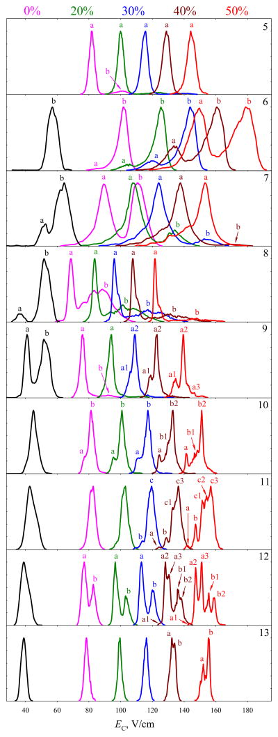
As usual with a denaturing solvent,6,15,16 ESI has generated protonated Ub ions with z = 5 – 13. The FAIMS spectra as a function of He content and benchmarks obtained7 employing a cylindrical unit at ED = 20 kV/cm with N2 are shown in Figure 1 (the spectra from cylindrical and planar units at ED = 20 kV/cm were compared previously).29 Raising ED has expectedly moved all spectra to higher EC, but changes go beyond that because of improved resolution and stronger field heating: by eq (1), the ΔT value at ED = 28 kV/cm is nearly double that at 20 kV/cm. With N2 gas, Ub5+ and Ub6+ still exhibit a single dominant peak: these low-charged species adopt stable compact shapes unaffected by moderate heating.6,15,16 For z = 7 – 9, stronger Coulomb repulsion destabilizes such geometries and unwraps them via multiple partly folded intermediates, with the conformer distributions sensitive to the ion temperature (TI = T + ΔT) and experimental timescale.6,15 In FAIMS, unfolding of peptides or proteins is normally manifested by emergence and growth of lower-EC features.15,16,26 Indeed, the present ratio of elongated a and more compact b conformers for z = 7 – 9 is much shifted toward a compared to the benchmarks,7 with b almost vanishing for Ub9+ (Figure 1). For z = 10 – 13, the protein was fully unfolded7 at lower ED and remains unfolded, but improved resolving power leads to the emergence of feature a on the low-EC side for Ub10+ and partial separation of two peaks for Ub12+. As the latter was seen (although less well) using the same planar analyzer at ED = 20 kV/cm,29 this resolution gain arises from both higher ED and the intrinsic advantage of planar geometry.29
Fig. 1.
Normalized FAIMS spectra measured for ubiquitin ions with z = 5 – 13 (top to bottom, as marked) using ED = 28 kV/cm and He/N2 mixtures with 0 – 50% He (color-coded on the top). The benchmarks obtained using a cylindrical FAIMS analyzer with ED = 20 kV/cm and N2 in Ref. [7] are in black. Selected conformers are labeled to help following the structural transitions.
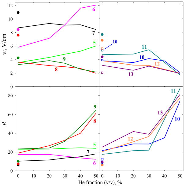
The peak widths (fwhm) for z = 5 and 8 – 13 fall in the range of w = 3.1 – 4.7 V/cm, while the much larger w = 5.8 V/cm for Ub6+ and w = 8.7 V/cm for Ub7+ indicate congestion of unresolved geometries (Figure 2). For each z, these values are smaller than those with the cylindrical FAIMS unit7 and overall compare to those with same planar device29 at lower ED = 20 kV/cm (Figure 2), which makes sense as w should not materially depend on ED (with planar gaps).30 The resolving power broadly increases over both benchmarks because of higher EC, to R = 10 – 25 from 6 – 10 for cylindrical unit7 and 8 – 20 for this planar device29 at ED = 20 kV/cm. However, the result is still within or close to the limits previously demonstrated for FAIMS or DT IMS. With planar FAIMS analyzers, the peak widths scale approximately as (Kz)−1/2: the features are narrower for species of greater mobility and/or charge.30,31 The mobilities of Ub ions with z = 6 – 13 in N2 at room temperature are15 are K ~ 0.85 – 1.3 cm2/(Vs), which is close to the values for 2+ peptides such as nine-residue bradykinin (1060 Da) with measured17 K ~ 1.3 cm2/(Vs) and 15-residue τ-phosphopeptides (1603 Da) with K ~ 1.0 cm2/(Vs) estimated from the established mass/mobility trend line.10 The features for these peptides in same FAIMS regime have18,23 w ~ 1.6 – 2.2 V/cm. When adjusted for the higher charge of Ub ions, those values would translate into w = 0.6 – 1.4 V/cm overall and 0.6 – 1.1 V/cm for z = 8 – 13. Hence the present peaks for Ub with z ≥ 8 are broader than the instrumental limits by 3 – 7 times.
Fig. 2.
Peak widths and resolving power values for the major features in ubiquitin ion spectra with z = 5 – 9 (left column) and z = 10 – 13 (right column): lines and circles for the data in Fig. 1 with planar and cylindrical FAIMS devices, respectively; squares for the data obtained (for z = 10 – 13) using a planar analyzer with ED = 20 kV/cm and N2 in Ref. [29].
As for peptides,18,22–24 raising the He fraction moves all spectra to yet higher EC while promoting unfolding (Figure 1). While Ub5+ exhibits no elongated structures at lower EC up to the maximum He content, their fractional abundances increase monotonically from ~2% to ~50% for Ub6+ and from ~50% to ~100% for Ub7+ and Ub8+. As predicted in theory and verified for peptides and other ions,18–20,22–24 addition of He raises K values and thus (with planar FAIMS devices) narrows the peak for any defined species, typically by ~20 – 30% on the way from N2 to 1:1 He/N2 as the peptide mobilities increase by ~50 – 60%. Here, the widths decrease for z = 8 – 13 by a greater 1.5 – 2.8 times (albeit not uniformly), stay flat for z = 7, and increase by 1.7 – 2.1 times for z = 5 and 6 (Figure 2). These data mean that the number and diversity of unresolved conformers expand for Ub5+ and Ub6+ that begin unfolding in the relevant TI range, but decrease for higher z as unfolded structures anneal to fewer delineated basins. As EC values increase while peaks narrow, the resolving power rapidly improves and reaches ~60 for z = 8 and 9 and ~80 for z = 10 – 13 (Figure 2). These are the highest R metrics reported for proteins using FAIMS, which allows distinguishing up to 5 conformers (seen for z = 11 and 12). Yet, the peak widths of 1.7 – 2.2 V/cm for z = 8 – 13 (at 50% He) are 2 – 3 times greater than those for above 2+ peptides18,23 (w = 1.5 – 1.8 V/cm) upon adjustment to z = 8 – 13. Thus even the narrowest features here still comprise multiple conformers, although to a lesser extent than those found using N2.
Comparisons of conformer distributions observed for z = 6 – 10 in FAIMS and 2-D FAIMS/DTIMS with those encountered in DTIMS upon thermal heating of ions6 permit gauging TI values in FAIMS.15 At ED = 20 kV/cm, thus estimated TI was15 68 – 76 °C. The conformer populations in present spectra at ED = 28 kV/cm with N2 (Figure 1) closely mirror those measured6 in DTIMS at T = 76 °C for Ub6+ (with a slight onset of unfolding in a), but T = 97 °C for Ub7+ (with equally abundant folded and unfolded families), Ub8+ (with a sharp primary peak for unfolded conformers and substantial broad feature for more compact species), and Ub9+ (with no folded geometries). The populations in FAIMS at 50% He best match those in DTIMS at T = 117 – 132 °C, with (partly) folded features competing with the elongated family for Ub6+, dropping to a significant shoulder for Ub7+, and totally vanishing for Ub8+.
Further, one may apply eq (1) to calculate15,18 time-averaged TI over the waveform cycle (<TI>) and maximum TI at the peak (Tmax). The TI values matched to DTIMS data fall between thus computed <TI> and Tmax for “compact” structures in all regimes (Table 1), supporting the argument that unfolding of those structures (and, generally, macromolecular transformations) in FAIMS is largely controlled by the maximum rather than average ion temperature over the waveform cycle.15 That TI at ED = 20 kV/cm amounts to ~75 – 90 % of Tmax has been rationalized15 by noting that Tmax applies only at the waveform peak, while isomerization requires time (especially for large molecules) and thus the ion temperature must be averaged over a finite segment around the maximum E. The shortfall grows at ED = 28 kV/cm (where TI falls to ~65 – 75% of Tmax), which is hard to explain in the same way as identical waveform profiles should lead to similar adjustment factors regardless of the amplitude. However, the real TI is determined by the instantaneous mobility of an isomerizing ion, not its initial geometry. Thus substituting the K values for compact shapes into eq (1) may be reasonable at lower temperatures where unfolding begins, but not for extensively denatured proteins at higher temperatures (Figure 1). As ion temperatures are proportional to K2, unfolding (which typically increases cross sections, thus decreasing mobility) may substantially attenuate the field heating. Indeed, the Tmax values computed for unfolded Ub geometries agree with the measurements well (Table 1).
Table 1.
Calculated and measured temperatures (°C) for ubiquitin ions in FAIMS
| Conditions | Mean TI (measured)a | For compact geometriesb | For elongated geometriesb | ||
|---|---|---|---|---|---|
| <TI> | Tmax | <TI> | Tmax | ||
| ED = 20 kV/cm, N2 | 70 | 34 – 38 | 78 – 93 | 30 – 33 | 58 – 74 |
| ED = 28 kV/cm, N2 | 92 | 45 – 48 | 129 – 142 | 38 – 44 | 93 – 123 |
| ED = 28 kV/cm, He/N2 | 125 | 54 – 58 | 167 – 185 | 43 – 52 | 118 – 159 |
Weighted averages of TI from matching DTIMS data for all charge states with multiple peaks.
The temperatures in N2 calculated assuming the mobilities measured (Ref. [15]) at peak apexes for z = 6 – 10, that is K0 = 1.02 – 1.18 cm2/(Vs) for “compact” families and 0.83 – 0.99 cm2/(Vs) for elongated ones. The mobilities in 1:1 He/N2 were estimated from the values in N2 and He via the Blanc’s law (Ref. [18]).
If the drastic peak narrowing upon He addition reflects a reduced conformational diversity upon annealing induced by stronger field heating, one may ask why no such narrowing is evident in the DT IMS data obtained6 at similar T = 117 – 132 °C. An obvious explanation is that present analyses take much longer (~200 ms) than those DT IMS separations (~10 – 20 ms),6,32 permitting a more complete annealing. Another possibility is that conformers that substantially evolve and change EC during FAIMS filtering (beyond the onset stage of ~10 ms) are removed by “self-cleaning”, leaving more stable and better-defined conformers to be observed.15 A similar mechanism supposedly eliminates isomerizing conformers in overtone mobility spectrometry (OMS) filtering, where high resolving powers are also achievable with longer analyses.33
Conclusions
In the first application of high-resolution FAIMS analyses to proteins using He/N2 gas, we have reached a resolving power up to ~80 or roughly four times the previous maximum. This allowed resolution of heretofore “co-eluting” conformers, up to five for unfolded ubiquitin ions. We expect more conformers distinguished for larger proteins that tend to occupy a wider separation space,7,15 especially those above ~30 kDa covering enormous EC ranges (presumably because of dipole alignment).28 The field heating of protein ions to the maximum temperatures of ~120 – 180 °C causes major isomerization, and, importantly, apparent annealing of unfolded geometries into fewer and/or closer deep wells on the energy surface. However, the observed peak widths are still limited by conformational multiplicity. Replacing He in He/N2 mixtures with H2 permits light gas fractions up to ~85%, which generally improves resolution for peptides (including larger ones).19,22 A similar benefit for proteins is likely.
Acknowledgments
We thank Dr. Keqi Tang and Ron Moore for experimental help and Prof. David Clemmer for insightful discussions. This work was supported by NIH NIGMS (8 P41 GM103493-10) and US DoE OBER (Panomics Project), and carried out in the Environmental Molecular Sciences Laboratory, a DoE national scientific user facility at PNNL.
References
- 1.Eiceman GA, Karpas Z. Ion Mobility Spectrometry. CRC Press; Boca Raton, FL: 2005. [Google Scholar]
- 2.Shvartsburg AA. Differential Ion Mobility Spectrometry: Nonlinear Ion Transport and Fundamentals of FAIMS. CRC Press; Boca Raton, FL: 2008. [Google Scholar]
- 3.Hoaglund-Hyzer CS, Counterman AE, Clemmer DE. Chem Rev. 1999;99:3037– 3079. doi: 10.1021/cr980139g. [DOI] [PubMed] [Google Scholar]
- 4.Purves RW, Guevremont R. Anal Chem. 1999;71:2346–2357. doi: 10.1021/ac981380y. [DOI] [PubMed] [Google Scholar]
- 5.Hudgins RR, Woenckhaus J, Jarrold MF. Int J Mass Spectrom Ion Processes. 1997;165/166:497–507. [Google Scholar]
- 6.Li J, Taraszka JA, Counterman AE, Clemmer DE. Int J Mass Spectrom. 1999;185/186/187:37–47. [Google Scholar]
- 7.Shvartsburg AA, Li F, Tang K, Smith RD. Anal Chem. 2006;78:3304–3315. doi: 10.1021/ac060283z. ibid 8575. [DOI] [PubMed] [Google Scholar]
- 8.Ruotolo BT, Giles K, Campuzano I, Sandercock AM, Bateman RH, Robinson CV. Science. 2005;310:1658–1661. doi: 10.1126/science.1120177. [DOI] [PubMed] [Google Scholar]
- 9.Uetrecht C, Rose RJ, van Duijn E, Lorenzen K, Heck AJR. Chem Soc Rev. 2010;39:1633–1655. doi: 10.1039/b914002f. [DOI] [PubMed] [Google Scholar]
- 10.Srebalus CA, Li J, Marshall WS, Clemmer DE. Anal Chem. 1999;71:3918–3927. doi: 10.1021/ac9903757. [DOI] [PubMed] [Google Scholar]
- 11.Koeniger SL, Merenbloom SI, Clemmer DE. J Phys Chem B. 2006;110:7017–7021. doi: 10.1021/jp056165h. [DOI] [PubMed] [Google Scholar]
- 12.Koeniger SL, Clemmer DE. J Am Soc Mass Spectrom. 2007;18:322–331. doi: 10.1016/j.jasms.2006.09.025. [DOI] [PubMed] [Google Scholar]
- 13.Wu C, Siems WF, Klasmeier J, Hill HH. Anal Chem. 2000;72:391–395. doi: 10.1021/ac990601c. [DOI] [PubMed] [Google Scholar]
- 14.Ibrahim Y, Shvartsburg AA, Smith RD, Belov ME. Anal Chem. 2011;83:5617–5623. doi: 10.1021/ac200719n. [DOI] [PMC free article] [PubMed] [Google Scholar]
- 15.Shvartsburg AA, Li F, Tang K, Smith RD. Anal Chem. 2007;79:1523–1528. doi: 10.1021/ac061306c. [DOI] [PubMed] [Google Scholar]
- 16.Robinson EW, Shvartsburg AA, Tang K, Smith RD. Anal Chem. 2008;80:7508–7515. doi: 10.1021/ac800655d. [DOI] [PMC free article] [PubMed] [Google Scholar]
- 17.Baker ES, Clowers BH, Li F, Tang K, Tolmachev AV, Prior DC, Belov ME, Smith RD. J Am Soc Mass Spectrom. 2007;18:1176–1187. doi: 10.1016/j.jasms.2007.03.031. [DOI] [PMC free article] [PubMed] [Google Scholar]
- 18.Shvartsburg AA, Prior DC, Tang K, Smith RD. Anal Chem. 2010;82:7649–7655. doi: 10.1021/ac101413k. [DOI] [PMC free article] [PubMed] [Google Scholar]
- 19.Shvartsburg AA, Smith RD. Anal Chem. 2011;83:9159–9166. doi: 10.1021/ac202386w. [DOI] [PMC free article] [PubMed] [Google Scholar]
- 20.Shvartsburg AA, Smith RD. Anal Chem. 2011;83:23–29. doi: 10.1021/ac202386w. [DOI] [PMC free article] [PubMed] [Google Scholar]
- 21.Shvartsburg AA, Mashkevich SV, Smith RD. J Phys Chem A. 2006;110:2663–2673. doi: 10.1021/jp055349t. [DOI] [PMC free article] [PubMed] [Google Scholar]
- 22.Shvartsburg AA, Zheng Y, Smith RD, Kelleher NL. Anal Chem. 2012;84:6317–6320. doi: 10.1021/ac301541r. [DOI] [PMC free article] [PubMed] [Google Scholar]
- 23.Shvartsburg AA, Singer D, Smith RD, Hoffmann R. Anal Chem. 2011;83:5078–5085. doi: 10.1021/ac200985s. [DOI] [PMC free article] [PubMed] [Google Scholar]
- 24.Shvartsburg AA, Creese AJ, Smith RD, Cooper HJ. Anal Chem. 2011;83:6918–6923. doi: 10.1021/ac201640d. [DOI] [PMC free article] [PubMed] [Google Scholar]
- 25.Purves RW, Barnett DA, Guevremont R. Int J Mass Spectrom. 2000;197:163–177. doi: 10.1002/1096-9888(200008)35:8<976::AID-JMS25>3.0.CO;2-Q. [DOI] [PubMed] [Google Scholar]
- 26.Purves RW, Barnett DA, Ells B, Guevremont R. J Am Soc Mass Spectrom. 2000;11:738–745. doi: 10.1016/S1044-0305(00)00136-7. [DOI] [PubMed] [Google Scholar]
- 27.Robinson EW, Williams ER. J Am Soc Mass Spectrom. 2005;16:1427–1437. doi: 10.1016/j.jasms.2005.04.007. [DOI] [PMC free article] [PubMed] [Google Scholar]
- 28.Shvartsburg AA, Bryskiewicz T, Purves RW, Tang K, Guevremont R, Smith RD. J Phys Chem B. 2006;110:21966–21980. doi: 10.1021/jp062573p. [DOI] [PubMed] [Google Scholar]
- 29.Shvartsburg AA, Li F, Tang K, Smith RD. Anal Chem. 2006;78:3706–3714. doi: 10.1021/ac052020v. [DOI] [PMC free article] [PubMed] [Google Scholar]
- 30.Shvartsburg AA, Smith RD. J Am Soc Mass Spectrom. 2007;18:1672–1681. doi: 10.1016/j.jasms.2007.06.013. [DOI] [PubMed] [Google Scholar]
- 31.Krylov EV, Nazarov EG, Miller RA. Int J Mass Spectrom. 2007;266:76–85. [Google Scholar]
- 32.Counterman AE, Valentine SJ, Srebalus CA, Henderson SC, Hoaglund CS, Clemmer DE. J Am Soc Mass Spectrom. 1998;9:743–759. doi: 10.1016/S1044-0305(98)00052-X. [DOI] [PubMed] [Google Scholar]
- 33.Lee S, Ewing MA, Nachtigall FM, Kurulugama RT, Valentine SJ, Clemmer DE. J Phys Chem B. 2010;114:12406– 12415. doi: 10.1021/jp1060123. [DOI] [PMC free article] [PubMed] [Google Scholar]


